Loading
PDBj
MenuPDBj@FacebookPDBj@TwitterPDBj@YouTubewwPDB FoundationwwPDB
RCSB PDBPDBeBMRBAdv. SearchSearch help
PDB: 82 results

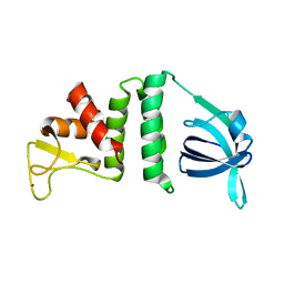
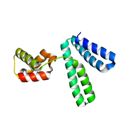
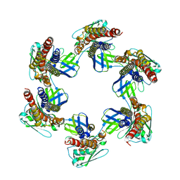
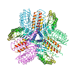
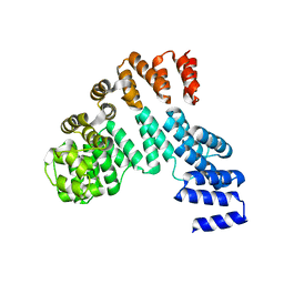
7X1K
DownloadVisualize
BU of 7x1k by Molmil
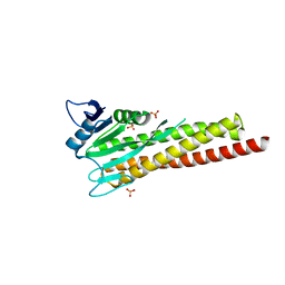
Crystal structure of the flagellar expression regulator DegU from Listeria monocytogenes
Descriptor: Chemotaxis protein CheY
Authors:Oh, H.B, Lee, S, Yoon, S.I.
Deposit date:2022-02-24
Release date:2022-08-10
Last modified:2023-11-29
Method:X-RAY DIFFRACTION (2.39 Å)
Cite:Structural and biochemical analyses of the flagellar expression regulator DegU from Listeria monocytogenes.
Sci Rep, 12, 2022
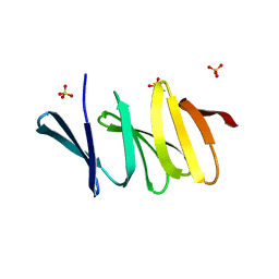
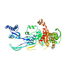
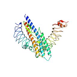
7X9R
DownloadVisualize
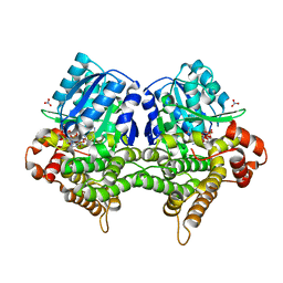
BU of 7x9r by Molmil
Crystal structure of the antirepressor GmaR
Descriptor: Glycosyl transferase family 2
Authors:Cho, S.Y, Na, H.W, Oh, H.B, Kwak, Y.M, Song, W.S, Park, S.C, Yoon, S.I.
Deposit date:2022-03-16
Release date:2022-11-09
Last modified:2024-05-29
Method:X-RAY DIFFRACTION (2.25 Å)
Cite:Structural basis of flagellar motility regulation by the MogR repressor and the GmaR antirepressor in Listeria monocytogenes.
Nucleic Acids Res., 50, 2022
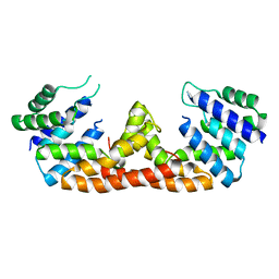
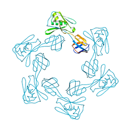
7X9S
DownloadVisualize
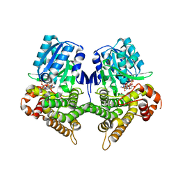
BU of 7x9s by Molmil
Crystal structure of a complex between the antirepressor GmaR and the transcriptional repressor MogR
Descriptor: GmaR, Motility gene repressor MogR
Authors:Cho, S.Y, Na, H.W, Oh, H.B, Kwak, Y.M, Song, W.S, Park, S.C, Yoon, S.I.
Deposit date:2022-03-16
Release date:2022-11-23
Last modified:2023-11-29
Method:X-RAY DIFFRACTION (3.11 Å)
Cite:Structural basis of flagellar motility regulation by the MogR repressor and the GmaR antirepressor in Listeria monocytogenes.
Nucleic Acids Res., 50, 2022
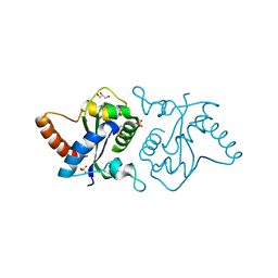
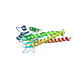
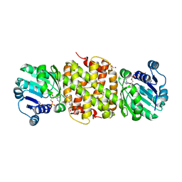
7YLF
DownloadVisualize
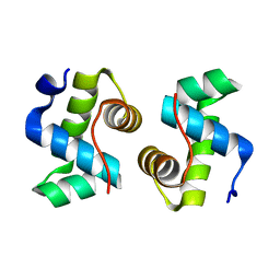
BU of 7ylf by Molmil
Crystal structure of the chicken Toll-like receptor 15 TIR domain (2-mercaptoethanol adduct)
Descriptor: GLYCEROL, SULFATE ION, Toll-like receptor 15
Authors:Ko, K.Y, Song, W.S, Yoon, S.I.
Deposit date:2022-07-26
Release date:2023-05-31
Last modified:2023-11-29
Method:X-RAY DIFFRACTION (1.9 Å)
Cite:Structural analysis of the Toll-like receptor 15 TIR domain.
Iucrj, 10, 2023
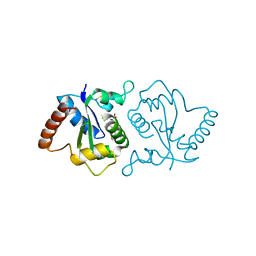
7YLG
DownloadVisualize
BU of 7ylg by Molmil
Crystal structure of the chicken Toll-like receptor 15 TIR domain (glutathione adduct)
Descriptor: GLUTATHIONE, Toll-like receptor 2
Authors:Ko, K.Y, Song, W.S, Yoon, S.I.
Deposit date:2022-07-26
Release date:2023-05-31
Last modified:2023-11-29
Method:X-RAY DIFFRACTION (1.8 Å)
Cite:Structural analysis of the Toll-like receptor 15 TIR domain.
Iucrj, 10, 2023
8H2C
DownloadVisualize
BU of 8h2c by Molmil
Crystal structure of the pseudaminic acid synthase PseI from Campylobacter jejuni
Descriptor: MANGANESE (II) ION, Pseudaminic acid synthase
Authors:Song, W.S, Park, M.A, Ki, D.U, Yoon, S.I.
Deposit date:2022-10-05
Release date:2022-11-09
Last modified:2024-04-03
Method:X-RAY DIFFRACTION (2.9 Å)
Cite:Structural analysis of the pseudaminic acid synthase PseI from Campylobacter jejuni.
Biochem.Biophys.Res.Commun., 635, 2022
7YMO
DownloadVisualize
BU of 7ymo by Molmil
Crystal structure of the recombination mediator protein RecO from Campylobacter jejuni
Descriptor: Recombination protein RecO, ZINC ION
Authors:Lee, S, Oh, H.B, Yoon, S.I.
Deposit date:2022-07-29
Release date:2022-08-31
Last modified:2024-05-29
Method:X-RAY DIFFRACTION (1.8 Å)
Cite:Crystal Structure of the Recombination Mediator Protein RecO from Campylobacter jejuni and Its Interaction with DNA and a Zinc Ion.
Int J Mol Sci, 23, 2022
7XFP
DownloadVisualize
BU of 7xfp by Molmil
Crystal structure of Helicobacter pylori IceA2
Descriptor: GLYCEROL, IceA2 protein, SULFATE ION
Authors:Cho, H.Y, Song, W.S, Yoon, S.I.
Deposit date:2022-04-02
Release date:2022-12-21
Last modified:2024-05-29
Method:X-RAY DIFFRACTION (1.87 Å)
Cite:Structural analysis of the virulence gene protein IceA2 from Helicobacter pylori.
Biochem.Biophys.Res.Commun., 612, 2022
7W1F
DownloadVisualize
BU of 7w1f by Molmil
Crystal structure of the dNTP triphosphohydrolase PA1124 from Pseudomonas aeruginosa
Descriptor: NICKEL (II) ION, Probable deoxyguanosinetriphosphate triphosphohydrolase
Authors:Oh, H.B, Song, W.S, Lee, K.C, Park, S.C, Yoon, S.I.
Deposit date:2021-11-19
Release date:2022-03-23
Last modified:2023-11-29
Method:X-RAY DIFFRACTION (2.9 Å)
Cite:Structural analysis of the dNTP triphosphohydrolase PA1124 from Pseudomonas aeruginosa.
Biochem.Biophys.Res.Commun., 589, 2022
8XJG
DownloadVisualize
BU of 8xjg by Molmil
Crystal structure of the YqeY protein from Vibrio parahaemolyticus
Descriptor: YqeY, ZINC ION
Authors:Kim, S.Y, Yoon, S.I.
Deposit date:2023-12-21
Release date:2024-05-22
Method:X-RAY DIFFRACTION (1.7 Å)
Cite:Structural analysis of the YqeY proteins from Campylobacter jejuni and Vibrio parahaemolyticus.
Biochem.Biophys.Res.Commun., 695, 2024
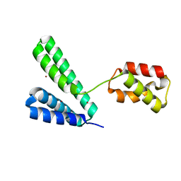
8XJE
DownloadVisualize
BU of 8xje by Molmil
Crystal structure of the YqeY protein from Campylobacter jejuni
Descriptor: YqeY
Authors:Kim, S.Y, Yoon, S.I.
Deposit date:2023-12-21
Release date:2024-05-22
Method:X-RAY DIFFRACTION (2.1 Å)
Cite:Structural analysis of the YqeY proteins from Campylobacter jejuni and Vibrio parahaemolyticus.
Biochem.Biophys.Res.Commun., 695, 2024
8YJX
DownloadVisualize
BU of 8yjx by Molmil
Crystal structure of penicillin-binding protein 2 (PBP2) from Campylobacter jejuni
Descriptor: Penicillin-binding protein 2, ZINC ION
Authors:Choi, H.J, Ki, D.W, Yoon, S.I.
Deposit date:2024-03-03
Release date:2024-04-17
Method:X-RAY DIFFRACTION (2.95 Å)
Cite:Structural and biochemical analysis of penicillin-binding protein 2 from Campylobacter jejuni.
Biochem.Biophys.Res.Commun., 710, 2024
5DQV
DownloadVisualize
BU of 5dqv by Molmil
The crystal structure of Bacillus subtilis YpgQ
Descriptor: NICKEL (II) ION, Uncharacterized protein
Authors:Jeon, Y.J, Song, W.S, Yoon, S.I.
Deposit date:2015-09-15
Release date:2016-04-27
Last modified:2024-03-20
Method:X-RAY DIFFRACTION (2 Å)
Cite:Structural and biochemical characterization of bacterial YpgQ protein reveals a metal-dependent nucleotide pyrophosphohydrolase
J.Struct.Biol., 195, 2016
8ZAK
DownloadVisualize
BU of 8zak by Molmil
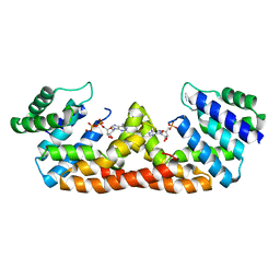
Crystal structure of the channel protein CorA from Campylobacter jejuni in complex with Ni2+
Descriptor: Magnesium transport protein CorA, NICKEL (II) ION, SULFATE ION
Authors:Ahn, S.Y, Yoon, S.I.
Deposit date:2024-04-25
Release date:2024-06-19
Method:X-RAY DIFFRACTION (2.4 Å)
Cite:Structural and biochemical analysis of the unique interactions of the Campylobacter jejuni CorA channel protein with divalent cations.
Biochem.Biophys.Res.Commun., 723, 2024
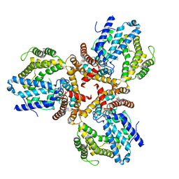
8ZAH
DownloadVisualize
BU of 8zah by Molmil
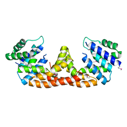
Crystal structure of the channel protein CorA from Campylobacter jejuni
Descriptor: Magnesium transport protein CorA, SULFATE ION
Authors:Ahn, S.Y, Yoon, S.I.
Deposit date:2024-04-25
Release date:2024-06-19
Method:X-RAY DIFFRACTION (2.1 Å)
Cite:Structural and biochemical analysis of the unique interactions of the Campylobacter jejuni CorA channel protein with divalent cations.
Biochem.Biophys.Res.Commun., 723, 2024
5H5T
DownloadVisualize
BU of 5h5t by Molmil
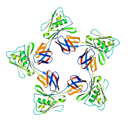
Crystal structure of the flagellar cap protein FliD D2-D3 domains from Salmonella Typhimurium
Descriptor: Flagellar hook-associated protein 2
Authors:Song, W.S, Cho, S.Y, Hong, H.J, Yoon, S.I.
Deposit date:2016-11-09
Release date:2017-02-22
Last modified:2023-11-08
Method:X-RAY DIFFRACTION (2.5 Å)
Cite:Self-Oligomerizing Structure of the Flagellar Cap Protein FliD and Its Implication in Filament Assembly.
J. Mol. Biol., 429, 2017
5DQW
DownloadVisualize
BU of 5dqw by Molmil
The crystal structure of Bacillus subtilis YpgQ in complex with ADP
Descriptor: ADENOSINE-5'-DIPHOSPHATE, NICKEL (II) ION, Uncharacterized protein
Authors:Jeon, Y.J, Song, W.S, Yoon, S.I.
Deposit date:2015-09-15
Release date:2016-04-27
Last modified:2023-11-08
Method:X-RAY DIFFRACTION (2.15 Å)
Cite:Structural and biochemical characterization of bacterial YpgQ protein reveals a metal-dependent nucleotide pyrophosphohydrolase
J.Struct.Biol., 195, 2016
5IHY
DownloadVisualize
BU of 5ihy by Molmil
The crystal structure of Bacillus subtilis SeMet-YpgQ
Descriptor: NICKEL (II) ION, Uncharacterized protein
Authors:Jeon, Y.J, Song, W.S, Yoon, S.I.
Deposit date:2016-03-01
Release date:2016-04-27
Last modified:2016-06-15
Method:X-RAY DIFFRACTION (2.5 Å)
Cite:Structural and biochemical characterization of bacterial YpgQ protein reveals a metal-dependent nucleotide pyrophosphohydrolase
J.Struct.Biol., 195, 2016
5H5V
DownloadVisualize
BU of 5h5v by Molmil
Crystal structure of the flagellar cap protein FliD D1-D2-D3 domains from Escherichia coli
Descriptor: Flagellar hook-associated protein 2
Authors:Song, W.S, Cho, S.Y, Hong, H.J, Yoon, S.I.
Deposit date:2016-11-09
Release date:2017-02-22
Last modified:2017-04-05
Method:X-RAY DIFFRACTION (3 Å)
Cite:Self-Oligomerizing Structure of the Flagellar Cap Protein FliD and Its Implication in Filament Assembly.
J. Mol. Biol., 429, 2017
5GY2
DownloadVisualize
BU of 5gy2 by Molmil
Crystal structure of a complex between Bacillus subtilis flagellin and zebrafish Toll-like receptor 5
Descriptor: 2-acetamido-2-deoxy-beta-D-glucopyranose, 2-acetamido-2-deoxy-beta-D-glucopyranose-(1-4)-2-acetamido-2-deoxy-beta-D-glucopyranose, Flagellin, ...
Authors:Song, W.S, Yoon, S.I.
Deposit date:2016-09-21
Release date:2017-02-15
Last modified:2023-11-08
Method:X-RAY DIFFRACTION (2.1 Å)
Cite:A conserved TLR5 binding and activation hot spot on flagellin
Sci Rep, 7, 2017
5H5W
DownloadVisualize
BU of 5h5w by Molmil
Crystal structure of the flagellar cap protein FliD D2-D3 domains from Escherichia coli
Descriptor: Flagellar hook-associated protein 2
Authors:Song, W.S, Cho, S.Y, Hong, H.J, Yoon, S.I.
Deposit date:2016-11-09
Release date:2017-02-22
Last modified:2023-11-08
Method:X-RAY DIFFRACTION (2.15 Å)
Cite:Self-Oligomerizing Structure of the Flagellar Cap Protein FliD and Its Implication in Filament Assembly.
J. Mol. Biol., 429, 2017
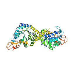
5JE8
DownloadVisualize
BU of 5je8 by Molmil
The crystal structure of Bacillus cereus 3-hydroxyisobutyrate dehydrogenase in complex with NAD
Descriptor: 3-hydroxyisobutyrate dehydrogenase, 4-(2-HYDROXYETHYL)-1-PIPERAZINE ETHANESULFONIC ACID, GLYCEROL, ...
Authors:Park, S.C, Yoon, S.I.
Deposit date:2016-04-18
Release date:2016-05-11
Last modified:2023-11-08
Method:X-RAY DIFFRACTION (2.1 Å)
Cite:Structural and biochemical characterization of the Bacillus cereus 3-hydroxyisobutyrate dehydrogenase
Biochem.Biophys.Res.Commun., 474, 2016
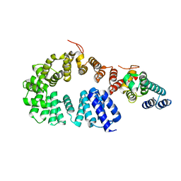
3UN9
DownloadVisualize
BU of 3un9 by Molmil
Crystal structure of an immune receptor
Descriptor: NLR family member X1, PLATINUM (II) ION
Authors:Hong, M, Yoon, S.I, Wilson, I.A.
Deposit date:2011-11-15
Release date:2012-03-28
Last modified:2024-02-28
Method:X-RAY DIFFRACTION (2.65 Å)
Cite:Structure and Functional Characterization of the RNA-Binding Element of the NLRX1 Innate Immune Modulator.
Immunity, 36, 2012
7W9Z
DownloadVisualize
BU of 7w9z by Molmil
Crystal structure of Bacillus subtilis YugJ in complex with NADP and nitrate
Descriptor: Iron-containing alcohol dehydrogenase, NADP NICOTINAMIDE-ADENINE-DINUCLEOTIDE PHOSPHATE, NITRATE ION
Authors:Cho, H.Y, Nam, M.S, Hong, H.J, Song, W.S, Yoon, S.I.
Deposit date:2021-12-11
Release date:2022-12-07
Last modified:2023-11-29
Method:X-RAY DIFFRACTION (1.65 Å)
Cite:Structural and Biochemical Analysis of the Furan Aldehyde Reductase YugJ from Bacillus subtilis.
Int J Mol Sci, 23, 2022
7W9Y
DownloadVisualize
BU of 7w9y by Molmil
Crystal structure of Bacillus subtilis YugJ in complex with NADP and nickel
Descriptor: Iron-containing alcohol dehydrogenase, NADP NICOTINAMIDE-ADENINE-DINUCLEOTIDE PHOSPHATE, NICKEL (II) ION
Authors:Cho, H.Y, Nam, M.S, Hong, H.J, Song, W.S, Yoon, S.I.
Deposit date:2021-12-11
Release date:2022-12-07
Last modified:2023-11-29
Method:X-RAY DIFFRACTION (1.93 Å)
Cite:Structural and Biochemical Analysis of the Furan Aldehyde Reductase YugJ from Bacillus subtilis.
Int J Mol Sci, 23, 2022

222926

数据于2024-07-24公开中

PDB statisticsPDBj update infoContact PDBjnumon