Loading
PDBj
MenuPDBj@FacebookPDBj@TwitterPDBj@YouTubewwPDB FoundationwwPDB
RCSB PDBPDBeBMRBAdv. SearchSearch help
PDB: 82 results

8XJE
DownloadVisualize
BU of 8xje by Molmil
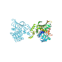
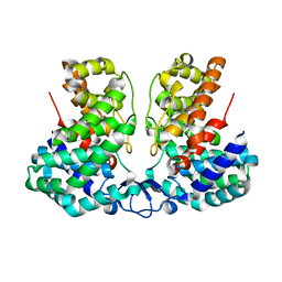
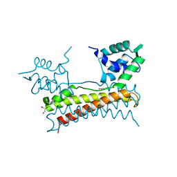
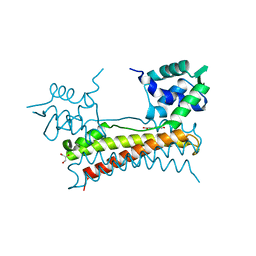
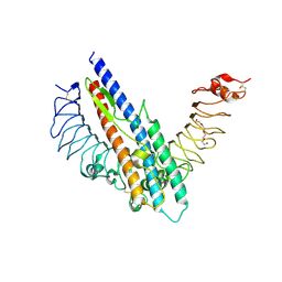
Crystal structure of the YqeY protein from Campylobacter jejuni
Descriptor: YqeY
Authors:Kim, S.Y, Yoon, S.I.
Deposit date:2023-12-21
Release date:2024-05-22
Method:X-RAY DIFFRACTION (2.1 Å)
Cite:Structural analysis of the YqeY proteins from Campylobacter jejuni and Vibrio parahaemolyticus.
Biochem.Biophys.Res.Commun., 695, 2024
8YJX
DownloadVisualize
BU of 8yjx by Molmil
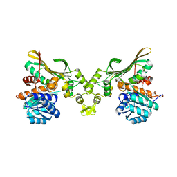
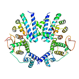
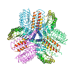
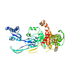
Crystal structure of penicillin-binding protein 2 (PBP2) from Campylobacter jejuni
Descriptor: Penicillin-binding protein 2, ZINC ION
Authors:Choi, H.J, Ki, D.W, Yoon, S.I.
Deposit date:2024-03-03
Release date:2024-04-17
Method:X-RAY DIFFRACTION (2.95 Å)
Cite:Structural and biochemical analysis of penicillin-binding protein 2 from Campylobacter jejuni.
Biochem.Biophys.Res.Commun., 710, 2024
8ZAH
DownloadVisualize
BU of 8zah by Molmil
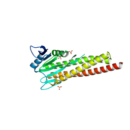
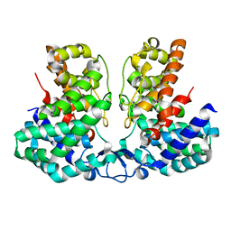
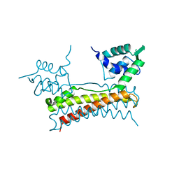
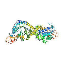
Crystal structure of the channel protein CorA from Campylobacter jejuni
Descriptor: Magnesium transport protein CorA, SULFATE ION
Authors:Ahn, S.Y, Yoon, S.I.
Deposit date:2024-04-25
Release date:2024-06-19
Method:X-RAY DIFFRACTION (2.1 Å)
Cite:Structural and biochemical analysis of the unique interactions of the Campylobacter jejuni CorA channel protein with divalent cations.
Biochem.Biophys.Res.Commun., 723, 2024
8ZAK
DownloadVisualize
BU of 8zak by Molmil
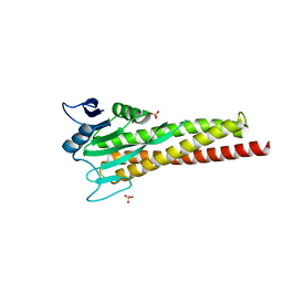
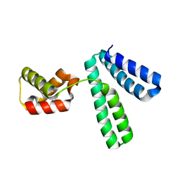
Crystal structure of the channel protein CorA from Campylobacter jejuni in complex with Ni2+
Descriptor: Magnesium transport protein CorA, NICKEL (II) ION, SULFATE ION
Authors:Ahn, S.Y, Yoon, S.I.
Deposit date:2024-04-25
Release date:2024-06-19
Method:X-RAY DIFFRACTION (2.4 Å)
Cite:Structural and biochemical analysis of the unique interactions of the Campylobacter jejuni CorA channel protein with divalent cations.
Biochem.Biophys.Res.Commun., 723, 2024
4XPK
DownloadVisualize
BU of 4xpk by Molmil
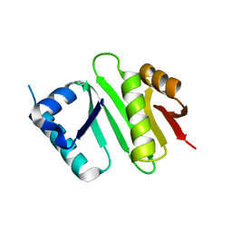
The crystal structure of Campylobacter jejuni N-acetyltransferase PseH
Descriptor: N-Acetyltransferase, PseH
Authors:Song, W.S, Nam, M.S, Namgung, B, Yoon, S.I.
Deposit date:2015-01-17
Release date:2015-03-18
Last modified:2023-11-08
Method:X-RAY DIFFRACTION (1.95 Å)
Cite:Structural analysis of PseH, the Campylobacter jejuni N-acetyltransferase involved in bacterial O-linked glycosylation.
Biochem.Biophys.Res.Commun., 458, 2015
4XPL
DownloadVisualize
BU of 4xpl by Molmil
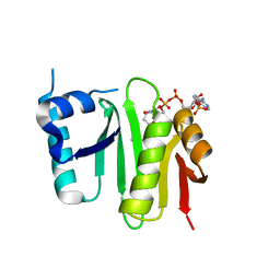
The crystal structure of Campylobacter jejuni N-acetyltransferase PseH in complex with acetyl coenzyme A
Descriptor: ACETYL COENZYME *A, N-Acetyltransferase, PseH
Authors:Song, W.S, Nam, M.S, Namgung, B, Yoon, S.I.
Deposit date:2015-01-17
Release date:2015-03-18
Last modified:2023-11-08
Method:X-RAY DIFFRACTION (1.95 Å)
Cite:Structural analysis of PseH, the Campylobacter jejuni N-acetyltransferase involved in bacterial O-linked glycosylation.
Biochem.Biophys.Res.Commun., 458, 2015
8H50
DownloadVisualize
BU of 8h50 by Molmil
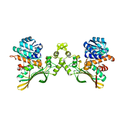
Crystal structure of carboxyspermidine dehydrogenase from Helicobacter pylori in space group C2221
Descriptor: Saccharopine dehydrogenase
Authors:Ko, K.Y, Park, S.C, Cho, S.Y, Yoon, S.I.
Deposit date:2022-10-11
Release date:2022-11-09
Last modified:2023-10-25
Method:X-RAY DIFFRACTION (2.9 Å)
Cite:Structural analysis of carboxyspermidine dehydrogenase from Helicobacter pylori.
Biochem.Biophys.Res.Commun., 635, 2022
8H52
DownloadVisualize
BU of 8h52 by Molmil
Crystal structure of Helicobacter pylori carboxyspermidine dehydrogenase in complex with NADP
Descriptor: NADP NICOTINAMIDE-ADENINE-DINUCLEOTIDE PHOSPHATE, Saccharopine dehydrogenase
Authors:Ko, K.Y, Park, S.C, Cho, S.Y, Yoon, S.I.
Deposit date:2022-10-11
Release date:2022-11-09
Last modified:2023-10-25
Method:X-RAY DIFFRACTION (3.1 Å)
Cite:Structural analysis of carboxyspermidine dehydrogenase from Helicobacter pylori.
Biochem.Biophys.Res.Commun., 635, 2022
8H4Z
DownloadVisualize
BU of 8h4z by Molmil
Crystal structure of carboxyspermidine dehydrogenase from Helicobacter pylori in space group P21212
Descriptor: Saccharopine dehydrogenase
Authors:Ko, K.Y, Park, S.C, Cho, S.Y, Yoon, S.I.
Deposit date:2022-10-11
Release date:2022-11-09
Last modified:2024-04-03
Method:X-RAY DIFFRACTION (2.2 Å)
Cite:Structural analysis of carboxyspermidine dehydrogenase from Helicobacter pylori.
Biochem.Biophys.Res.Commun., 635, 2022
8IEU
DownloadVisualize
BU of 8ieu by Molmil
Crystal structure of the DUF2891 family protein CJ0554 from Campylobacter jejuni in space group P41212
Descriptor: DUF2891 domain-containing protein
Authors:Kim, S.Y, Cho, H.Y, Yoon, S.I.
Deposit date:2023-02-16
Release date:2023-05-31
Last modified:2024-05-29
Method:X-RAY DIFFRACTION (2.3 Å)
Cite:Unique dimeric structure of the DUF2891 family protein CJ0554 from Campylobacter jejuni.
Biochem.Biophys.Res.Commun., 655, 2023
8IEV
DownloadVisualize
BU of 8iev by Molmil
Crystal structure of the DUF2891 family protein CJ0554 from Campylobacter jejuni in space group C2
Descriptor: DUF2891 domain-containing protein
Authors:Kim, S.Y, Cho, H.Y, Yoon, S.I.
Deposit date:2023-02-16
Release date:2023-05-31
Last modified:2024-05-29
Method:X-RAY DIFFRACTION (2.08 Å)
Cite:Unique dimeric structure of the DUF2891 family protein CJ0554 from Campylobacter jejuni.
Biochem.Biophys.Res.Commun., 655, 2023
8J56
DownloadVisualize
BU of 8j56 by Molmil
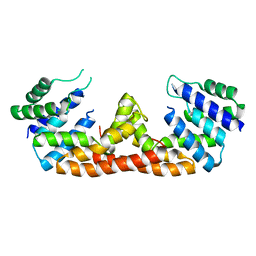
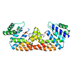
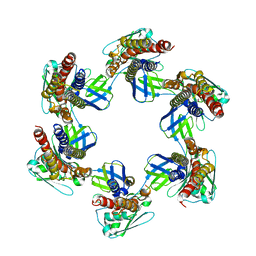
Crystal structure of the FlhDC complex from Cupriavidus necator
Descriptor: Flagellar transcriptional regulator FlhC, Flagellar transcriptional regulator FlhD, ZINC ION
Authors:Cho, S.Y, Oh, H.B, Yoon, S.I.
Deposit date:2023-04-21
Release date:2024-02-28
Method:X-RAY DIFFRACTION (3.5 Å)
Cite:Hexameric structure of the flagellar master regulator FlhDC from Cupriavidus necator and its interaction with flagellar promoter DNA.
Biochem.Biophys.Res.Commun., 672, 2023
5X12
DownloadVisualize
BU of 5x12 by Molmil
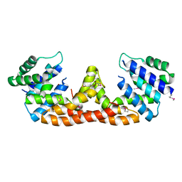
Crystal structure of Bacillus subtilis PadR
Descriptor: Transcriptional regulator
Authors:Park, S.C, Kwak, Y.M, Song, W.S, Hong, M, Yoon, S.I.
Deposit date:2017-01-24
Release date:2017-11-22
Last modified:2024-03-20
Method:X-RAY DIFFRACTION (1.7 Å)
Cite:Structural basis of effector and operator recognition by the phenolic acid-responsive transcriptional regulator PadR
Nucleic Acids Res., 45, 2017
5X11
DownloadVisualize
BU of 5x11 by Molmil
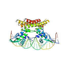
Crystal structure of Bacillus subtilis PadR in complex with operator DNA
Descriptor: DNA (28-MER), Transcriptional regulator
Authors:Park, S.C, Kwak, Y.M, Song, W.S, Hong, M, Yoon, S.I.
Deposit date:2017-01-24
Release date:2017-11-22
Last modified:2023-11-22
Method:X-RAY DIFFRACTION (2.65 Å)
Cite:Structural basis of effector and operator recognition by the phenolic acid-responsive transcriptional regulator PadR
Nucleic Acids Res., 45, 2017
5X14
DownloadVisualize
BU of 5x14 by Molmil
Crystal structure of Bacillus subtilis PadR in complex with ferulic acid
Descriptor: 3-(4-HYDROXY-3-METHOXYPHENYL)-2-PROPENOIC ACID, GLYCEROL, Transcriptional regulator
Authors:Park, S.C, Kwak, Y.M, Song, W.S, Hong, M, Yoon, S.I.
Deposit date:2017-01-24
Release date:2017-11-22
Last modified:2023-11-22
Method:X-RAY DIFFRACTION (1.68 Å)
Cite:Structural basis of effector and operator recognition by the phenolic acid-responsive transcriptional regulator PadR
Nucleic Acids Res., 45, 2017
5X13
DownloadVisualize
BU of 5x13 by Molmil
Crystal structure of Bacillus subtilis PadR in complex with p-coumaric acid
Descriptor: 4'-HYDROXYCINNAMIC ACID, GLYCEROL, Transcriptional regulator
Authors:Park, S.C, Kwak, Y.M, Song, W.S, Hong, M, Yoon, S.I.
Deposit date:2017-01-24
Release date:2017-11-22
Last modified:2023-11-22
Method:X-RAY DIFFRACTION (1.7 Å)
Cite:Structural basis of effector and operator recognition by the phenolic acid-responsive transcriptional regulator PadR
Nucleic Acids Res., 45, 2017
5DQV
DownloadVisualize
BU of 5dqv by Molmil
The crystal structure of Bacillus subtilis YpgQ
Descriptor: NICKEL (II) ION, Uncharacterized protein
Authors:Jeon, Y.J, Song, W.S, Yoon, S.I.
Deposit date:2015-09-15
Release date:2016-04-27
Last modified:2024-03-20
Method:X-RAY DIFFRACTION (2 Å)
Cite:Structural and biochemical characterization of bacterial YpgQ protein reveals a metal-dependent nucleotide pyrophosphohydrolase
J.Struct.Biol., 195, 2016
5DQW
DownloadVisualize
BU of 5dqw by Molmil
The crystal structure of Bacillus subtilis YpgQ in complex with ADP
Descriptor: ADENOSINE-5'-DIPHOSPHATE, NICKEL (II) ION, Uncharacterized protein
Authors:Jeon, Y.J, Song, W.S, Yoon, S.I.
Deposit date:2015-09-15
Release date:2016-04-27
Last modified:2023-11-08
Method:X-RAY DIFFRACTION (2.15 Å)
Cite:Structural and biochemical characterization of bacterial YpgQ protein reveals a metal-dependent nucleotide pyrophosphohydrolase
J.Struct.Biol., 195, 2016
5H5V
DownloadVisualize
BU of 5h5v by Molmil
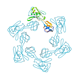
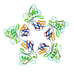
Crystal structure of the flagellar cap protein FliD D1-D2-D3 domains from Escherichia coli
Descriptor: Flagellar hook-associated protein 2
Authors:Song, W.S, Cho, S.Y, Hong, H.J, Yoon, S.I.
Deposit date:2016-11-09
Release date:2017-02-22
Last modified:2024-11-06
Method:X-RAY DIFFRACTION (3 Å)
Cite:Self-Oligomerizing Structure of the Flagellar Cap Protein FliD and Its Implication in Filament Assembly.
J. Mol. Biol., 429, 2017
5H5W
DownloadVisualize
BU of 5h5w by Molmil
Crystal structure of the flagellar cap protein FliD D2-D3 domains from Escherichia coli
Descriptor: Flagellar hook-associated protein 2
Authors:Song, W.S, Cho, S.Y, Hong, H.J, Yoon, S.I.
Deposit date:2016-11-09
Release date:2017-02-22
Last modified:2023-11-08
Method:X-RAY DIFFRACTION (2.15 Å)
Cite:Self-Oligomerizing Structure of the Flagellar Cap Protein FliD and Its Implication in Filament Assembly.
J. Mol. Biol., 429, 2017
5H5T
DownloadVisualize
BU of 5h5t by Molmil
Crystal structure of the flagellar cap protein FliD D2-D3 domains from Salmonella Typhimurium
Descriptor: Flagellar hook-associated protein 2
Authors:Song, W.S, Cho, S.Y, Hong, H.J, Yoon, S.I.
Deposit date:2016-11-09
Release date:2017-02-22
Last modified:2023-11-08
Method:X-RAY DIFFRACTION (2.5 Å)
Cite:Self-Oligomerizing Structure of the Flagellar Cap Protein FliD and Its Implication in Filament Assembly.
J. Mol. Biol., 429, 2017
5IHY
DownloadVisualize
BU of 5ihy by Molmil
The crystal structure of Bacillus subtilis SeMet-YpgQ
Descriptor: NICKEL (II) ION, Uncharacterized protein
Authors:Jeon, Y.J, Song, W.S, Yoon, S.I.
Deposit date:2016-03-01
Release date:2016-04-27
Last modified:2016-06-15
Method:X-RAY DIFFRACTION (2.5 Å)
Cite:Structural and biochemical characterization of bacterial YpgQ protein reveals a metal-dependent nucleotide pyrophosphohydrolase
J.Struct.Biol., 195, 2016
5GY2
DownloadVisualize
BU of 5gy2 by Molmil
Crystal structure of a complex between Bacillus subtilis flagellin and zebrafish Toll-like receptor 5
Descriptor: 2-acetamido-2-deoxy-beta-D-glucopyranose, 2-acetamido-2-deoxy-beta-D-glucopyranose-(1-4)-2-acetamido-2-deoxy-beta-D-glucopyranose, Flagellin, ...
Authors:Song, W.S, Yoon, S.I.
Deposit date:2016-09-21
Release date:2017-02-15
Last modified:2024-11-06
Method:X-RAY DIFFRACTION (2.1 Å)
Cite:A conserved TLR5 binding and activation hot spot on flagellin
Sci Rep, 7, 2017
3UN9
DownloadVisualize
BU of 3un9 by Molmil
Crystal structure of an immune receptor
Descriptor: NLR family member X1, PLATINUM (II) ION
Authors:Hong, M, Yoon, S.I, Wilson, I.A.
Deposit date:2011-11-15
Release date:2012-03-28
Last modified:2024-02-28
Method:X-RAY DIFFRACTION (2.65 Å)
Cite:Structure and Functional Characterization of the RNA-Binding Element of the NLRX1 Innate Immune Modulator.
Immunity, 36, 2012
8H2C
DownloadVisualize
BU of 8h2c by Molmil
Crystal structure of the pseudaminic acid synthase PseI from Campylobacter jejuni
Descriptor: MANGANESE (II) ION, Pseudaminic acid synthase
Authors:Song, W.S, Park, M.A, Ki, D.U, Yoon, S.I.
Deposit date:2022-10-05
Release date:2022-11-09
Last modified:2024-04-03
Method:X-RAY DIFFRACTION (2.9 Å)
Cite:Structural analysis of the pseudaminic acid synthase PseI from Campylobacter jejuni.
Biochem.Biophys.Res.Commun., 635, 2022

227111

건을2024-11-06부터공개중

PDB statisticsPDBj update infoContact PDBjnumon