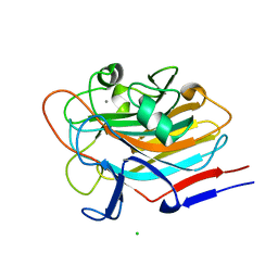
4WVE
 
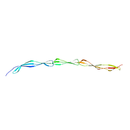 | |
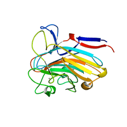
5DBL
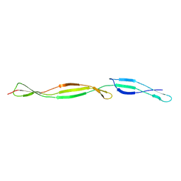 | |
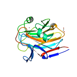
7AVH
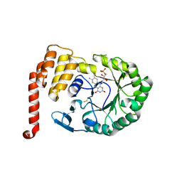
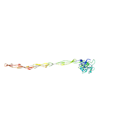
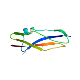
 | | Streptococcal High Identity Repeats in Tandem (SHIRT) domains 3-4 from cell surface protein SGO_0707 | | 分子名称: | 2-AMINO-2-HYDROXYMETHYL-PROPANE-1,3-DIOL, CHLORIDE ION, LPXTG cell wall surface protein | | 著者 | Whelan, F, Jenkins, H.T, Potts, J.R. | | 登録日 | 2020-11-05 | | 公開日 | 2021-06-09 | | 最終更新日 | 2024-05-01 | | 実験手法 | X-RAY DIFFRACTION (1.35 Å) | | 主引用文献 | Periscope Proteins are variable-length regulators of bacterial cell surface interactions.
Proc.Natl.Acad.Sci.USA, 118, 2021
|
|
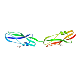
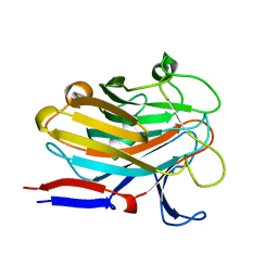
7AVJ
 | |
4XP7
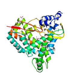
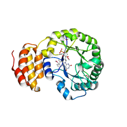
 | | Crystal structure of Human tRNA dihydrouridine synthase 2 | | 分子名称: | 1-DEOXY-1-(7,8-DIMETHYL-2,4-DIOXO-3,4-DIHYDRO-2H-BENZO[G]PTERIDIN-1-ID-10(5H)-YL)-5-O-PHOSPHONATO-D-RIBITOL, tRNA-dihydrouridine(20) synthase [NAD(P)+]-like | | 著者 | Whelan, F, Jenkins, H.T, Griffiths, S, Byrne, R.T, Dodson, E.J, Antson, A.A. | | 登録日 | 2015-01-16 | | 公開日 | 2015-01-28 | | 最終更新日 | 2024-05-01 | | 実験手法 | X-RAY DIFFRACTION (1.9 Å) | | 主引用文献 | From bacterial to human dihydrouridine synthase: automated structure determination.
Acta Crystallogr.,Sect.D, 71, 2015
|
|
6S5X
 | | Structure of RibR, the most N-terminal Rib domain from Group B Streptococcus species Streptococcus agalactiae | | 分子名称: | Group B streptococcal R4 surface protein, SODIUM ION | | 著者 | Whelan, F, Turkenburg, J.P, Griffiths, S.C, Bateman, A, Potts, J.R. | | 登録日 | 2019-07-02 | | 公開日 | 2019-12-11 | | 最終更新日 | 2024-06-19 | | 実験手法 | X-RAY DIFFRACTION (1.7 Å) | | 主引用文献 | Defining the remarkable structural malleability of a bacterial surface protein Rib domain implicated in infection.
Proc.Natl.Acad.Sci.USA, 116, 2019
|
|
6S5Y
 | |
6S5Z
 | | Structure of Rib R28N from Streptococcus pyogenes | | 分子名称: | SODIUM ION, Surface protein R28 | | 著者 | Whelan, F, Griffiths, S.C, Whittingham, J.L, Bateman, A, Potts, J.R. | | 登録日 | 2019-07-02 | | 公開日 | 2019-12-11 | | 最終更新日 | 2024-06-19 | | 実験手法 | X-RAY DIFFRACTION (1.8 Å) | | 主引用文献 | Defining the remarkable structural malleability of a bacterial surface protein Rib domain implicated in infection.
Proc.Natl.Acad.Sci.USA, 116, 2019
|
|
8DEO
 | | Structure of AAP A domain and B-repeats (residues 351-813) from Staphylococcus epidermidis | | 分子名称: | Accumulation associated protein, CALCIUM ION, CHLORIDE ION | | 著者 | Harris, G, Whelan, F, Clark, L, Turkenburg, J.P, Potts, J.R. | | 登録日 | 2022-06-21 | | 公開日 | 2023-05-03 | | 最終更新日 | 2023-10-25 | | 実験手法 | X-RAY DIFFRACTION (2.3 Å) | | 主引用文献 | Staphylococcal Periscope proteins Aap, SasG, and Pls project noncanonical legume-like lectin adhesin domains from the bacterial surface.
J.Biol.Chem., 299, 2023
|
|
8U1I
 | | Crystal structure of SyoA bound to 4-methylsyringol | | 分子名称: | 2,6-dimethoxy-4-methylphenol, Cytochrome P450, NITRATE ION, ... | | 著者 | Harlington, A.C, Shearwin, K.E, Bell, S.G, Whelan, F. | | 登録日 | 2023-09-01 | | 公開日 | 2024-07-24 | | 実験手法 | X-RAY DIFFRACTION (1.12 Å) | | 主引用文献 | Structural insights into the S-lignin O-demethylase SyoA
To Be Published
|
|
8U09
 | | Crystal structure of substrate-free SyoA | | 分子名称: | ACETATE ION, Cytochrome P450, GLYCEROL, ... | | 著者 | Harlington, A.H, Shearwin, K.E, Bell, S.G, Whelan, F. | | 登録日 | 2023-08-28 | | 公開日 | 2024-07-24 | | 実験手法 | X-RAY DIFFRACTION (1.98 Å) | | 主引用文献 | Structural insights into the S-lignin O-demethylase SyoA
To Be Published
|
|
8U19
 | | Crystal structure of SyoA bound to syringol | | 分子名称: | 2,6-dimethoxyphenol, Cytochrome P450, MAGNESIUM ION, ... | | 著者 | Harlington, A.C, Shearwin, K.E, Bell, S.G, Whelan, F. | | 登録日 | 2023-08-31 | | 公開日 | 2024-07-24 | | 実験手法 | X-RAY DIFFRACTION (1.26 Å) | | 主引用文献 | Structural insights into the S-lignin O-demethylase SyoA
To Be Published
|
|
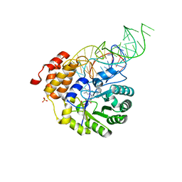
4BFA
 | | Crystal structure of E. coli dihydrouridine synthase C (DusC) | | 分子名称: | 1,2-ETHANEDIOL, FLAVIN MONONUCLEOTIDE, NITRATE ION, ... | | 著者 | Byrne, R.T, Whelan, F, Konevega, A, Aziz, N, Rodnina, M, Antson, A.A. | | 登録日 | 2013-03-16 | | 公開日 | 2013-11-06 | | 最終更新日 | 2023-12-20 | | 実験手法 | X-RAY DIFFRACTION (1.65 Å) | | 主引用文献 | Major Reorientation of tRNA Substrates Defines Specificity of Dihydrouridine Synthases.
Proc.Natl.Acad.Sci.USA, 112, 2015
|
|
4BF9
 | | Crystal structure of E. coli dihydrouridine synthase C (DusC) (selenomethionine derivative) | | 分子名称: | FLAVIN MONONUCLEOTIDE, TRNA-DIHYDROURIDINE SYNTHASE C | | 著者 | Byrne, R.T, Whelan, F, Konevega, A, Aziz, N, Rodnina, M, Antson, A.A. | | 登録日 | 2013-03-16 | | 公開日 | 2013-11-06 | | 最終更新日 | 2015-05-27 | | 実験手法 | X-RAY DIFFRACTION (2.6 Å) | | 主引用文献 | Major Reorientation of tRNA Substrates Defines Specificity of Dihydrouridine Synthases.
Proc.Natl.Acad.Sci.USA, 112, 2015
|
|
5A9K
 | | Structural basis for DNA strand separation by a hexameric replicative helicase | | 分子名称: | MAGNESIUM ION, PHOSPHATE ION, REPLICATION PROTEIN E1 | | 著者 | Chaban, Y, Stead, J.A, Ryzhenkova, K, Whelan, F, Lamber, K, Antson, F, Sanders, C.M, Orlova, E.V. | | 登録日 | 2015-07-21 | | 公開日 | 2015-08-26 | | 最終更新日 | 2024-05-08 | | 実験手法 | ELECTRON MICROSCOPY (19 Å) | | 主引用文献 | Structural Basis for DNA Strand Separation by a Hexameric Replicative Helicase.
Nucleic Acids Res., 43, 2015
|
|
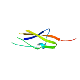
7AVK
 | | Streptococcal High Identity Repeats in Tandem (SHIRT) domain 10 from cell surface protein SGO_0707 | | 分子名称: | LPXTG cell wall surface protein, isothiocyanate | | 著者 | Degut, C, Gilburt, J, Whelan, F, Jenkins, H.T, Potts, J.R. | | 登録日 | 2020-11-05 | | 公開日 | 2021-06-09 | | 最終更新日 | 2024-05-01 | | 実験手法 | X-RAY DIFFRACTION (0.82 Å) | | 主引用文献 | Periscope Proteins are variable-length regulators of bacterial cell surface interactions.
Proc.Natl.Acad.Sci.USA, 118, 2021
|
|
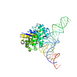
4YCP
 | | E. coli dihydrouridine synthase C (DusC) in complex with tRNATrp | | 分子名称: | FLAVIN MONONUCLEOTIDE, MAGNESIUM ION, SULFATE ION, ... | | 著者 | Byrne, R.T, Jenkins, H.T, Peters, D.T, Whelan, F, Stowell, J, Aziz, N, Kasatsky, P, Rodnina, M.V, Koonin, E.V, Konevega, A.L, Antson, A.A. | | 登録日 | 2015-02-20 | | 公開日 | 2015-04-22 | | 最終更新日 | 2024-01-10 | | 実験手法 | X-RAY DIFFRACTION (2.55 Å) | | 主引用文献 | Major reorientation of tRNA substrates defines specificity of dihydrouridine synthases.
Proc.Natl.Acad.Sci.USA, 112, 2015
|
|
4YCO
 | | E. coli dihydrouridine synthase C (DusC) in complex with tRNAPhe | | 分子名称: | FLAVIN MONONUCLEOTIDE, MAGNESIUM ION, MANGANESE (II) ION, ... | | 著者 | Byrne, R.T, Jenkins, H.T, Peters, D.T, Whelan, F, Stowell, J, Aziz, N, Kasatsky, P, Rodnina, M.V, Koonin, E.V, Konevega, A.L, Antson, A.A. | | 登録日 | 2015-02-20 | | 公開日 | 2015-04-22 | | 最終更新日 | 2024-01-10 | | 実験手法 | X-RAY DIFFRACTION (2.1 Å) | | 主引用文献 | Major reorientation of tRNA substrates defines specificity of dihydrouridine synthases.
Proc.Natl.Acad.Sci.USA, 112, 2015
|
|
7SIE
 | | Structure of AAP A-domain (residues 351-605) from Staphylococcus epidermidis | | 分子名称: | Accumulation associated protein, CALCIUM ION, CHLORIDE ION | | 著者 | Atkin, K.E, Brentnall, A.S, Dodson, E.J, Whelan, F, Clark, L, Turkenburg, J.P, Potts, J.R. | | 登録日 | 2021-10-13 | | 公開日 | 2022-10-19 | | 最終更新日 | 2024-05-22 | | 実験手法 | X-RAY DIFFRACTION (1.3 Å) | | 主引用文献 | Staphylococcal Periscope proteins Aap, SasG, and Pls project noncanonical legume-like lectin adhesin domains from the bacterial surface.
J.Biol.Chem., 299, 2023
|
|
7SJK
 | | Structure of PLS A-domain (residues 391-656) from Staphylococcus aureus | | 分子名称: | CALCIUM ION, Pls Plasmin sensitive surface protein | | 著者 | Clark, L, Whelan, F, Atkin, K.E, Brentnall, A.S, Dodson, E.J, Turkenburg, J.P, Potts, J.R. | | 登録日 | 2021-10-18 | | 公開日 | 2022-10-26 | | 最終更新日 | 2023-10-25 | | 実験手法 | X-RAY DIFFRACTION (1.208 Å) | | 主引用文献 | Staphylococcal Periscope proteins Aap, SasG, and Pls project noncanonical legume-like lectin adhesin domains from the bacterial surface.
J.Biol.Chem., 299, 2023
|
|
7SMH
 | | Structure of SASG A-domain (residues 163-419) from Staphylococcus aureus | | 分子名称: | 1,2-ETHANEDIOL, CALCIUM ION, Surface protein G | | 著者 | Atkin, K.E, Whelan, F, Brentnall, A.S, Dodson, E.J, Turkenburg, J.P, Potts, J.R. | | 登録日 | 2021-10-25 | | 公開日 | 2022-11-02 | | 最終更新日 | 2023-10-25 | | 実験手法 | X-RAY DIFFRACTION (1.65 Å) | | 主引用文献 | Staphylococcal Periscope proteins Aap, SasG, and Pls project noncanonical legume-like lectin adhesin domains from the bacterial surface.
J.Biol.Chem., 299, 2023
|
|
7SP2
 | | Structure of PLS A-domain (residues 391-656; 513-518 deletion mutant) from Staphylococcus aureus | | 分子名称: | CALCIUM ION, Plasmin Sensitive Protein Pls | | 著者 | Clark, L, Whelan, F, Atkin, K.E, Brentnall, A.S, Dodson, E.J, Turkenburg, J.P, Potts, J.R. | | 登録日 | 2021-11-02 | | 公開日 | 2022-11-09 | | 最終更新日 | 2023-10-25 | | 実験手法 | X-RAY DIFFRACTION (2.75 Å) | | 主引用文献 | Structure of PLS A-domain (residues 391-65) from Staphylococcus aureus
Not Published
|
|
6S5W
 | | Structure of Rib domain 'Rib Long' from Lactobacillus acidophilus | | 分子名称: | SODIUM ION, SULFATE ION, Surface protein | | 著者 | Griffiths, S.C, Cooper, R.E.M, Whelan, F, Whittingham, J.L, Bateman, A, Potts, J.R. | | 登録日 | 2019-07-02 | | 公開日 | 2019-12-11 | | 最終更新日 | 2024-06-19 | | 実験手法 | X-RAY DIFFRACTION (1.07 Å) | | 主引用文献 | Defining the remarkable structural malleability of a bacterial surface protein Rib domain implicated in infection.
Proc.Natl.Acad.Sci.USA, 116, 2019
|
|


