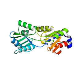
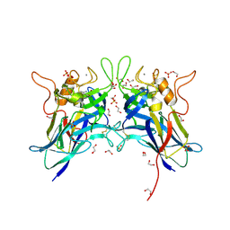
3SKY
 
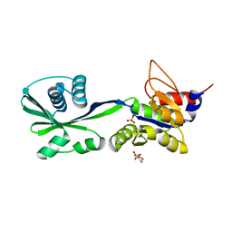 | | 2.1A crystal structure of the phosphate bound ATP binding domain of Archaeoglobus fulgidus COPB | | 分子名称: | 3[N-MORPHOLINO]PROPANE SULFONIC ACID, Copper-exporting P-type ATPase B, PHOSPHATE ION | | 著者 | Jayakanthan, S, Roberts, S.A, Weichsel, A, Arguello, J.M, McEvoy, M.M. | | 登録日 | 2011-06-23 | | 公開日 | 2012-06-20 | | 最終更新日 | 2024-02-28 | | 実験手法 | X-RAY DIFFRACTION (2.1 Å) | | 主引用文献 | Conformations of the apo-, substrate-bound and phosphate-bound ATP-binding domain of the Cu(II) ATPase CopB illustrate coupling of domain movement to the catalytic cycle.
Biosci.Rep., 32, 2012
|
|
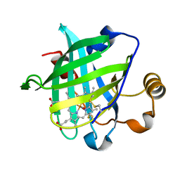
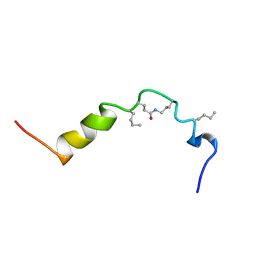
4GJ4
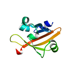 | |
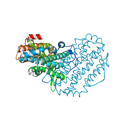
1QA1
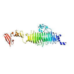 | | TAILSPIKE PROTEIN, MUTANT V331G | | 分子名称: | TAILSPIKE PROTEIN | | 著者 | Baxa, U, Steinbacher, S, Weintraub, A, Huber, R, Seckler, R. | | 登録日 | 1999-04-10 | | 公開日 | 2000-01-12 | | 最終更新日 | 2024-02-21 | | 実験手法 | X-RAY DIFFRACTION (2 Å) | | 主引用文献 | Mutations improving the folding of phage P22 tailspike protein affect its receptor binding activity.
J.Mol.Biol., 293, 1999
|
|
1QA3
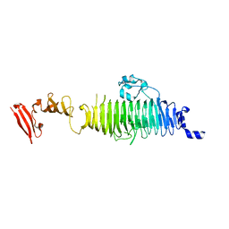 | | TAILSPIKE PROTEIN, MUTANT A334I | | 分子名称: | TAILSPIKE PROTEIN | | 著者 | Baxa, U, Steinbacher, S, Weintraub, A, Huber, R, Seckler, R. | | 登録日 | 1999-04-10 | | 公開日 | 2000-01-12 | | 最終更新日 | 2024-02-21 | | 実験手法 | X-RAY DIFFRACTION (2 Å) | | 主引用文献 | Mutations improving the folding of phage P22 tailspike protein affect its receptor binding activity.
J.Mol.Biol., 293, 1999
|
|
1CLW
 | | TAILSPIKE PROTEIN FROM PHAGE P22, V331A MUTANT | | 分子名称: | TAILSPIKE PROTEIN | | 著者 | Steinbacher, S, Baxa, U, Weintraub, A, Huber, R, Seckler, R. | | 登録日 | 1999-05-04 | | 公開日 | 1999-11-05 | | 最終更新日 | 2023-08-09 | | 実験手法 | X-RAY DIFFRACTION (2 Å) | | 主引用文献 | Mutations improving the folding of phage P22 tailspike protein affect its receptor binding activity.
J.Mol.Biol., 293, 1999
|
|
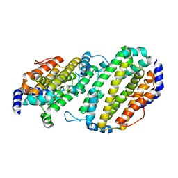
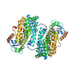
3SKX
 | | Crystal structure of the ATP binding domain of Archaeoglobus fulgidus COPB | | 分子名称: | ACETATE ION, Copper-exporting P-type ATPase B | | 著者 | Jayakanthan, S, Roberts, S.A, Weichsel, A, Arguello, J.M, McEvoy, M.M. | | 登録日 | 2011-06-23 | | 公開日 | 2012-06-20 | | 最終更新日 | 2024-02-28 | | 実験手法 | X-RAY DIFFRACTION (1.59 Å) | | 主引用文献 | Conformations of the apo-, substrate-bound and phosphate-bound ATP-binding domain of the Cu(II) ATPase CopB illustrate coupling of domain movement to the catalytic cycle.
Biosci.Rep., 32, 2012
|
|
2B7J
 | |
2B7K
 | |
2B8E
 | |
1YWC
 | | Structure of the ferrous CO complex of NP4 from Rhodnius Prolixus at pH 7.0 | | 分子名称: | CARBON MONOXIDE, PROTOPORPHYRIN IX CONTAINING FE, nitrophorin 4 | | 著者 | Maes, E.M, Weichsel, A, Roberts, S.A, Montfort, W.R. | | 登録日 | 2005-02-17 | | 公開日 | 2005-10-04 | | 最終更新日 | 2023-08-23 | | 実験手法 | X-RAY DIFFRACTION (1 Å) | | 主引用文献 | Ultrahigh Resolution Structures of Nitrophorin 4: Heme Distortion in Ferrous CO and NO Complexes
Biochemistry, 44, 2005
|
|
2ALX
 | | Ribonucleotide Reductase R2 from Escherichia coli in space group P6(1)22 | | 分子名称: | MANGANESE (II) ION, MERCURY (II) ION, Ribonucleoside-diphosphate reductase 1 | | 著者 | Sommerhalter, M, Saleh, L, Bollinger Jr, J.M, Rosenzweig, A.C. | | 登録日 | 2005-08-08 | | 公開日 | 2005-11-29 | | 最終更新日 | 2023-08-23 | | 実験手法 | X-RAY DIFFRACTION (2.6 Å) | | 主引用文献 | Structure of Escherichia coli ribonucleotide reductase R2 in space group P6122.
Acta Crystallogr.,Sect.D, 61, 2005
|
|
1QA2
 | | TAILSPIKE PROTEIN, MUTANT A334V | | 分子名称: | TAILSPIKE PROTEIN | | 著者 | Baxa, U, Steinbacher, S, Weintraub, A, Huber, R, Seckler, R. | | 登録日 | 1999-04-10 | | 公開日 | 2000-01-12 | | 最終更新日 | 2024-02-14 | | 実験手法 | X-RAY DIFFRACTION (2 Å) | | 主引用文献 | Mutations improving the folding of phage P22 tailspike protein affect its receptor binding activity.
J.Mol.Biol., 293, 1999
|
|
1SMQ
 | | Structure of the Ribonucleotide Reductase Rnr2 Homodimer from Saccharomyces cerevisiae | | 分子名称: | Ribonucleoside-diphosphate reductase small chain 1 | | 著者 | Sommerhalter, M, Voegtli, W.C, Perlstein, D.L, Ge, J, Stubbe, J, Rosenzweig, A.C. | | 登録日 | 2004-03-09 | | 公開日 | 2004-08-10 | | 最終更新日 | 2023-08-23 | | 実験手法 | X-RAY DIFFRACTION (3.1 Å) | | 主引用文献 | Structures of the yeast ribonucleotide reductase Rnr2 and Rnr4 homodimers.
Biochemistry, 43, 2004
|
|
1HTH
 | | The solution structure of cyclic human parathyroid hormone fragment 1-34, NMR, 10 structures | | 分子名称: | CYCLIC PARATHYROID HORMONE | | 著者 | Roesch, P, Seidel, G, Schaefer, W, Esswein, A, Hofmann, E. | | 登録日 | 1997-04-09 | | 公開日 | 1997-10-15 | | 最終更新日 | 2021-11-03 | | 実験手法 | SOLUTION NMR | | 主引用文献 | The structure of human parathyroid hormone-related protein(1-34) in near-physiological solution.
Febs Lett., 444, 1999
|
|
7L6G
 | | MbnP from Methylosinus trichosporium | | 分子名称: | 1,2-ETHANEDIOL, CALCIUM ION, COPPER (II) ION, ... | | 著者 | Manesis, A.C, Rosenzweig, A.C. | | 登録日 | 2020-12-23 | | 公開日 | 2021-05-26 | | 最終更新日 | 2021-07-14 | | 実験手法 | X-RAY DIFFRACTION (2.04 Å) | | 主引用文献 | Copper binding by a unique family of metalloproteins is dependent on kynurenine formation.
Proc.Natl.Acad.Sci.USA, 118, 2021
|
|
3N3B
 | | Ribonucleotide Reductase Dimanganese(II)-NrdF from Escherichia coli in Complex with Reduced NrdI with a Trapped Peroxide | | 分子名称: | FLAVIN MONONUCLEOTIDE, HYDROGEN PEROXIDE, MANGANESE (II) ION, ... | | 著者 | Boal, A.K, Cotruvo Jr, J.A, Stubbe, J, Rosenzweig, A.C. | | 登録日 | 2010-05-19 | | 公開日 | 2010-08-18 | | 最終更新日 | 2023-09-06 | | 実験手法 | X-RAY DIFFRACTION (2.36 Å) | | 主引用文献 | Structural basis for activation of class Ib ribonucleotide reductase.
Science, 329, 2010
|
|
3N37
 | | Ribonucleotide Reductase Dimanganese(II)-NrdF from Escherichia coli | | 分子名称: | GLYCEROL, MANGANESE (II) ION, Ribonucleoside-diphosphate reductase 2 subunit beta | | 著者 | Boal, A.K, Cotruvo Jr, J.A, Stubbe, J, Rosenzweig, A.C. | | 登録日 | 2010-05-19 | | 公開日 | 2010-08-18 | | 最終更新日 | 2023-09-06 | | 実験手法 | X-RAY DIFFRACTION (1.65 Å) | | 主引用文献 | Structural basis for activation of class Ib ribonucleotide reductase.
Science, 329, 2010
|
|
3N38
 | | Ribonucleotide Reductase NrdF from Escherichia coli Soaked with Ferrous Ions | | 分子名称: | FE (II) ION, Ribonucleoside-diphosphate reductase 2 subunit beta | | 著者 | Boal, A.K, Cotruvo Jr, J.A, Stubbe, J, Rosenzweig, A.C. | | 登録日 | 2010-05-19 | | 公開日 | 2010-08-18 | | 最終更新日 | 2023-09-06 | | 実験手法 | X-RAY DIFFRACTION (1.9 Å) | | 主引用文献 | Structural basis for activation of class Ib ribonucleotide reductase.
Science, 329, 2010
|
|
3N3A
 | | Ribonucleotide Reductase Dimanganese(II)-NrdF from Escherichia coli in Complex with Reduced NrdI | | 分子名称: | FLAVIN MONONUCLEOTIDE, MANGANESE (II) ION, Protein nrdI, ... | | 著者 | Boal, A.K, Cotruvo Jr, J.A, Stubbe, J, Rosenzweig, A.C. | | 登録日 | 2010-05-19 | | 公開日 | 2010-08-18 | | 最終更新日 | 2023-09-06 | | 実験手法 | X-RAY DIFFRACTION (1.99 Å) | | 主引用文献 | Structural basis for activation of class Ib ribonucleotide reductase.
Science, 329, 2010
|
|
3N39
 | | Ribonucleotide Reductase Dimanganese(II)-NrdF from Escherichia coli in Complex with NrdI | | 分子名称: | FLAVIN MONONUCLEOTIDE, MANGANESE (II) ION, Protein nrdI, ... | | 著者 | Boal, A.K, Cotruvo Jr, J.A, Stubbe, J, Rosenzweig, A.C. | | 登録日 | 2010-05-19 | | 公開日 | 2010-08-18 | | 最終更新日 | 2023-09-06 | | 実験手法 | X-RAY DIFFRACTION (2.5 Å) | | 主引用文献 | Structural basis for activation of class Ib ribonucleotide reductase.
Science, 329, 2010
|
|
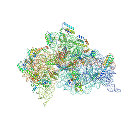
2VQF
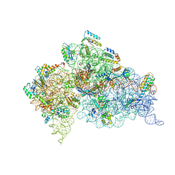 | | Modified uridines with C5-methylene substituents at the first position of the tRNA anticodon stabilize U-G wobble pairing during decoding | | 分子名称: | 16S RRNA, 30S RIBOSOMAL PROTEIN S10, 30S RIBOSOMAL PROTEIN S11, ... | | 著者 | Kurata, S, Weixlbaumer, A, Ohtsuki, T, Shimazaki, T, Wada, T, Kirino, Y, Takai, K, Watanabe, K, Ramakrishnan, V, Suzuki, T. | | 登録日 | 2008-03-14 | | 公開日 | 2008-04-29 | | 最終更新日 | 2023-12-13 | | 実験手法 | X-RAY DIFFRACTION (2.9 Å) | | 主引用文献 | Modified Uridines with C5-Methylene Substituents at the First Position of the tRNA Anticodon Stabilize U.G Wobble Pairing During Decoding.
J.Biol.Chem., 283, 2008
|
|
2VQE
 | | Modified uridines with C5-methylene substituents at the first position of the tRNA anticodon stabilize U-G wobble pairing during decoding | | 分子名称: | 16S RRNA, 30S RIBOSOMAL PROTEIN S10, 30S RIBOSOMAL PROTEIN S11, ... | | 著者 | Kurata, S, Weixlbaumer, A, Ohtsuki, T, Shimazaki, T, Wada, T, Kirino, Y, Takai, K, Watanabe, K, Ramakrishnan, V, Suzuki, T. | | 登録日 | 2008-03-13 | | 公開日 | 2008-04-29 | | 最終更新日 | 2023-12-13 | | 実験手法 | X-RAY DIFFRACTION (2.5 Å) | | 主引用文献 | Modified Uridines with C5-Methylene Substituents at the First Position of the tRNA Anticodon Stabilize U.G Wobble Pairing During Decoding.
J.Biol.Chem., 283, 2008
|
|
3FGC
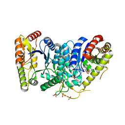 | | Crystal Structure of the Bacterial Luciferase:Flavin Complex Reveals the Basis of Intersubunit Communication | | 分子名称: | Alkanal monooxygenase alpha chain, Alkanal monooxygenase beta chain, FLAVIN MONONUCLEOTIDE, ... | | 著者 | Campbell, Z.T, Weichsel, A, Montfort, W.R, Baldwin, T.O. | | 登録日 | 2008-12-05 | | 公開日 | 2009-05-26 | | 最終更新日 | 2023-09-06 | | 実験手法 | X-RAY DIFFRACTION (2.3 Å) | | 主引用文献 | Crystal structure of the bacterial luciferase/flavin complex provides insight into the function of the beta subunit.
Biochemistry, 48, 2009
|
|
3FDC
 | | Crystal Structure of Avidin | | 分子名称: | Avidin, iron(II) tetracyano-5-(2-Oxo-hexahydro-thieno[3,4-d]imidazol-6-yl)-pentanoic acid (4'-methyl-[2,2']bipyridinyl-4-ylmethyl)-amide | | 著者 | Barker, K.D, Sazinsky, M.H, Eckermann, A.L, Abajian, C, Hartings, M.R, Rosenzweig, A.C, Meade, T.J. | | 登録日 | 2008-11-25 | | 公開日 | 2009-12-01 | | 最終更新日 | 2023-09-06 | | 実験手法 | X-RAY DIFFRACTION (3.1 Å) | | 主引用文献 | Protein Binding and the Electronic Properties of Iron(II) Complexes: An Electrochemical and Optical Investigation of Outer Sphere Effects.
Bioconjug.Chem., 20, 2009
|
|
3FLL
 | | Crystal structure of E55Q mutant of nitrophorin 4 | | 分子名称: | AMMONIA, Nitrophorin-4, PROTOPORPHYRIN IX CONTAINING FE | | 著者 | Montfort, W.R, Weichsel, A. | | 登録日 | 2008-12-18 | | 公開日 | 2009-02-10 | | 最終更新日 | 2023-09-06 | | 実験手法 | X-RAY DIFFRACTION (1.5 Å) | | 主引用文献 | Effect of mutation of carboxyl side-chain amino acids near the heme on the midpoint potentials and ligand binding constants of nitrophorin 2 and its NO, histamine, and imidazole complexes.
J.Am.Chem.Soc., 131, 2009
|
|


