Loading
PDBj
MenuPDBj@FacebookPDBj@TwitterPDBj@YouTubewwPDB FoundationwwPDB
RCSB PDBPDBeBMRBAdv. SearchSearch help
PDB: 5 results

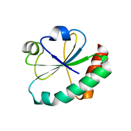
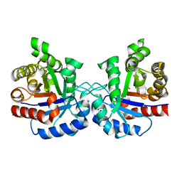
4IOT
DownloadVisualize
BU of 4iot by Molmil
High-resolution Structure of Triosephosphate isomerase from E. coli
Descriptor: SULFATE ION, Triosephosphate isomerase
Authors:Vinaik, R, Kozlov, G, Gehring, K, Montreal-Kingston Bacterial Structural Genomics Initiative (BSGI)
Deposit date:2013-01-08
Release date:2013-01-23
Last modified:2023-09-20
Method:X-RAY DIFFRACTION (1.85 Å)
Cite:Triosephosphate isomerase is a common crystallization contaminant of soluble His-tagged proteins produced in Escherichia coli.
Acta Crystallogr.,Sect.F, 69, 2013
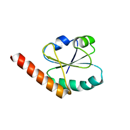
4GWR
DownloadVisualize
BU of 4gwr by Molmil
Crystal Structure of the second catalytic domain of protein disulfide isomerase P5
Descriptor: Protein disulfide-isomerase A6
Authors:Vinaik, R, Kozlov, G, Gehring, K.
Deposit date:2012-09-03
Release date:2013-09-04
Last modified:2023-11-08
Method:X-RAY DIFFRACTION (1.81 Å)
Cite:To be published
To be Published, 2013
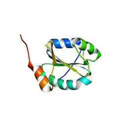
4EF0
DownloadVisualize
BU of 4ef0 by Molmil
Crystal Structure of the first catalytic domain of protein disulfide isomerase P5
Descriptor: Protein disulfide-isomerase A6
Authors:Vinaik, R, Kozlov, G, Gehring, K.
Deposit date:2012-03-28
Release date:2013-04-03
Last modified:2023-09-13
Method:X-RAY DIFFRACTION (1.5 Å)
Cite:Crystal Structure of the first catalytic domain of protein disulfide isomerase P5
To be Published
4I6X
DownloadVisualize
BU of 4i6x by Molmil
Crystal Structure of Non-catalyic Domain of Protein Disulfide Isomerase-related (PDIr) Protein
Descriptor: Protein disulfide-isomerase A5
Authors:Kozlov, G, Vinaik, R, Gehring, K.
Deposit date:2012-11-30
Release date:2013-04-03
Last modified:2024-11-06
Method:X-RAY DIFFRACTION (1.5 Å)
Cite:Structure of the Non-Catalytic Domain of the Protein Disulfide Isomerase-Related Protein (PDIR) Reveals Function in Protein Binding.
Plos One, 8, 2013
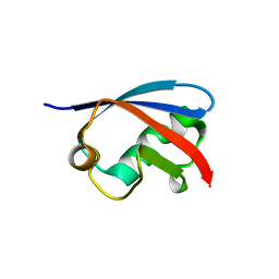
4EEW
DownloadVisualize
BU of 4eew by Molmil
Crystal structure of the Ubl domain of BAG6
Descriptor: Large proline-rich protein BAG6
Authors:Kozlov, G, LePage, K, Vinaik, R, Gehring, K.
Deposit date:2012-03-28
Release date:2012-04-11
Last modified:2023-09-13
Method:X-RAY DIFFRACTION (1.3 Å)
Cite:Crystal structure of the Ubl domain of BAG6
To be Published

227111

数据于2024-11-06公开中

PDB statisticsPDBj update infoContact PDBjnumon