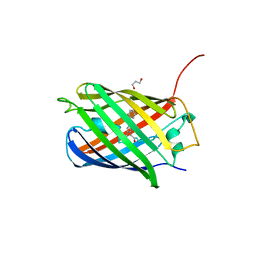
3FVE
 
 | | Crystal structure of diaminopimelate epimerase Mycobacterium tuberculosis DapF | | 分子名称: | 2,3-DIHYDROXY-1,4-DITHIOBUTANE, Diaminopimelate epimerase, GLYCEROL | | 著者 | Usha, V, Dover, L.G, Roper, D.I, Futterer, K, Besra, G.S. | | 登録日 | 2009-01-15 | | 公開日 | 2009-01-27 | | 最終更新日 | 2023-09-06 | | 実験手法 | X-RAY DIFFRACTION (2.6 Å) | | 主引用文献 | Structure of the diaminopimelate epimerase DapF from Mycobacterium tuberculosis
Acta Crystallogr.,Sect.D, 65, 2009
|
|
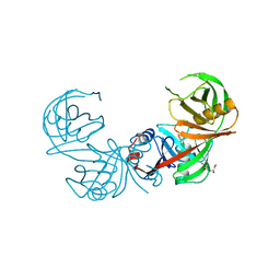
7RBW
 | | Structure of Biliverdin-binding Serpin of Boana punctata (polka-dot tree frog) | | 分子名称: | BILIVERDINE IX ALPHA, Biliverdin bindin serpin | | 著者 | Fedorov, E, Manoilov, K.Y, Verkhusha, V, Almo, S.C, Ghosh, A. | | 登録日 | 2021-07-06 | | 公開日 | 2021-11-24 | | 最終更新日 | 2023-10-18 | | 実験手法 | X-RAY DIFFRACTION (2.05 Å) | | 主引用文献 | Structural and Functional Characterization of a Biliverdin-Binding Near-Infrared Fluorescent Protein From the Serpin Superfamily.
J.Mol.Biol., 434, 2021
|
|
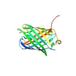
3KCT
 | | CRYSTAL STRUCTURE OF PAmCherry1 in the photoactivated state | | 分子名称: | PAmCherry1 protein | | 著者 | Malashkevich, V.N, Subach, F.V, Zencheck, W.D, Xiao, H, Filonov, G.S, Almo, S.C, Verkhusha, V.V. | | 登録日 | 2009-10-21 | | 公開日 | 2009-11-17 | | 最終更新日 | 2018-01-24 | | 実験手法 | X-RAY DIFFRACTION (1.65 Å) | | 主引用文献 | Photoactivation mechanism of PAmCherry based on crystal structures of the protein in the dark and fluorescent states.
Proc.Natl.Acad.Sci.USA, 106, 2009
|
|
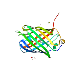
3KCS
 | | Crystal structure of PAmCherry1 in the dark state | | 分子名称: | PAmCherry1 protein | | 著者 | Malashkevich, V.N, Subach, F.V, Zencheck, W.D, Xiao, H, Filonov, G.S, Almo, S.C, Verkhusha, V.V. | | 登録日 | 2009-10-21 | | 公開日 | 2009-11-17 | | 最終更新日 | 2018-01-24 | | 実験手法 | X-RAY DIFFRACTION (1.5 Å) | | 主引用文献 | Photoactivation mechanism of PAmCherry based on crystal structures of the protein in the dark and fluorescent states.
Proc.Natl.Acad.Sci.USA, 106, 2009
|
|
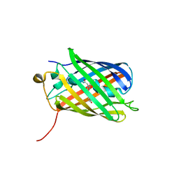
3M22
 | | Crystal structure of TagRFP fluorescent protein | | 分子名称: | TagRFP | | 著者 | Malashkevich, V.N, Subach, O.M, Ramagopal, U.A, Almo, S.C, Verkhusha, V.V. | | 登録日 | 2010-03-06 | | 公開日 | 2010-05-12 | | 最終更新日 | 2017-11-08 | | 実験手法 | X-RAY DIFFRACTION (2.2 Å) | | 主引用文献 | Structural characterization of acylimine-containing blue and red chromophores in mTagBFP and TagRFP fluorescent proteins.
Chem.Biol., 17, 2010
|
|
3M24
 | | Crystal structure of TagBFP fluorescent protein | | 分子名称: | 2,3-DIHYDROXY-1,4-DITHIOBUTANE, CHLORIDE ION, GLYCEROL, ... | | 著者 | Malashkevich, V.N, Subach, O.M, Almo, S.C, Verkhusha, V.V. | | 登録日 | 2010-03-06 | | 公開日 | 2010-05-26 | | 最終更新日 | 2023-11-15 | | 実験手法 | X-RAY DIFFRACTION (2.2 Å) | | 主引用文献 | Structural characterization of acylimine-containing blue and red chromophores in mTagBFP and TagRFP fluorescent proteins.
Chem.Biol., 17, 2010
|
|
3NT9
 | |
3NT3
 | | CRYSTAL STRUCTURE OF LSSmKate2 red fluorescent proteins with large Stokes shift | | 分子名称: | GLYCEROL, LSSmKate2 red fluorescent protein | | 著者 | Malashkevich, V.N, Piatkevich, K, Almo, S.C, Verkhusha, V. | | 登録日 | 2010-07-02 | | 公開日 | 2010-08-18 | | 最終更新日 | 2023-11-22 | | 実験手法 | X-RAY DIFFRACTION (1.5 Å) | | 主引用文献 | Engineering ESPT Pathways Based on Structural Analysis of LSSmKate Red Fluorescent Proteins with Large Stokes Shift.
J.Am.Chem.Soc., 132, 2010
|
|
4KGF
 | | Crystal structure of near-infrared fluorescent protein with an extended stokes shift, ph 8.0 | | 分子名称: | CHLORIDE ION, TagRFP675, red fluorescent protein | | 著者 | Malashkevich, V.N, Piatkevich, K, Almo, S.C, Verkhusha, V, New York Structural Genomics Research Consortium (NYSGRC) | | 登録日 | 2013-04-29 | | 公開日 | 2013-05-08 | | 最終更新日 | 2023-12-06 | | 実験手法 | X-RAY DIFFRACTION (2.3 Å) | | 主引用文献 | Extended Stokes shift in fluorescent proteins: chromophore-protein interactions in a near-infrared TagRFP675 variant.
Sci Rep, 3, 2013
|
|
4KGE
 | | Crystal structure of near-infrared fluorescent protein with an extended stokes shift, pH 4.5 | | 分子名称: | CHLORIDE ION, TagRFP675, red fluorescent protein | | 著者 | Malashkevich, V.N, Piatkevich, K, Almo, S.C, Verkhusha, V, New York Structural Genomics Research Consortium (NYSGRC) | | 登録日 | 2013-04-29 | | 公開日 | 2013-05-08 | | 最終更新日 | 2023-12-06 | | 実験手法 | X-RAY DIFFRACTION (2.3 Å) | | 主引用文献 | Extended Stokes shift in fluorescent proteins: chromophore-protein interactions in a near-infrared TagRFP675 variant.
Sci Rep, 3, 2013
|
|
3H1O
 | | The Structure of Fluorescent Protein FP480 | | 分子名称: | Fluorescent protein FP480, GLYCEROL | | 著者 | Pletnev, S, Morozova, K.S, Verkhusha, V.V, Dauter, Z. | | 登録日 | 2009-04-13 | | 公開日 | 2009-09-08 | | 最終更新日 | 2017-11-01 | | 実験手法 | X-RAY DIFFRACTION (2 Å) | | 主引用文献 | Rotational order-disorder structure of fluorescent protein FP480
Acta Crystallogr.,Sect.D, 65, 2009
|
|
3H1R
 | | Order-disorder structure of fluorescent protein FP480 | | 分子名称: | Fluorescent protein FP480 | | 著者 | Pletnev, S, Morozova, K.S, Verkhusha, V.V, Dauter, Z. | | 登録日 | 2009-04-13 | | 公開日 | 2009-09-08 | | 最終更新日 | 2017-11-01 | | 実験手法 | X-RAY DIFFRACTION (2.41 Å) | | 主引用文献 | Rotational order-disorder structure of fluorescent protein FP480
Acta Crystallogr.,Sect.D, 65, 2009
|
|
4NWS
 | |


