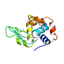
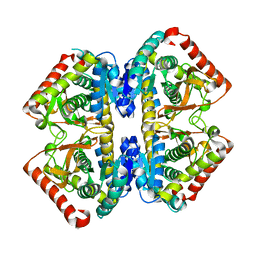
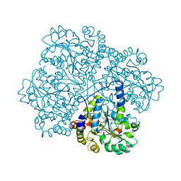
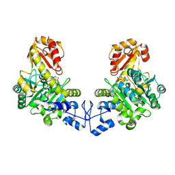
3WQB
 
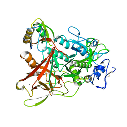 | | Crystal structure of aeromonas sobria serine protease (ASP) and the chaperone (ORF2) complex | | 分子名称: | CALCIUM ION, Extracellular serine protease, Open reading frame 2 | | 著者 | Kobayashi, H, Yoshida, T, Miyakawa, T, Kato, R, Tashiro, M, Yamanaka, H, Tanokura, M, Tsuge, H. | | 登録日 | 2014-01-24 | | 公開日 | 2015-03-25 | | 最終更新日 | 2023-11-08 | | 実験手法 | X-RAY DIFFRACTION (1.41 Å) | | 主引用文献 | Structural Basis for Action of the External Chaperone for a Propeptide-deficient Serine Protease from Aeromonas sobria.
J.Biol.Chem., 290, 2015
|
|
3WI0
 | |
3WI1
 | |
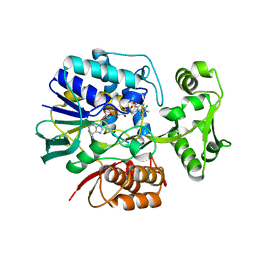
5ZJ4
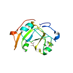 | | Guanine-specific ADP-ribosyltransferase | | 分子名称: | (4S)-2-METHYL-2,4-PENTANEDIOL, ADP-ribosyltransferase | | 著者 | Yoshida, T, Tsuge, H. | | 登録日 | 2018-03-19 | | 公開日 | 2018-08-08 | | 最終更新日 | 2023-11-22 | | 実験手法 | X-RAY DIFFRACTION (1.49739277 Å) | | 主引用文献 | Substrate N2atom recognition mechanism in pierisin family DNA-targeting, guanine-specific ADP-ribosyltransferase ScARP.
J. Biol. Chem., 293, 2018
|
|
5YIN
 | |
5GTT
 | | Crystal structure of C. perfringens iota-like enterotoxin CPILE-a | | 分子名称: | 1,2-ETHANEDIOL, Binary enterotoxin of Clostridium perfringens component a | | 著者 | Toniti, W, Yoshida, T, Tsurumura, T, Irikura, D, Tsuge, H. | | 登録日 | 2016-08-23 | | 公開日 | 2017-03-01 | | 最終更新日 | 2024-03-20 | | 実験手法 | X-RAY DIFFRACTION (2.011 Å) | | 主引用文献 | Crystal structure and structure-based mutagenesis of actin-specific ADP-ribosylating toxin CPILE-a as novel enterotoxin
PLoS ONE, 12, 2017
|
|
5ZJ5
 | | Guanine-specific ADP-ribosyltransferase with NADH and GDP | | 分子名称: | 1,4-DIHYDRONICOTINAMIDE ADENINE DINUCLEOTIDE, ADP-ribosyltransferase, GUANOSINE-5'-DIPHOSPHATE | | 著者 | Yoshida, T, Tsuge, H. | | 登録日 | 2018-03-19 | | 公開日 | 2018-08-08 | | 最終更新日 | 2023-11-22 | | 実験手法 | X-RAY DIFFRACTION (1.568112 Å) | | 主引用文献 | Substrate N2atom recognition mechanism in pierisin family DNA-targeting, guanine-specific ADP-ribosyltransferase ScARP.
J. Biol. Chem., 293, 2018
|
|
2DCH
 | | Crystal structure of archaeal intron-encoded homing endonuclease I-Tsp061I | | 分子名称: | CHLORIDE ION, SULFATE ION, putative homing endonuclease | | 著者 | Nakayama, H, Tsuge, H, Shimamura, T, Miyano, M, Nomura, N, Sako, Y. | | 登録日 | 2006-01-06 | | 公開日 | 2006-07-06 | | 最終更新日 | 2024-03-13 | | 実験手法 | X-RAY DIFFRACTION (2.06 Å) | | 主引用文献 | Structure of a hyperthermophilic archaeal homing endonuclease, I-Tsp061I: contribution of cross-domain polar networks to thermostability.
J.Mol.Biol., 365, 2007
|
|
2D4A
 | | Structure of the malate dehydrogenase from Aeropyrum pernix | | 分子名称: | Malate dehydrogenase | | 著者 | Kawakami, R, Sakuraba, H, Tsuge, H, Ohshima, T. | | 登録日 | 2005-10-12 | | 公開日 | 2006-11-14 | | 最終更新日 | 2024-03-13 | | 実験手法 | X-RAY DIFFRACTION (2.87 Å) | | 主引用文献 | Refolding, characterization and crystal structure of (S)-malate dehydrogenase from the hyperthermophilic archaeon Aeropyrum pernix.
Biochim.Biophys.Acta, 1794, 2009
|
|
2DC1
 | | Crystal Structure Of L-Aspartate Dehydrogenase From Hyperthermophilic Archaeon Archaeoglobus fulgidus | | 分子名称: | CITRIC ACID, L-aspartate dehydrogenase, NICOTINAMIDE-ADENINE-DINUCLEOTIDE | | 著者 | Yoneda, K, Sakuraba, H, Tsuge, H, Ohshima, T. | | 登録日 | 2005-12-19 | | 公開日 | 2006-12-26 | | 最終更新日 | 2024-03-13 | | 実験手法 | X-RAY DIFFRACTION (1.9 Å) | | 主引用文献 | Crystal structure of archaeal highly thermostable L-aspartate dehydrogenase/NAD/citrate ternary complex.
Febs J., 274, 2007
|
|
3VXJ
 | | Dye-decolorizing peroxidase (DyP) complex with 2,6-dimethoxyphenol | | 分子名称: | 2,6-dimethoxyphenol, 2-acetamido-2-deoxy-beta-D-glucopyranose, DIMETHYL SULFOXIDE, ... | | 著者 | Sugano, Y, Yoshida, T, Tsuge, H. | | 登録日 | 2012-09-14 | | 公開日 | 2012-11-07 | | 最終更新日 | 2023-11-08 | | 実験手法 | X-RAY DIFFRACTION (1.39 Å) | | 主引用文献 | Dye-decolorizing peroxidase (DyP) complex with 2,6-dimethoxyphenol
to be published
|
|
2DDT
 | | Crystal structure of sphingomyelinase from Bacillus cereus with magnesium ion | | 分子名称: | 2-(N-MORPHOLINO)-ETHANESULFONIC ACID, MAGNESIUM ION, SULFATE ION, ... | | 著者 | Ago, H, Oda, M, Tsuge, H, Katunuma, N, Miyano, M, Sakurai, J, RIKEN Structural Genomics/Proteomics Initiative (RSGI) | | 登録日 | 2006-02-02 | | 公開日 | 2006-05-02 | | 最終更新日 | 2024-03-13 | | 実験手法 | X-RAY DIFFRACTION (1.8 Å) | | 主引用文献 | Structural basis of the sphingomyelin phosphodiesterase activity in neutral sphingomyelinase from Bacillus cereus.
J.Biol.Chem., 281, 2006
|
|
2DDS
 | | Crystal structure of sphingomyelinase from Bacillus cereus with cobalt ion | | 分子名称: | COBALT (II) ION, Sphingomyelin phosphodiesterase | | 著者 | Ago, H, Oda, M, Takahashi, M, Tsuge, H, Ochi, S, Katunuma, N, Miyano, M, Sakurai, J, RIKEN Structural Genomics/Proteomics Initiative (RSGI) | | 登録日 | 2006-02-02 | | 公開日 | 2006-05-02 | | 最終更新日 | 2011-07-13 | | 実験手法 | X-RAY DIFFRACTION (1.8 Å) | | 主引用文献 | Structural Basis of the Sphingomyelin Phosphodiesterase Activity in Neutral Sphingomyelinase from Bacillus cereus.
J.Biol.Chem., 281, 2006
|
|
2DDR
 | | Crystal structure of sphingomyelinase from Bacillus cereus with calcium ion | | 分子名称: | CALCIUM ION, Sphingomyelin phosphodiesterase | | 著者 | Ago, H, Oda, M, Takahashi, M, Tsuge, H, Ochi, S, Katunuma, N, Miyano, M, Sakurai, J, RIKEN Structural Genomics/Proteomics Initiative (RSGI) | | 登録日 | 2006-02-02 | | 公開日 | 2006-05-02 | | 最終更新日 | 2011-07-13 | | 実験手法 | X-RAY DIFFRACTION (1.4 Å) | | 主引用文献 | Structural Basis of the Sphingomyelin Phosphodiesterase Activity in Neutral Sphingomyelinase from Bacillus cereus.
J.Biol.Chem., 281, 2006
|
|
5WTZ
 | | Crystal structure of C. perfringens iota-like enterotoxin CPILE-a with NAD+ | | 分子名称: | Binary enterotoxin of Clostridium perfringens component a, NICOTINAMIDE-ADENINE-DINUCLEOTIDE | | 著者 | Toniti, W, Yoshida, T, Tsurumura, T, Irikura, D, Tsuge, H. | | 登録日 | 2016-12-15 | | 公開日 | 2017-03-01 | | 最終更新日 | 2023-11-08 | | 実験手法 | X-RAY DIFFRACTION (1.803 Å) | | 主引用文献 | Crystal structure and structure-based mutagenesis of actin-specific ADP-ribosylating toxin CPILE-a as novel enterotoxin
PLoS ONE, 12, 2017
|
|
5WU0
 | | Crystal structure of C. perfringens iota-like enterotoxin CPILE-a with NADH | | 分子名称: | 1,4-DIHYDRONICOTINAMIDE ADENINE DINUCLEOTIDE, Binary enterotoxin of Clostridium perfringens component a | | 著者 | Toniti, W, Yoshida, T, Tsurumura, T, Irikura, D, Tsuge, H. | | 登録日 | 2016-12-15 | | 公開日 | 2017-03-01 | | 最終更新日 | 2023-11-08 | | 実験手法 | X-RAY DIFFRACTION (2.251 Å) | | 主引用文献 | Crystal structure and structure-based mutagenesis of actin-specific ADP-ribosylating toxin CPILE-a as novel enterotoxin
PLoS ONE, 12, 2017
|
|
2E5V
 | | Crystal structure of L-Aspartate Oxidase from hyperthermophilic archaeon Sulfolobus tokodaii | | 分子名称: | CHLORIDE ION, FLAVIN-ADENINE DINUCLEOTIDE, L-aspartate oxidase | | 著者 | Yoneda, K, Sakuraba, H, Asai, I, Tsuge, H, Katunuma, N, Ohshima, T. | | 登録日 | 2006-12-25 | | 公開日 | 2008-01-01 | | 最終更新日 | 2024-03-13 | | 実験手法 | X-RAY DIFFRACTION (2.09 Å) | | 主引用文献 | Structure of l-aspartate oxidase from the hyperthermophilic archaeon Sulfolobus tokodaii
Biochim.Biophys.Acta, 1784, 2008
|
|
2CB1
 | | Crystal Structure of O-actetyl Homoserine Sulfhydrylase From Thermus Thermophilus HB8,OAH2. | | 分子名称: | O-ACETYL HOMOSERINE SULFHYDRYLASE, PYRIDOXAL-5'-PHOSPHATE | | 著者 | Imagawa, T, Utsunomiya, H, Tsuge, H, Ebihara, A, Kanagawa, M, Nakagawa, N, Kuroishi, C, Agari, Y, Kuramitsu, S, Yokoyama, S. | | 登録日 | 2005-12-28 | | 公開日 | 2007-01-03 | | 最終更新日 | 2023-12-13 | | 実験手法 | X-RAY DIFFRACTION (2 Å) | | 主引用文献 | The Crystal Structure of O-Acetyl Homoserine Sulfhydrylase
To be Published
|
|
2E48
 | | Crystal Structure of Human D-Amino Acid Oxidase: Substrate-Free Holoenzyme | | 分子名称: | D-amino-acid oxidase, FLAVIN-ADENINE DINUCLEOTIDE | | 著者 | Kawazoe, T, Tsuge, H, Imagawa, T, Fukui, K. | | 登録日 | 2006-12-05 | | 公開日 | 2007-03-06 | | 最終更新日 | 2023-10-25 | | 実験手法 | X-RAY DIFFRACTION (2.9 Å) | | 主引用文献 | Structural basis of d-DOPA oxidation by d-amino acid oxidase: Alternative pathway for dopamine biosynthesis.
Biochem.Biophys.Res.Commun., 355, 2007
|
|
2E82
 | | Crystal structure of human D-amino acid oxidase complexed with imino-DOPA | | 分子名称: | (2E)-3-(3,4-DIHYDROXYPHENYL)-2-IMINOPROPANOIC ACID, D-amino-acid oxidase, FLAVIN-ADENINE DINUCLEOTIDE | | 著者 | Kawazoe, T, Tsuge, H, Imagawa, T, Kuramitsu, S, Fukui, K. | | 登録日 | 2007-01-16 | | 公開日 | 2007-03-06 | | 最終更新日 | 2023-10-25 | | 実験手法 | X-RAY DIFFRACTION (2.7 Å) | | 主引用文献 | Structural basis of d-DOPA oxidation by d-amino acid oxidase: Alternative pathway for dopamine biosynthesis.
Biochem.Biophys.Res.Commun., 355, 2007
|
|
2E4A
 | | Crystal Structure of Human D-Amino Acid Oxidase in complex with o-aminobenzoate | | 分子名称: | 2-AMINOBENZOIC ACID, D-amino-acid oxidase, FLAVIN-ADENINE DINUCLEOTIDE | | 著者 | Kawazoe, T, Tsuge, H, Imagawa, T, Fukui, K. | | 登録日 | 2006-12-05 | | 公開日 | 2007-03-06 | | 最終更新日 | 2023-11-15 | | 実験手法 | X-RAY DIFFRACTION (2.6 Å) | | 主引用文献 | Structural basis of d-DOPA oxidation by d-amino acid oxidase: Alternative pathway for dopamine biosynthesis.
Biochem.Biophys.Res.Commun., 355, 2007
|
|
2CTZ
 | | Crystal structure of o-acetyl homoserine sulfhydrylase from Thermus thermophilus HB8 | | 分子名称: | O-acetyl-L-homoserine sulfhydrylase, PYRIDOXAL-5'-PHOSPHATE | | 著者 | Imagawa, T, Kousumi, Y, Tsuge, H, Utsunomiya, H, Ebihara, A, Nakagawa, N, Yokoyama, S, Kuramitsu, S, RIKEN Structural Genomics/Proteomics Initiative (RSGI) | | 登録日 | 2005-05-24 | | 公開日 | 2005-11-24 | | 最終更新日 | 2011-07-13 | | 実験手法 | X-RAY DIFFRACTION (2.6 Å) | | 主引用文献 | Crystal structure of o-acetyl homoserine sulfhydrylase from Thermus thermophilus HB8
To be Published
|
|
2E49
 | | Crystal Structure of Human D-Amino Acid Oxidase in Complex with Imino-Serine | | 分子名称: | 3-hydroxy-2-iminopropanoic acid, D-amino-acid oxidase, FLAVIN-ADENINE DINUCLEOTIDE | | 著者 | Kawazoe, T, Tsuge, H, Imagawa, T, Fukui, K. | | 登録日 | 2006-12-05 | | 公開日 | 2007-03-06 | | 最終更新日 | 2023-10-25 | | 実験手法 | X-RAY DIFFRACTION (3.2 Å) | | 主引用文献 | Structural basis of d-DOPA oxidation by d-amino acid oxidase: Alternative pathway for dopamine biosynthesis.
Biochem.Biophys.Res.Commun., 355, 2007
|
|
2DU8
 | | Crystal structure of human D-amino acid oxidase | | 分子名称: | BENZOIC ACID, D-amino-acid oxidase, FLAVIN-ADENINE DINUCLEOTIDE | | 著者 | Kawazoe, T, Tsuge, H, Fukui, K. | | 登録日 | 2006-07-20 | | 公開日 | 2006-11-21 | | 最終更新日 | 2023-10-25 | | 実験手法 | X-RAY DIFFRACTION (2.5 Å) | | 主引用文献 | Crystal structure of human D-amino acid oxidase: Context-dependent variability of the backbone conformation of the VAAGL hydrophobic stretch located at the si-face of the flavin ring
Protein Sci., 15, 2006
|
|


