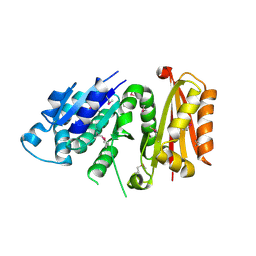
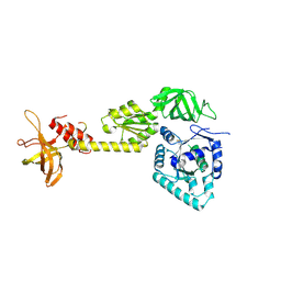
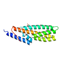
3WGN
 
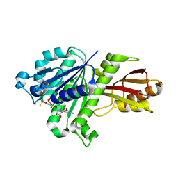 | | STAPHYLOCOCCUS AUREUS FTSZ bound with GTP-gamma-S | | 分子名称: | 5'-GUANOSINE-DIPHOSPHATE-MONOTHIOPHOSPHATE, Cell division protein FtsZ | | 著者 | Matsui, T, Mogi, N, Tanaka, I, Yao, M. | | 登録日 | 2013-08-06 | | 公開日 | 2013-12-25 | | 最終更新日 | 2023-11-08 | | 実験手法 | X-RAY DIFFRACTION (2.606 Å) | | 主引用文献 | Structural change in FtsZ Induced by intermolecular interactions between bound GTP and the T7 loop
J.Biol.Chem., 289, 2014
|
|
2ZZG
 | | Crystal structure of alanyl-tRNA synthetase in complex with 5''-O-(N-(L-alanyl)-sulfamyoxyl) adenine without oligomerization domain | | 分子名称: | '5'-O-(N-(L-ALANYL)-SULFAMOYL)ADENOSINE, Alanyl-tRNA synthetase, ZINC ION | | 著者 | Sokabe, M, Ose, T, Tokunaga, K, Nakamura, A, Nureki, O, Yao, M, Tanaka, I. | | 登録日 | 2009-02-10 | | 公開日 | 2009-07-21 | | 最終更新日 | 2023-11-01 | | 実験手法 | X-RAY DIFFRACTION (3.1 Å) | | 主引用文献 | The structure of alanyl-tRNA synthetase with editing domain.
Proc.Natl.Acad.Sci.USA, 106, 2009
|
|
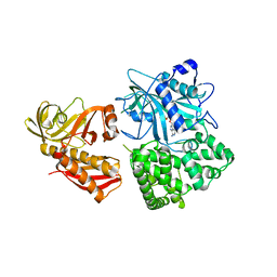
3A24
 | | Crystal structure of BT1871 retaining glycosidase | | 分子名称: | 2-(N-MORPHOLINO)-ETHANESULFONIC ACID, CALCIUM ION, alpha-galactosidase | | 著者 | Okuyama, M, Kitamura, M, Hondoh, H, Tanaka, I, Yao, M. | | 登録日 | 2009-04-28 | | 公開日 | 2009-10-06 | | 最終更新日 | 2023-11-01 | | 実験手法 | X-RAY DIFFRACTION (2.3 Å) | | 主引用文献 | Catalytic mechanism of retaining alpha-galactosidase belonging to glycoside hydrolase family 97.
J.Mol.Biol., 392, 2009
|
|
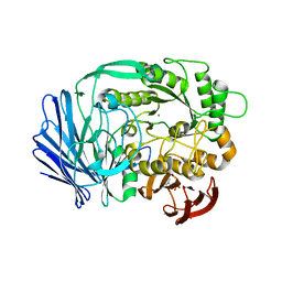
3VOA
 | | Staphylococcus aureus FtsZ 12-316 GDP-form | | 分子名称: | CALCIUM ION, Cell division protein FtsZ, GUANOSINE-5'-DIPHOSPHATE | | 著者 | Yamane, J, Matsui, T, Mogi, N, Yao, M, Tanaka, I. | | 登録日 | 2012-01-20 | | 公開日 | 2012-08-29 | | 最終更新日 | 2023-11-08 | | 実験手法 | X-RAY DIFFRACTION (1.73 Å) | | 主引用文献 | Structural reorganization of the bacterial cell-division protein FtsZ from Staphylococcus aureus
Acta Crystallogr.,Sect.D, 68, 2012
|
|
2ZOZ
 | | Crystal structure of the ethidium-bound form of the multi-drug binding transcriptional repressor CgmR | | 分子名称: | ETHIDIUM, GLYCEROL, SULFATE ION, ... | | 著者 | Itou, H, Shirakihara, Y, Tanaka, I. | | 登録日 | 2008-06-20 | | 公開日 | 2008-07-08 | | 最終更新日 | 2023-11-01 | | 実験手法 | X-RAY DIFFRACTION (1.95 Å) | | 主引用文献 | Crystal Structures of the Multidrug Binding Repressor Corynebacteriumglutamicum CgmR in Complex with Inducers and with an Operator
J.Mol.Biol., 403, 2010
|
|
2ZKL
 | | Crystal Structure of capsular polysaccharide assembling protein CapF from staphylococcus aureus | | 分子名称: | Capsular polysaccharide synthesis enzyme Cap5F, GLYCEROL, ZINC ION | | 著者 | Miyafusa, T, Tanaka, Y, Yao, M, Tanaka, I, Tsumoto, K. | | 登録日 | 2008-03-25 | | 公開日 | 2009-03-31 | | 最終更新日 | 2024-03-13 | | 実験手法 | X-RAY DIFFRACTION (2.61 Å) | | 主引用文献 | Crystal structure of capsular polysaccharide assembling protein from Staphylococcus aureus
to be published
|
|
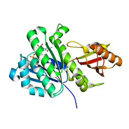
3VO9
 | | Staphylococcus aureus FtsZ apo-form (SeMet) | | 分子名称: | Cell division protein FtsZ | | 著者 | Matsui, T, Yamane, J, Mogi, N, Yao, M, Tanaka, I. | | 登録日 | 2012-01-20 | | 公開日 | 2012-08-29 | | 最終更新日 | 2013-08-14 | | 実験手法 | X-RAY DIFFRACTION (2.706 Å) | | 主引用文献 | Structural reorganization of the bacterial cell-division protein FtsZ from Staphylococcus aureus
Acta Crystallogr.,Sect.D, 68, 2012
|
|
3A8B
 | |
2R97
 | | Crystal structure of E. coli WrbA in complex with FMN | | 分子名称: | FLAVIN MONONUCLEOTIDE, Flavoprotein WrbA | | 著者 | Kuta Smatanova, I, Wolfova, J, Brynda, J, Mesters, J.R, Grandori, R, Carey, J. | | 登録日 | 2007-09-12 | | 公開日 | 2008-09-23 | | 最終更新日 | 2023-08-30 | | 実験手法 | X-RAY DIFFRACTION (2 Å) | | 主引用文献 | Structural organization of WrbA in apo- and holoprotein crystals.
Biochim.Biophys.Acta, 1794, 2009
|
|
2ZXU
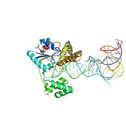 | | Crystal structure of tRNA modification enzyme MiaA in the complex with tRNA(Phe) and DMASPP | | 分子名称: | DIMETHYLALLYL S-THIOLODIPHOSPHATE, MAGNESIUM ION, tRNA delta(2)-isopentenylpyrophosphate transferase, ... | | 著者 | Sakai, J, Yao, M, Chimnaronk, S, Tanaka, I. | | 登録日 | 2009-01-07 | | 公開日 | 2009-05-26 | | 最終更新日 | 2023-11-01 | | 実験手法 | X-RAY DIFFRACTION (2.75 Å) | | 主引用文献 | Snapshots of dynamics in synthesizing N(6)-isopentenyladenosine at the tRNA anticodon
Biochemistry, 48, 2009
|
|
3B0V
 | | tRNA-dihydrouridine synthase from Thermus thermophilus in complex with tRNA | | 分子名称: | FLAVIN MONONUCLEOTIDE, tRNA, tRNA-dihydrouridine synthase | | 著者 | Yu, F, Tanaka, Y, Yamashita, K, Nakamura, A, Yao, M, Tanaka, I. | | 登録日 | 2011-06-14 | | 公開日 | 2011-12-14 | | 最終更新日 | 2017-10-11 | | 実験手法 | X-RAY DIFFRACTION (3.51 Å) | | 主引用文献 | Molecular basis of dihydrouridine formation on tRNA
Proc.Natl.Acad.Sci.USA, 108, 2011
|
|
3WBK
 | | crystal structure analysis of eukaryotic translation initiation factor 5B and 1A complex | | 分子名称: | Eukaryotic translation initiation factor 1A, Eukaryotic translation initiation factor 5B | | 著者 | Zheng, A, Yamamoto, R, Ose, T, Yu, J, Tanaka, I, Yao, M. | | 登録日 | 2013-05-20 | | 公開日 | 2014-11-19 | | 最終更新日 | 2023-11-08 | | 実験手法 | X-RAY DIFFRACTION (3.3 Å) | | 主引用文献 | X-ray structures of eIF5B and the eIF5B-eIF1A complex: the conformational flexibility of eIF5B is restricted on the ribosome by interaction with eIF1A
Acta Crystallogr.,Sect.D, 70, 2014
|
|
3WA1
 | | Crystal structure of BinB: A receptor binding component of the binary toxin from Lysinibacillus sphaericus | | 分子名称: | BinB protein | | 著者 | Srisucharitpanit, K, Yao, M, Chimnaronk, S, Promdonkoy, B, Boonserm, P, Tanaka, I. | | 登録日 | 2013-04-22 | | 公開日 | 2014-07-02 | | 最終更新日 | 2014-10-08 | | 実験手法 | X-RAY DIFFRACTION (1.75 Å) | | 主引用文献 | Crystal structure of BinB: A receptor binding component of the binary toxin from Lysinibacillus sphaericus
Proteins, 82, 2014
|
|
1UCA
 | | Crystal structure of the Ribonuclease MC1 from bitter gourd seeds complexed with 2'-UMP | | 分子名称: | PHOSPHORIC ACID MONO-[2-(2,4-DIOXO-3,4-DIHYDRO-2H-PYRIMIDIN-1-YL)-4-HYDROXY-5-HYDROXYMETHYL-TETRAHYDRO-FURAN-3-YL] ESTER, Ribonuclease MC | | 著者 | Suzuki, A, Yao, M, Tanaka, I, Numata, T, Kikukawa, S, Yamasaki, N, Kimura, M. | | 登録日 | 2003-04-10 | | 公開日 | 2003-04-29 | | 最終更新日 | 2023-10-25 | | 実験手法 | X-RAY DIFFRACTION (1.48 Å) | | 主引用文献 | Crystal structures of the ribonuclease MC1 from bitter gourd seeds, complexed with 2'-UMP or 3'-UMP, reveal structural basis for uridine specificity
Biochem.Biophys.Res.Commun., 275, 2000
|
|
1UCD
 | | Crystal structure of Ribonuclease MC1 from bitter gourd seeds complexed with 5'-UMP | | 分子名称: | Ribonuclease MC, URACIL, URIDINE-5'-MONOPHOSPHATE | | 著者 | Suzuki, A, Numata, T, Yao, M, Kimura, M, Tanaka, I. | | 登録日 | 2003-04-10 | | 公開日 | 2004-05-18 | | 最終更新日 | 2023-10-25 | | 実験手法 | X-RAY DIFFRACTION (1.3 Å) | | 主引用文献 | Structure of RNase MC1 from bitter gourd seeds in complex with 5'UMP
To be published
|
|
5B4C
 | | Crystal structure of H10N mutant of LpxH with manganese | | 分子名称: | GLYCEROL, MANGANESE (II) ION, UDP-2,3-diacylglucosamine hydrolase | | 著者 | Okada, C, Wakabayashi, H, Yao, M, Tanaka, I. | | 登録日 | 2016-04-03 | | 公開日 | 2016-09-28 | | 最終更新日 | 2023-11-08 | | 実験手法 | X-RAY DIFFRACTION (1.96 Å) | | 主引用文献 | Crystal structures of the UDP-diacylglucosamine pyrophosphohydrase LpxH from Pseudomonas aeruginosa
Sci Rep, 6, 2016
|
|
5B49
 | | Crystal structure of LpxH with manganese from Pseudomonas aeruginosa | | 分子名称: | (R)-((2R,3S,4R,5R,6R)-3-HYDROXY-2-(HYDROXYMETHYL)-5-((R)-3-HYDROXYTETRADECANAMIDO)-6-(PHOSPHONOOXY)TETRAHYDRO-2H-PYRAN-4-YL) 3-HYDROXYTETRADECANOATE, GLYCEROL, MANGANESE (II) ION, ... | | 著者 | Okada, C, Wakabayashi, H, Yao, M, Tanaka, I. | | 登録日 | 2016-04-03 | | 公開日 | 2016-09-28 | | 最終更新日 | 2024-03-20 | | 実験手法 | X-RAY DIFFRACTION (1.65 Å) | | 主引用文献 | Crystal structures of the UDP-diacylglucosamine pyrophosphohydrase LpxH from Pseudomonas aeruginosa
Sci Rep, 6, 2016
|
|
2RG1
 | | Crystal structure of E. coli WrbA apoprotein | | 分子名称: | CHLORIDE ION, Flavoprotein WrbA | | 著者 | Kuta Smatanova, I, Wolfova, J, Brynda, J, Lapkouski, M, Mesters, J.R, Grandori, R, Carey, J. | | 登録日 | 2007-10-02 | | 公開日 | 2008-10-14 | | 最終更新日 | 2023-08-30 | | 実験手法 | X-RAY DIFFRACTION (1.85 Å) | | 主引用文献 | Structural organization of WrbA in apo- and holoprotein crystals.
Biochim.Biophys.Acta, 1794, 2009
|
|
4P1Y
 | | Crystal structure of staphylococcal gamma-hemolysin prepore | | 分子名称: | Gamma-hemolysin component A, Gamma-hemolysin component B | | 著者 | Yamashita, D, Tanaka, Y, Tanaka, I, Yao, M. | | 登録日 | 2014-02-28 | | 公開日 | 2014-10-01 | | 最終更新日 | 2023-12-27 | | 実験手法 | X-RAY DIFFRACTION (2.992 Å) | | 主引用文献 | Molecular basis of transmembrane beta-barrel formation of staphylococcal pore-forming toxins.
Nat Commun, 5, 2014
|
|
4F84
 | | Structure analysis of Geranyl diphosphate methyltransferase in complex with SAM | | 分子名称: | Geranyl diphosphate 2-C-methyltransferase, S-ADENOSYLMETHIONINE | | 著者 | Ariyawutthiphan, O, Ose, T, Minami, A, Gao, Y.G, Yao, M, Oikawa, H, Tanaka, I. | | 登録日 | 2012-05-17 | | 公開日 | 2012-10-31 | | 最終更新日 | 2023-11-08 | | 実験手法 | X-RAY DIFFRACTION (2.2 Å) | | 主引用文献 | Structure analysis of geranyl pyrophosphate methyltransferase and the proposed reaction mechanism of SAM-dependent C-methylation
Acta Crystallogr.,Sect.D, 68, 2012
|
|
4P24
 | | pore forming toxin | | 分子名称: | (4S)-2-METHYL-2,4-PENTANEDIOL, Alpha-hemolysin | | 著者 | Sugawara, T, Yamashita, D, Tanaka, Y, Tanaka, I, Yao, M. | | 登録日 | 2014-03-01 | | 公開日 | 2015-03-11 | | 最終更新日 | 2023-12-27 | | 実験手法 | X-RAY DIFFRACTION (3.1 Å) | | 主引用文献 | Structural basis for pore-forming mechanism of staphylococcal alpha-hemolysin.
Toxicon, 108, 2015
|
|
4PAC
 | | Crystal Structure of Histidine-containing Phosphotransfer Protein AHP2 from Arabidopsis thaliana | | 分子名称: | 2-(N-MORPHOLINO)-ETHANESULFONIC ACID, Histidine-containing phosphotransfer protein 2, IMIDAZOLE | | 著者 | Degtjarik, O, Dopitova, R, Puehringer, S, Weiss, M.S, Janda, L, Hejatko, J, Kuta-Smatanova, I. | | 登録日 | 2014-04-08 | | 公開日 | 2015-05-20 | | 最終更新日 | 2024-05-08 | | 実験手法 | X-RAY DIFFRACTION (2.53 Å) | | 主引用文献 | Crystal structure of AHP2 from Arabidopsis thaliana
To Be Published
|
|
2ZPA
 | | Crystal Structure of tRNA(Met) Cytidine Acetyltransferase | | 分子名称: | ACETYL COENZYME *A, ADENOSINE-5'-DIPHOSPHATE, SULFATE ION, ... | | 著者 | Chimnaronk, S, Manita, T, Yao, M, Tanaka, I. | | 登録日 | 2008-07-08 | | 公開日 | 2009-04-07 | | 最終更新日 | 2011-07-13 | | 実験手法 | X-RAY DIFFRACTION (2.35 Å) | | 主引用文献 | RNA helicase module in an acetyltransferase that modifies a specific tRNA anticodon
Embo J., 28, 2009
|
|
4HZG
 | | Structure of haloalkane dehalogenase DhaA from Rhodococcus rhodochrous | | 分子名称: | CHLORIDE ION, Haloalkane dehalogenase | | 著者 | Stsiapanava, A, Weiss, M.S, Mesters, J.R, Kuta Smatanova, I. | | 登録日 | 2012-11-15 | | 公開日 | 2013-10-02 | | 最終更新日 | 2023-09-20 | | 実験手法 | X-RAY DIFFRACTION (1.95 Å) | | 主引用文献 | Crystallographic analysis of 1,2,3-trichloropropane biodegradation by the haloalkane dehalogenase DhaA31.
Acta Crystallogr.,Sect.D, 70, 2014
|
|
7FCW
 | | X-ray structure of H2O-solvent lysozyme | | 分子名称: | CHLORIDE ION, Lysozyme C, NICKEL (II) ION | | 著者 | Chatake, T, Tanaka, I, Kusaka, K, Fujiwara, S. | | 登録日 | 2021-07-15 | | 公開日 | 2022-04-06 | | 最終更新日 | 2023-11-29 | | 実験手法 | X-RAY DIFFRACTION (1.43 Å) | | 主引用文献 | Protonation states of hen egg-white lysozyme observed using D/H contrast neutron crystallography.
Acta Crystallogr D Struct Biol, 78, 2022
|
|


