6IFB
 
 | |
4APR
 | |
6IFA
 | |
5APR
 | |
6APR
 | |
5Y2J
 | |
5Y2E
 | |
5Y2H
 | |
3APR
 | |
5Y9R
 | |
5YQ8
 | |
2O1J
 | |
5YS4
 | |
2APR
 | |
7EXO
 | |
7ELV
 | |
7BT8
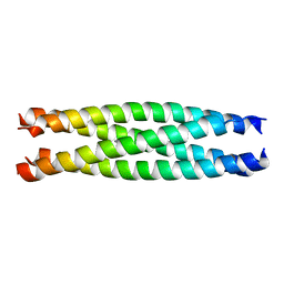
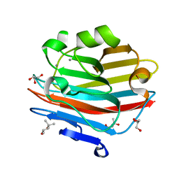
 | | Mevo lectin complex with mannotriose | | 分子名称: | 1,2-ETHANEDIOL, GLYCEROL, alpha-D-mannopyranose-(1-3)-[alpha-D-mannopyranose-(1-6)]beta-D-mannopyranose, ... | | 著者 | Sivaji, N, Suguna, K, Surolia, A, Vijayan, M. | | 登録日 | 2020-03-31 | | 公開日 | 2021-02-03 | | 最終更新日 | 2023-11-29 | | 実験手法 | X-RAY DIFFRACTION (2.7 Å) | | 主引用文献 | Structural and related studies on Mevo lectin from Methanococcus voltae A3: the first thorough characterization of an archeal lectin and its interactions.
Glycobiology, 31, 2021
|
|
5D0E
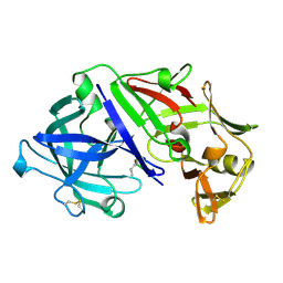
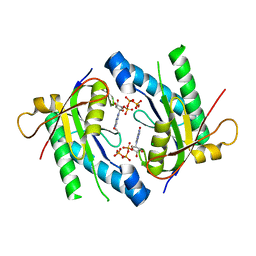 | | Crystal Structure of an adenylyl cyclase Ma1120-Cat in complex with GTP and calcium from Mycobacterium avium | | 分子名称: | CALCIUM ION, CHLORIDE ION, Cyclase, ... | | 著者 | Bharambe, N.G, Barathy, D.V, Suguna, K. | | 登録日 | 2015-08-03 | | 公開日 | 2016-08-10 | | 最終更新日 | 2023-11-08 | | 実験手法 | X-RAY DIFFRACTION (1.48 Å) | | 主引用文献 | Substrate specificity determinants of class III nucleotidyl cyclases
Febs J., 283, 2016
|
|
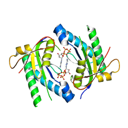
5D0H
 | |
2ZMN
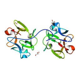
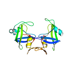
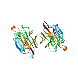 | | Crystal Structure of basic winged bean lectin in complex with Gal-alpha- 1,6 Glc | | 分子名称: | 2-acetamido-2-deoxy-beta-D-glucopyranose-(1-4)-2-acetamido-2-deoxy-beta-D-glucopyranose, Basic agglutinin, CALCIUM ION, ... | | 著者 | Kulkarni, K.A, Katiyar, S, Surolia, A, Vijayan, M, Suguna, K. | | 登録日 | 2008-04-19 | | 公開日 | 2008-07-29 | | 最終更新日 | 2023-11-01 | | 実験手法 | X-RAY DIFFRACTION (2.9 Å) | | 主引用文献 | Structure and sugar-specificity of basic winged-bean lectin: structures of new disaccharide complexes and a comparative study with other known disaccharide complexes of the lectin.
Acta Crystallogr.,Sect.D, 64, 2008
|
|
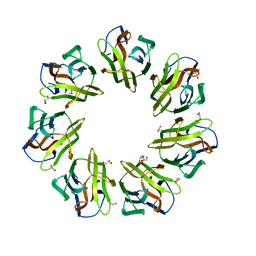
7W4B
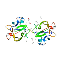
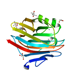
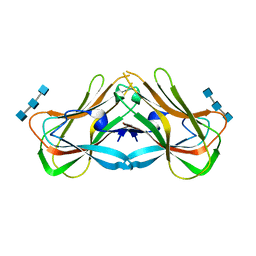 | | Phloem lectin (PP2) structure -complex with Chitotrise | | 分子名称: | 17 kDa phloem lectin, 2-acetamido-2-deoxy-beta-D-glucopyranose-(1-4)-2-acetamido-2-deoxy-beta-D-glucopyranose-(1-4)-2-acetamido-2-deoxy-beta-D-glucopyranose | | 著者 | Sivaji, N, Kishore, B.B, Suguna, K, Surolia, A. | | 登録日 | 2021-11-26 | | 公開日 | 2023-03-08 | | 最終更新日 | 2024-05-22 | | 実験手法 | X-RAY DIFFRACTION (2.5 Å) | | 主引用文献 | Structure and interactions of the phloem lectin (phloem protein 2) Cus17 from Cucumis sativus.
Structure, 31, 2023
|
|
2ZML
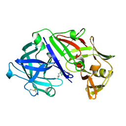
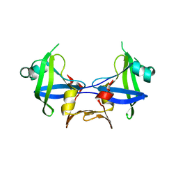
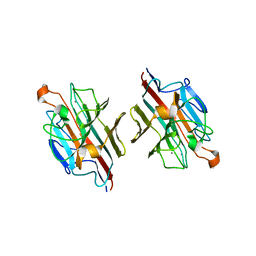 | | Crystal structure of basic winged bean lectin in complex with Gal-ALPHA 1,4 Gal | | 分子名称: | 2-acetamido-2-deoxy-beta-D-glucopyranose, Basic agglutinin, CALCIUM ION, ... | | 著者 | Kulkarni, K.A, Katiyar, S, Surolia, A, Vijayan, M, Suguna, K. | | 登録日 | 2008-04-19 | | 公開日 | 2008-07-29 | | 最終更新日 | 2023-11-01 | | 実験手法 | X-RAY DIFFRACTION (2.65 Å) | | 主引用文献 | Structure and sugar-specificity of basic winged-bean lectin: structures of new disaccharide complexes and a comparative study with other known disaccharide complexes of the lectin.
Acta Crystallogr.,Sect.D, 64, 2008
|
|
3R0E
 | |
1CR7
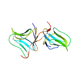
 | | PEANUT LECTIN-LACTOSE COMPLEX MONOCLINIC FORM | | 分子名称: | CALCIUM ION, LECTIN, MANGANESE (II) ION, ... | | 著者 | Ravishankar, R, Suguna, K, Surolia, A, Vijayan, M. | | 登録日 | 1999-08-14 | | 公開日 | 2001-04-21 | | 最終更新日 | 2023-08-09 | | 実験手法 | X-RAY DIFFRACTION (2.6 Å) | | 主引用文献 | Crystal structures of the peanut lectin-lactose complex at acidic pH: retention of unusual quaternary structure, empty and carbohydrate bound combining sites, molecular mimicry and crystal packing directed by interactions at the combining site.
Proteins, 43, 2001
|
|
1CQ9
 | | PEANUT LECTIN-TRICLINIC FORM | | 分子名称: | CALCIUM ION, MANGANESE (II) ION, PROTEIN (PEANUT LECTIN) | | 著者 | Ravishankar, R, Suguna, K, Surolia, A, Vijayan, M. | | 登録日 | 1999-08-06 | | 公開日 | 2002-05-01 | | 最終更新日 | 2023-08-09 | | 実験手法 | X-RAY DIFFRACTION (3.5 Å) | | 主引用文献 | Crystal structures of the peanut lectin-lactose complex at acidic pH: retention of unusual
quaternary structure, empty and carbohydrate bound combining sites, molecular mimicry
and crystal packing directed by interactions at the combining site.
Proteins, 43, 2001
|
|


