Loading
PDBj
MenuPDBj@FacebookPDBj@TwitterPDBj@YouTubewwPDB FoundationwwPDB
RCSB PDBPDBeBMRBAdv. SearchSearch help
PDB: 60 results

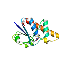
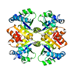
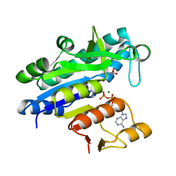
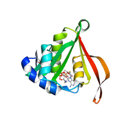
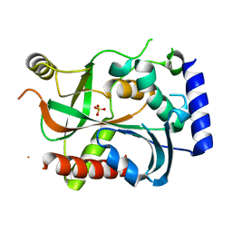
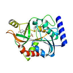
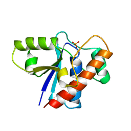
1PHR
DownloadVisualize
BU of 1phr by Molmil
THE CRYSTAL STRUCTURE OF A LOW MOLECULAR PHOSPHOTYROSINE PROTEIN PHOSPHATASE
Descriptor: LOW MOLECULAR WEIGHT PHOSPHOTYROSINE PROTEIN PHOSPHATASE, SULFATE ION
Authors:Su, X.-D, Taddei, N, Stefani, M, Ramponi, G, Nordlund, P.
Deposit date:1994-07-05
Release date:1995-07-31
Last modified:2024-02-14
Method:X-RAY DIFFRACTION (2.1 Å)
Cite:The crystal structure of a low-molecular-weight phosphotyrosine protein phosphatase.
Nature, 370, 1994
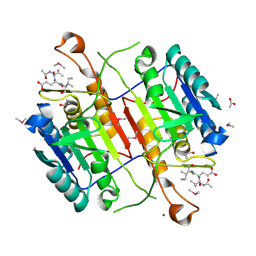
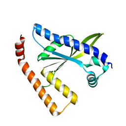
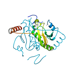
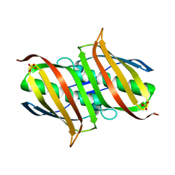
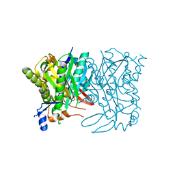
1JL3
DownloadVisualize
BU of 1jl3 by Molmil
Crystal Structure of B. subtilis ArsC
Descriptor: ARSENATE REDUCTASE, SULFATE ION
Authors:Su, X.-D, Bennett, M.S.
Deposit date:2001-07-15
Release date:2001-10-24
Last modified:2024-02-07
Method:X-RAY DIFFRACTION (1.6 Å)
Cite:Bacillus subtilis arsenate reductase is structurally and functionally similar to low molecular weight protein tyrosine phosphatases.
Proc.Natl.Acad.Sci.USA, 98, 2001
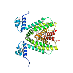
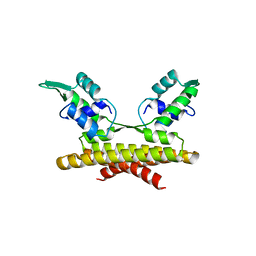
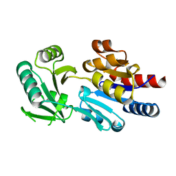
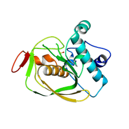
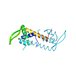
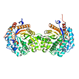
2F07
DownloadVisualize
BU of 2f07 by Molmil
Crystal Structure of YvdT from Bacillus subtilis
Descriptor: 2-[BIS-(2-HYDROXY-ETHYL)-AMINO]-2-HYDROXYMETHYL-PROPANE-1,3-DIOL, YvdT
Authors:Su, X.-D, Yu, Y.-M, Nan, J.
Deposit date:2005-11-12
Release date:2006-11-21
Last modified:2024-03-13
Method:X-RAY DIFFRACTION (2.3 Å)
Cite:Crystal Structure of YvdT from Bacillus subtilis
To be Published
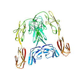
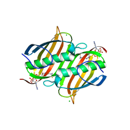
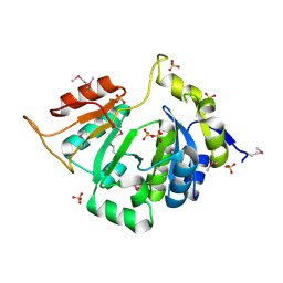
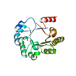
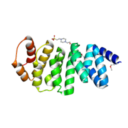
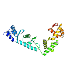
1BIH
DownloadVisualize
BU of 1bih by Molmil
CRYSTAL STRUCTURE OF THE INSECT IMMUNE PROTEIN HEMOLIN: A NEW DOMAIN ARRANGEMENT WITH IMPLICATIONS FOR HOMOPHILIC ADHESION
Descriptor: HEMOLIN, PHOSPHATE ION
Authors:Su, X.-D, Gastinel, L.N, Vaughn, D.E, Faye, I, Poon, P, Bjorkman, P.J.
Deposit date:1998-06-17
Release date:1998-10-14
Last modified:2018-03-07
Method:X-RAY DIFFRACTION (3.1 Å)
Cite:Crystal structure of hemolin: a horseshoe shape with implications for homophilic adhesion.
Science, 281, 1998
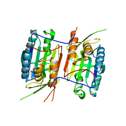
3NR2
DownloadVisualize
BU of 3nr2 by Molmil
Crystal structure of Caspase-6 zymogen
Descriptor: Caspase-6
Authors:Su, X.-D, Wang, X.-J, Liu, X, Mi, W, Wang, K.-T.
Deposit date:2010-06-30
Release date:2010-10-27
Last modified:2023-11-01
Method:X-RAY DIFFRACTION (2.9 Å)
Cite:Crystal structures of human caspase 6 reveal a new mechanism for intramolecular cleavage self-activation
Embo Rep., 11, 2010
3S2S
DownloadVisualize
BU of 3s2s by Molmil
The crystal structure of pyrazinamidase/nicotinamidase from streptococcus mutans UA159
Descriptor: CACODYLIC ACID, Putative pyrazinamidase/nicotinamidase, ZINC ION
Authors:Su, X.-D, Liu, X, Zhang, H.
Deposit date:2011-05-17
Release date:2012-05-16
Last modified:2024-03-20
Method:X-RAY DIFFRACTION (1.7 Å)
Cite:Get phases from arsenic anomalous scattering: de novo SAD phasing of two protein structures crystallized in cacodylate buffer
Plos One, 6, 2011
3S70
DownloadVisualize
BU of 3s70 by Molmil
Crystal structure of active caspase-6 bound with Ac-VEID-CHO solved by As-SAD
Descriptor: ACETATE ION, CACODYLATE ION, Caspase-6, ...
Authors:Su, X.-D, Liu, X, Wang, X.-J.
Deposit date:2011-05-26
Release date:2012-04-11
Last modified:2024-10-16
Method:X-RAY DIFFRACTION (1.625 Å)
Cite:Get phases from arsenic anomalous scattering: de novo SAD phasing of two protein structures crystallized in cacodylate buffer
Plos One, 6, 2011
3L9F
DownloadVisualize
BU of 3l9f by Molmil
The Crystal Structure of smu.1604c from Streptococcus mutans UA159
Descriptor: MAGNESIUM ION, Putative uncharacterized protein smu.1604c
Authors:Su, X.-D, Liu, X.
Deposit date:2010-01-05
Release date:2011-01-05
Last modified:2024-03-20
Method:X-RAY DIFFRACTION (1.8 Å)
Cite:The Crystal Structure of smu.1604c from Streptococcus mutans UA159
TO BE PUBLISHED
3LBE
DownloadVisualize
BU of 3lbe by Molmil
The Crystal Structure of smu.793 from Streptococcus mutans UA159 bound to acetyl CoA
Descriptor: CHLORIDE ION, COENZYME A, Putative uncharacterized protein smu.793
Authors:Su, X.-D, Hou, Q.M, Fan, X.X, Nan, J, Liu, X.
Deposit date:2010-01-08
Release date:2011-01-12
Last modified:2023-11-01
Method:X-RAY DIFFRACTION (1.7 Å)
Cite:The Crystal Structure of smu.793 from Streptococcus mutans UA159 bound to acetyl CoA
TO BE PUBLISHED
3L86
DownloadVisualize
BU of 3l86 by Molmil
The Crystal Structure of smu.665 from Streptococcus mutans UA159
Descriptor: ADENOSINE-5'-DIPHOSPHATE, Acetylglutamate kinase, MAGNESIUM ION, ...
Authors:Su, X.-D, Liu, X, Wu, C.W.
Deposit date:2009-12-30
Release date:2011-01-05
Last modified:2023-11-01
Method:X-RAY DIFFRACTION (2.06 Å)
Cite:The Crystal Structure of smu.665 from Streptococcus mutans UA159
TO BE PUBLISHED
3L9D
DownloadVisualize
BU of 3l9d by Molmil
The Crystal Structure of smu.1046c from Streptococcus mutans UA159
Descriptor: Putative GTP pyrophosphokinase
Authors:Su, X.-D, Huang, Y.H, Liu, X.
Deposit date:2010-01-05
Release date:2011-01-12
Last modified:2023-11-01
Method:X-RAY DIFFRACTION (2.484 Å)
Cite:The Crystal Structure of smu.1046c from Streptococcus mutans UA159
TO BE PUBLISHED
3L7Y
DownloadVisualize
BU of 3l7y by Molmil
The Crystal Structure of SMU.1108c from Streptococcus mutans UA159
Descriptor: MAGNESIUM ION, Putative uncharacterized protein smu.1108c
Authors:Su, X.-D, Feng, M.J, Liu, X.
Deposit date:2009-12-29
Release date:2010-12-08
Last modified:2023-11-01
Method:X-RAY DIFFRACTION (1.996 Å)
Cite:The Crystal Structure of SMU.1108c from Streptococcus mutans UA159
TO BE PUBLISHED
3L7V
DownloadVisualize
BU of 3l7v by Molmil
Crystal structure of a hypothetical protein smu.1377c from Streptococcus mutans UA159
Descriptor: Putative uncharacterized protein smu.1377c, SULFATE ION
Authors:Su, X.-D, Fu, T.-M, Liu, X.
Deposit date:2009-12-29
Release date:2010-07-21
Last modified:2024-10-30
Method:X-RAY DIFFRACTION (2.256 Å)
Cite:The structure of the hypothetical protein smu.1377c from Streptococcus mutans suggests a role in tRNA modification
Acta Crystallogr.,Sect.F, 66, 2010
3LD2
DownloadVisualize
BU of 3ld2 by Molmil
The Crystal Structure of smu.2055 from Streptococcus mutans UA159
Descriptor: COENZYME A, Putative acetyltransferase
Authors:Su, X.-D, Zhan, X.R, Gao, X.Z, Liu, X.
Deposit date:2010-01-12
Release date:2011-01-12
Last modified:2011-07-13
Method:X-RAY DIFFRACTION (2.5 Å)
Cite:The Crystal Structure of smu.2055 from Streptococcus mutans UA159
TO BE PUBLISHED
3L7X
DownloadVisualize
BU of 3l7x by Molmil
The Crystal Structure of SMU.412c from Streptococcus mutans UA159
Descriptor: Putative Hit-like protein involved in cell-cycle regulation, SODIUM ION, ZINC ION
Authors:Su, X.-D, Ye, Z.Y, Liu, X.
Deposit date:2009-12-29
Release date:2010-12-08
Last modified:2023-11-01
Method:X-RAY DIFFRACTION (1.699 Å)
Cite:The Crystal Structure of SMU.412c from Streptococcus mutans UA159
TO BE PUBLISHED
3L87
DownloadVisualize
BU of 3l87 by Molmil
The Crystal Structure of smu.143c from Streptococcus mutans UA159
Descriptor: FE (III) ION, Peptide deformylase
Authors:Su, X.-D, Cao, Q, Liu, X.
Deposit date:2009-12-30
Release date:2011-01-05
Last modified:2023-11-01
Method:X-RAY DIFFRACTION (2 Å)
Cite:The Crystal Structure of smu.143c from Streptococcus mutans UA159
TO BE PUBLISHED
3L9C
DownloadVisualize
BU of 3l9c by Molmil
The Crystal Structure of smu.777 from Streptococcus mutans UA159
Descriptor: 3-dehydroquinate dehydratase
Authors:Su, X.-D, Huang, Y.H, Liu, X.
Deposit date:2010-01-05
Release date:2011-01-05
Last modified:2023-11-01
Method:X-RAY DIFFRACTION (1.6 Å)
Cite:The Crystal Structure of smu.777 from Streptococcus mutans UA159
TO BE PUBLISHED
3LA8
DownloadVisualize
BU of 3la8 by Molmil
The Crystal Structure of smu.1229 from Streptococcus mutans UA159
Descriptor: Putative purine nucleoside phosphorylase, SULFATE ION
Authors:Su, X.-D, Hou, Q.M, Liu, X.
Deposit date:2010-01-06
Release date:2011-01-12
Last modified:2023-11-01
Method:X-RAY DIFFRACTION (1.8 Å)
Cite:The Crystal Structure of smu.1229 from Streptococcus mutans UA159
TO BE PUBLISHED
3LBB
DownloadVisualize
BU of 3lbb by Molmil
The Crystal Structure of smu.793 from Streptococcus mutans UA159
Descriptor: CHLORIDE ION, Putative uncharacterized protein smu.793, SULFATE ION
Authors:Su, X.-D, Hou, Q.M, Fan, X.X, Nan, J, Liu, X.
Deposit date:2010-01-08
Release date:2011-01-12
Last modified:2023-11-01
Method:X-RAY DIFFRACTION (2.1 Å)
Cite:The Crystal Structure of smu.793 from Streptococcus mutans UA159
TO BE PUBLISHED
3L7W
DownloadVisualize
BU of 3l7w by Molmil
The Crystal Structure of smu.1704 from Streptococcus mutans UA159
Descriptor: Putative uncharacterized protein SMU.1704
Authors:Su, X.-D, Liu, X, Fu, T.M.
Deposit date:2009-12-29
Release date:2010-12-08
Last modified:2024-10-30
Method:X-RAY DIFFRACTION (2.2 Å)
Cite:The Crystal Structure of smu.1704 from Streptococcus mutans UA159
TO BE PUBLISHED
3L9T
DownloadVisualize
BU of 3l9t by Molmil
The Crystal Structure of smu.31 from Streptococcus mutans UA159
Descriptor: 4-(2-HYDROXYETHYL)-1-PIPERAZINE ETHANESULFONIC ACID, Putative uncharacterized protein smu.31
Authors:Su, X.-D, Cao, Q, Liu, X.
Deposit date:2010-01-05
Release date:2011-01-05
Last modified:2011-07-13
Method:X-RAY DIFFRACTION (2.206 Å)
Cite:The Crystal Structure of smu.31 from Streptococcus mutans UA159
TO BE PUBLISHED
3LBA
DownloadVisualize
BU of 3lba by Molmil
The Crystal Structure of smu.1229 from Streptococcus mutans UA159 bound to hypoxanthine
Descriptor: HYPOXANTHINE, Putative purine nucleoside phosphorylase, SULFATE ION
Authors:Su, X.-D, Hou, Q.M, Wang, H.F, Liu, X.
Deposit date:2010-01-08
Release date:2011-01-12
Last modified:2024-03-20
Method:X-RAY DIFFRACTION (2.24 Å)
Cite:The Crystal Structure of smu.1229 from Streptococcus mutans UA159 bound to hypoxanthine
TO BE PUBLISHED
3LEH
DownloadVisualize
BU of 3leh by Molmil
The Crystal Structure of smu.943c from Streptococcus mutans UA159
Descriptor: NITRATE ION, Putative hydroxymethylglutaryl-CoA synthase
Authors:Su, X.-D, Liu, Y.H, Liu, X.
Deposit date:2010-01-14
Release date:2011-01-19
Last modified:2023-11-01
Method:X-RAY DIFFRACTION (1.7 Å)
Cite:The Crystal Structure of smu.943c from Streptococcus mutans UA159
TO BE PUBLISHED
1HXJ
DownloadVisualize
BU of 1hxj by Molmil
CRYSTAL STRUCTURE OF THE MAIZE ZM-P60.1 BETA-GLUCOSIDASE
Descriptor: BETA-GLUCOSIDASE
Authors:Vevodova, J, Su, X.-D, Marek, J, Brzobohaty, B.
Deposit date:2001-01-15
Release date:2003-01-21
Last modified:2023-08-09
Method:X-RAY DIFFRACTION (2.05 Å)
Cite:Insights into the functional architecture of the catalytic center of a maize beta-glucosidase Zm-p60.1
Plant Physiol., 127, 2001
1LVA
DownloadVisualize
BU of 1lva by Molmil
Crystal structure of a C-terminal fragment of Moorella thermoacetica elongation factor SelB
Descriptor: SULFATE ION, Selenocysteine-specific elongation factor, YTTRIUM ION
Authors:Selmer, M, Su, X.-D.
Deposit date:2002-05-28
Release date:2002-08-21
Last modified:2024-10-23
Method:X-RAY DIFFRACTION (2.12 Å)
Cite:Crystal structure of an mRNA-binding fragment of Moorella thermoacetica elongation factor SelB.
EMBO J., 21, 2002


123>

226707

건을2024-10-30부터공개중

PDB statisticsPDBj update infoContact PDBjnumon