Loading
PDBj
MenuPDBj@FacebookPDBj@TwitterPDBj@YouTubewwPDB FoundationwwPDB
RCSB PDBPDBeBMRBAdv. SearchSearch help
PDB: 198 results

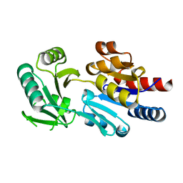
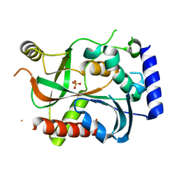
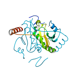
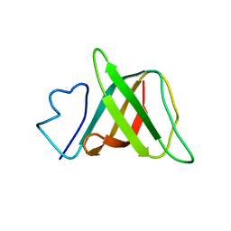
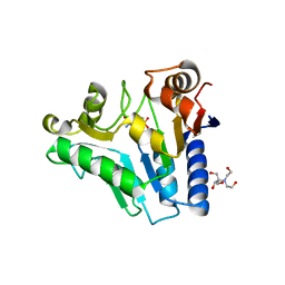
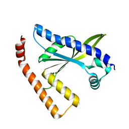
2HAJ
DownloadVisualize
BU of 2haj by Molmil
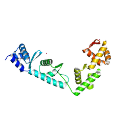
Solution structure of the helicase-binding domain of Escherichia coli primase
Descriptor: DNA primase
Authors:Su, X.C, Loscha, K.V, Dixon, N.E, Otting, G.
Deposit date:2006-06-13
Release date:2006-10-17
Last modified:2024-05-15
Method:SOLUTION NMR
Cite:Monomeric solution structure of the helicase-binding domain of Escherichia coli DnaG primase
Febs J., 273, 2006
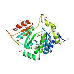
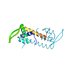
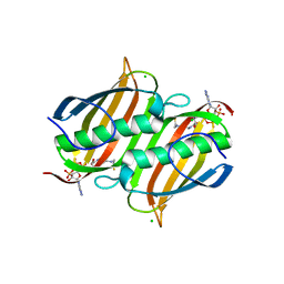
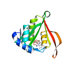
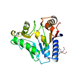
3LBE
DownloadVisualize
BU of 3lbe by Molmil
The Crystal Structure of smu.793 from Streptococcus mutans UA159 bound to acetyl CoA
Descriptor: CHLORIDE ION, COENZYME A, Putative uncharacterized protein smu.793
Authors:Su, X.-D, Hou, Q.M, Fan, X.X, Nan, J, Liu, X.
Deposit date:2010-01-08
Release date:2011-01-12
Last modified:2023-11-01
Method:X-RAY DIFFRACTION (1.7 Å)
Cite:The Crystal Structure of smu.793 from Streptococcus mutans UA159 bound to acetyl CoA
TO BE PUBLISHED
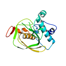
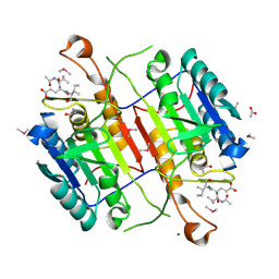
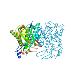
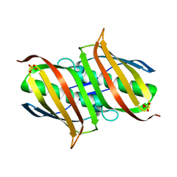
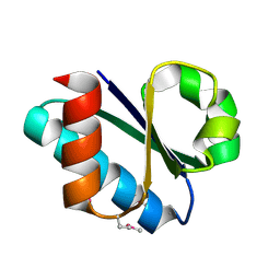
6LPD
DownloadVisualize
BU of 6lpd by Molmil
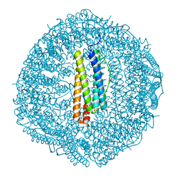
Phascolosoma esculenta
Descriptor: FE (II) ION, FE (III) ION, Ferritin
Authors:Su, X.R, Ming, T.H.
Deposit date:2020-01-09
Release date:2021-01-13
Last modified:2023-11-29
Method:X-RAY DIFFRACTION (1.65 Å)
Cite:Structural comparison of two ferritins from the marine invertebrate Phascolosoma esculenta.
Febs Open Bio, 11, 2021
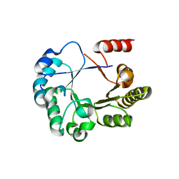
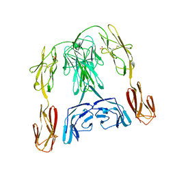
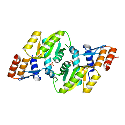
2PER
DownloadVisualize
BU of 2per by Molmil
Crystal Structure of Human Chloride Intracellular Channel protein 2
Descriptor: 5-MERCAPTO-2-NITRO-BENZOIC ACID, Chloride intracellular channel protein 2
Authors:Su, X.D, Liang, Y.H, Mi, W.
Deposit date:2007-04-03
Release date:2008-03-04
Last modified:2023-10-25
Method:X-RAY DIFFRACTION (2 Å)
Cite:The crystal structure of human chloride intracellular channel protein 2: A disulfide bond with functional implications.
Proteins, 71, 2008
3L9F
DownloadVisualize
BU of 3l9f by Molmil
The Crystal Structure of smu.1604c from Streptococcus mutans UA159
Descriptor: MAGNESIUM ION, Putative uncharacterized protein smu.1604c
Authors:Su, X.-D, Liu, X.
Deposit date:2010-01-05
Release date:2011-01-05
Last modified:2024-03-20
Method:X-RAY DIFFRACTION (1.8 Å)
Cite:The Crystal Structure of smu.1604c from Streptococcus mutans UA159
TO BE PUBLISHED
3L86
DownloadVisualize
BU of 3l86 by Molmil
The Crystal Structure of smu.665 from Streptococcus mutans UA159
Descriptor: ADENOSINE-5'-DIPHOSPHATE, Acetylglutamate kinase, MAGNESIUM ION, ...
Authors:Su, X.-D, Liu, X, Wu, C.W.
Deposit date:2009-12-30
Release date:2011-01-05
Last modified:2023-11-01
Method:X-RAY DIFFRACTION (2.06 Å)
Cite:The Crystal Structure of smu.665 from Streptococcus mutans UA159
TO BE PUBLISHED
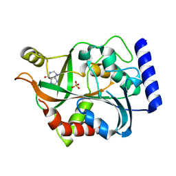
3L9D
DownloadVisualize
BU of 3l9d by Molmil
The Crystal Structure of smu.1046c from Streptococcus mutans UA159
Descriptor: Putative GTP pyrophosphokinase
Authors:Su, X.-D, Huang, Y.H, Liu, X.
Deposit date:2010-01-05
Release date:2011-01-12
Last modified:2023-11-01
Method:X-RAY DIFFRACTION (2.484 Å)
Cite:The Crystal Structure of smu.1046c from Streptococcus mutans UA159
TO BE PUBLISHED
3L7Y
DownloadVisualize
BU of 3l7y by Molmil
The Crystal Structure of SMU.1108c from Streptococcus mutans UA159
Descriptor: MAGNESIUM ION, Putative uncharacterized protein smu.1108c
Authors:Su, X.-D, Feng, M.J, Liu, X.
Deposit date:2009-12-29
Release date:2010-12-08
Last modified:2023-11-01
Method:X-RAY DIFFRACTION (1.996 Å)
Cite:The Crystal Structure of SMU.1108c from Streptococcus mutans UA159
TO BE PUBLISHED
3L7V
DownloadVisualize
BU of 3l7v by Molmil
Crystal structure of a hypothetical protein smu.1377c from Streptococcus mutans UA159
Descriptor: Putative uncharacterized protein smu.1377c, SULFATE ION
Authors:Su, X.-D, Fu, T.-M, Liu, X.
Deposit date:2009-12-29
Release date:2010-07-21
Last modified:2018-05-30
Method:X-RAY DIFFRACTION (2.256 Å)
Cite:The structure of the hypothetical protein smu.1377c from Streptococcus mutans suggests a role in tRNA modification
Acta Crystallogr.,Sect.F, 66, 2010
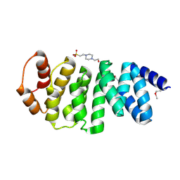
4MZF
DownloadVisualize
BU of 4mzf by Molmil
Crystal structure of human Spindlin1 bound to histone H3(K4me3-R8me2a) peptide
Descriptor: CHLORIDE ION, MAGNESIUM ION, Peptide from Histone H3.2, ...
Authors:Su, X, Ding, X, Li, H.
Deposit date:2013-09-30
Release date:2014-03-26
Method:X-RAY DIFFRACTION (2.098 Å)
Cite:Molecular basis underlying histone H3 lysine-arginine methylation pattern readout by Spin/Ssty repeats of Spindlin1
Genes Dev., 28, 2014
4MZH
DownloadVisualize
BU of 4mzh by Molmil
Crystal structure of human Spindlin1 bound to histone H3(K4me3-R8me2s) peptide
Descriptor: MAGNESIUM ION, Peptide from Histone H3.2, Spindlin-1
Authors:Su, X, Ding, X, Li, H.
Deposit date:2013-09-30
Release date:2014-03-26
Method:X-RAY DIFFRACTION (2.204 Å)
Cite:Molecular basis underlying histone H3 lysine-arginine methylation pattern readout by Spin/Ssty repeats of Spindlin1
Genes Dev., 28, 2014
3S2S
DownloadVisualize
BU of 3s2s by Molmil
The crystal structure of pyrazinamidase/nicotinamidase from streptococcus mutans UA159
Descriptor: CACODYLIC ACID, Putative pyrazinamidase/nicotinamidase, ZINC ION
Authors:Su, X.-D, Liu, X, Zhang, H.
Deposit date:2011-05-17
Release date:2012-05-16
Last modified:2024-03-20
Method:X-RAY DIFFRACTION (1.7 Å)
Cite:Get phases from arsenic anomalous scattering: de novo SAD phasing of two protein structures crystallized in cacodylate buffer
Plos One, 6, 2011
3S70
DownloadVisualize
BU of 3s70 by Molmil
Crystal structure of active caspase-6 bound with Ac-VEID-CHO solved by As-SAD
Descriptor: ACETATE ION, CACODYLATE ION, Caspase-6, ...
Authors:Su, X.-D, Liu, X, Wang, X.-J.
Deposit date:2011-05-26
Release date:2012-04-11
Last modified:2012-12-12
Method:X-RAY DIFFRACTION (1.625 Å)
Cite:Get phases from arsenic anomalous scattering: de novo SAD phasing of two protein structures crystallized in cacodylate buffer
Plos One, 6, 2011
4MZG
DownloadVisualize
BU of 4mzg by Molmil
Crystal structure of human Spindlin1 bound to histone H3K4me3 peptide
Descriptor: (4R)-2-METHYLPENTANE-2,4-DIOL, (4S)-2-METHYL-2,4-PENTANEDIOL, CHLORIDE ION, ...
Authors:Su, X, Ding, X, Li, H.
Deposit date:2013-09-30
Release date:2014-03-26
Method:X-RAY DIFFRACTION (1.698 Å)
Cite:Molecular basis underlying histone H3 lysine-arginine methylation pattern readout by Spin/Ssty repeats of Spindlin1
Genes Dev., 28, 2014
7EWQ
DownloadVisualize
BU of 7ewq by Molmil
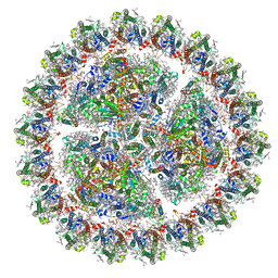
Structure of Mumps virus nucleocapsid ring
Descriptor: Nucleoprotein, RNA (5'-R(P*UP*UP*UP*UP*UP*U)-3')
Authors:Su, X, Shen, Q, Shan, H.
Deposit date:2021-05-25
Release date:2021-06-23
Last modified:2024-08-21
Method:ELECTRON MICROSCOPY (3.5 Å)
Cite:Structural plasticity of mumps virus nucleocapsids with cryo-EM structures.
Commun Biol, 4, 2021
5EMC
DownloadVisualize
BU of 5emc by Molmil
Transcription factor GRDBD and smGRE complex
Descriptor: DNA (5'-D(*CP*CP*AP*GP*AP*AP*(5CM)P*AP*TP*CP*AP*TP*GP*TP*TP*(5CM)P*TP*G)-3'), DNA (5'-D(*CP*CP*AP*GP*AP*AP*(5CM)P*AP*TP*GP*AP*TP*GP*TP*TP*(5CM)P*TP*G)-3'), Glucocorticoid receptor, ...
Authors:Su, X.D, Lian, T, Jin, J.
Deposit date:2015-11-06
Release date:2016-06-29
Last modified:2024-03-20
Method:X-RAY DIFFRACTION (2.3 Å)
Cite:The effects of cytosine methylation on general transcription factors
To Be Published
3UYJ
DownloadVisualize
BU of 3uyj by Molmil
Crystal structure of JMJD5 catalytic core domain in complex with nickle and alpha-KG
Descriptor: 2-OXOGLUTARIC ACID, Lysine-specific demethylase 8, NICKEL (II) ION
Authors:Su, X, Li, H.
Deposit date:2011-12-06
Release date:2011-12-21
Method:X-RAY DIFFRACTION (2.351 Å)
Cite:Crystal structure of JMJD5 catalytic core domain in complex with nickle and alpha-KG
To be Published
2RI1
DownloadVisualize
BU of 2ri1 by Molmil
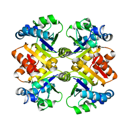
Crystal Structure of glucosamine 6-phosphate deaminase (NagB) with GlcN6P from S. mutans
Descriptor: 2-[BIS-(2-HYDROXY-ETHYL)-AMINO]-2-HYDROXYMETHYL-PROPANE-1,3-DIOL, 2-amino-2-deoxy-6-O-phosphono-alpha-D-glucopyranose, Glucosamine-6-phosphate deaminase
Authors:Liu, C, Li, D, Su, X.D.
Deposit date:2007-10-10
Release date:2008-03-25
Last modified:2024-03-13
Method:X-RAY DIFFRACTION (2.03 Å)
Cite:Ring-opening mechanism revealed by crystal structures of NagB and its ES intermediate complex
J.Mol.Biol., 379, 2008
2RI0
DownloadVisualize
BU of 2ri0 by Molmil
Crystal Structure of glucosamine 6-phosphate deaminase (NagB) from S. mutans
Descriptor: 2-[BIS-(2-HYDROXY-ETHYL)-AMINO]-2-HYDROXYMETHYL-PROPANE-1,3-DIOL, Glucosamine-6-phosphate deaminase, SODIUM ION
Authors:Li, D, Liu, C, Li, L.F, Su, X.D.
Deposit date:2007-10-10
Release date:2008-03-25
Last modified:2024-03-13
Method:X-RAY DIFFRACTION (1.6 Å)
Cite:Ring-opening mechanism revealed by crystal structures of NagB and its ES intermediate complex
J.Mol.Biol., 379, 2008
3CXS
DownloadVisualize
BU of 3cxs by Molmil
Crystal structure of human GNA1
Descriptor: Glucosamine 6-phosphate N-acetyltransferase
Authors:Wang, J, Liu, X, Li, L.-F, Su, X.-D.
Deposit date:2008-04-25
Release date:2008-09-16
Last modified:2023-11-01
Method:X-RAY DIFFRACTION (2.7 Å)
Cite:Acceptor substrate binding revealed by crystal structure of human glucosamine-6-phosphate N-acetyltransferase 1
Febs Lett., 582, 2008
3CZC
DownloadVisualize
BU of 3czc by Molmil
The Crystal Structure of a putative PTS IIB(PtxB) from Streptococcus mutans
Descriptor: RmpB
Authors:Lei, J, Su, X.D.
Deposit date:2008-04-29
Release date:2009-03-10
Last modified:2017-10-25
Method:X-RAY DIFFRACTION (2.02 Å)
Cite:Crystal structures of phosphotransferase system enzymes PtxB (IIB(Asc)) and PtxA (IIA(Asc)) from Streptococcus mutans
J.Mol.Biol., 386, 2009
3V6M
DownloadVisualize
BU of 3v6m by Molmil
Inhibition of caspase-6 activity by single mutation outside the active site
Descriptor: Caspase-6
Authors:Cao, Q, Wang, X.J, Liu, D.F, Li, L.F, Su, X.D.
Deposit date:2011-12-20
Release date:2012-03-28
Last modified:2023-11-08
Method:X-RAY DIFFRACTION (2.692 Å)
Cite:Inhibitory mechanism of caspase-6 phosphorylation revealed by crystal structures, molecular dynamics simulations, and biochemical assays
J.Biol.Chem., 287, 2012
3V6L
DownloadVisualize
BU of 3v6l by Molmil
Crystal Structure of caspase-6 inactivation mutation
Descriptor: Caspase-6
Authors:Cao, Q, Wang, X.J, Liu, D.F, Li, L.F, Su, X.D.
Deposit date:2011-12-20
Release date:2012-03-28
Last modified:2023-11-08
Method:X-RAY DIFFRACTION (2.2 Å)
Cite:Inhibitory mechanism of caspase-6 phosphorylation revealed by crystal structures, molecular dynamics simulations, and biochemical assays
J.Biol.Chem., 287, 2012
3L7N
DownloadVisualize
BU of 3l7n by Molmil
Crystal structure of SMU.1228c
Descriptor: Putative uncharacterized protein
Authors:Fan, X.-X, Wang, K.-T, Su, X.-D.
Deposit date:2009-12-29
Release date:2010-12-29
Last modified:2023-11-01
Method:X-RAY DIFFRACTION (2.7 Å)
Cite:Crystal structure of SMU.1228c
TO BE PUBLISHED
3L7Q
DownloadVisualize
BU of 3l7q by Molmil
Crystal structure of AldR from streptococcus mutans
Descriptor: Putative translation initiation inhibitor, aldR regulator-like protein
Authors:Fan, X.X, Wang, K.T, Su, X.D.
Deposit date:2009-12-29
Release date:2010-12-29
Last modified:2023-11-01
Method:X-RAY DIFFRACTION (2.5 Å)
Cite:Crystal structure of AldR from streptococcus mutans
To be Published

225399

건을2024-09-25부터공개중

PDB statisticsPDBj update infoContact PDBjnumon