Loading
PDBj
MenuPDBj@FacebookPDBj@TwitterPDBj@YouTubewwPDB FoundationwwPDB
RCSB PDBPDBeBMRBAdv. SearchSearch help
PDB: 11 results

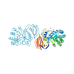
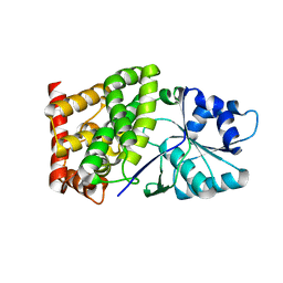
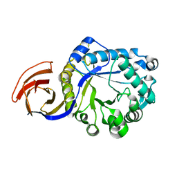
3KL3
DownloadVisualize
BU of 3kl3 by Molmil
Crystal structure of Ligand bound XynC
Descriptor: D-HISTIDINE, Glucuronoxylanase xynC, TETRAETHYLENE GLYCOL, ...
Authors:St John, F.J, Hurlbert, J.C, Pozharski, E.
Deposit date:2009-11-06
Release date:2010-12-08
Last modified:2023-09-06
Method:X-RAY DIFFRACTION (2.33 Å)
Cite:Ligand bound structures of a glycosyl hydrolase family 30 glucuronoxylan xylanohydrolase.
J.Mol.Biol., 407, 2011
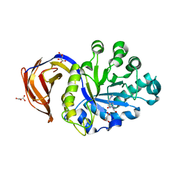
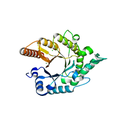
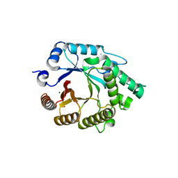
5CXP
DownloadVisualize
BU of 5cxp by Molmil
X-ray crystallographic protein structure of the glycoside hydrolase family 30 subfamily 8 xylanase, Xyn30A, from Clostridium acetobutylicum
Descriptor: CHLORIDE ION, HEXAETHYLENE GLYCOL, Possible xylan degradation enzyme (Glycosyl hydrolase family 30-like domain and Ricin B-like domain), ...
Authors:St John, F.J, Pozharski, E, Hurlbert, J.C.
Deposit date:2015-07-29
Release date:2016-08-10
Last modified:2023-09-27
Method:X-RAY DIFFRACTION (1.77 Å)
Cite:Crystal structure of a GH30 xylanase
To Be Published
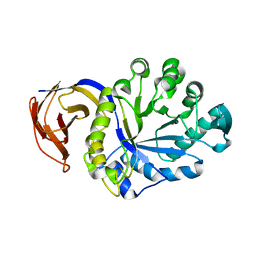
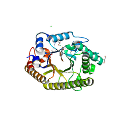
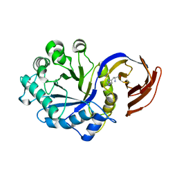
3KL0
DownloadVisualize
BU of 3kl0 by Molmil
Crystal structure of the glucuronoxylan xylanohydrolase XynC from Bacillus subtilis
Descriptor: D(-)-TARTARIC ACID, Glucuronoxylanase xynC, HISTIDINE, ...
Authors:St John, F.J, Hurlbert, J.C, Pozharski, E.
Deposit date:2009-11-06
Release date:2010-12-08
Last modified:2024-03-13
Method:X-RAY DIFFRACTION (1.64 Å)
Cite:Ligand bound structures of a glycosyl hydrolase family 30 glucuronoxylan xylanohydrolase.
J.Mol.Biol., 407, 2011
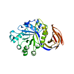
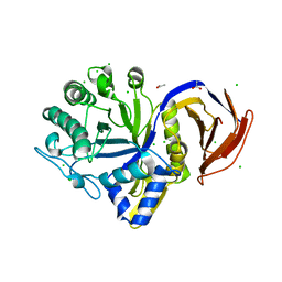
3KL5
DownloadVisualize
BU of 3kl5 by Molmil
Structure Analysis of a Xylanase From Glycosyl Hydrolase Family Thirty: Carbohydrate Ligand Complexes Reveal this Family of Enzymes Unique Mechanism of Substrate Specificity and Recognition
Descriptor: 4-O-methyl-alpha-D-glucopyranuronic acid-(1-2)-beta-D-xylopyranose-(1-4)-beta-D-xylopyranose, Glucuronoxylanase xynC
Authors:St John, F.J, Hurlbert, J.C, Pozharski, E.
Deposit date:2009-11-06
Release date:2010-12-08
Last modified:2023-09-06
Method:X-RAY DIFFRACTION (2.59 Å)
Cite:Ligand bound structures of a glycosyl hydrolase family 30 glucuronoxylan xylanohydrolase.
J.Mol.Biol., 407, 2011
3GTN
DownloadVisualize
BU of 3gtn by Molmil
Crystal Structure of XynC from Bacillus subtilis 168
Descriptor: Glucuronoxylanase xynC
Authors:St John, F.J, Hurlbert, J.C, Pozharski, E.
Deposit date:2009-03-27
Release date:2009-04-28
Last modified:2024-02-21
Method:X-RAY DIFFRACTION (2.68 Å)
Cite:Crystallization and crystallographic analysis of Bacillus subtilis xylanase C.
Acta Crystallogr.,Sect.F, 65, 2009
6CSJ
DownloadVisualize
BU of 6csj by Molmil
Structure of a Bacillus coagulans polyol dehydrogenase double mutant with an acquired D-lactate dehydrogenase activity
Descriptor: Glycerol dehydrogenase
Authors:Hurlbert, J.C, St.John, F.J.
Deposit date:2018-03-20
Release date:2019-07-03
Last modified:2023-10-04
Method:X-RAY DIFFRACTION (2.395 Å)
Cite:Kinetic characterization and structure analysis of an altered polyol dehydrogenase with d-lactate dehydrogenase activity.
Protein Sci., 29, 2020
3RO8
DownloadVisualize
BU of 3ro8 by Molmil
Crystal structure of the catalytic domain of XynA1 from Paenibacillus sp. JDR-2
Descriptor: CHLORIDE ION, Endo-1,4-beta-xylanase, MAGNESIUM ION
Authors:Pozharski, E, St John, F.J.
Deposit date:2011-04-25
Release date:2012-05-16
Last modified:2024-02-28
Method:X-RAY DIFFRACTION (1.34 Å)
Cite:Novel structural features of xylanase A1 from Paenibacillus sp. JDR-2.
J.Struct.Biol., 180, 2012
3RDK
DownloadVisualize
BU of 3rdk by Molmil
Protein crystal structure of xylanase A1 of Paenibacillus sp. JDR-2
Descriptor: 4-O-methyl-alpha-D-glucopyranuronic acid-(1-2)-beta-D-xylopyranose-(1-4)-beta-D-xylopyranose-(1-4)-alpha-D-xylopyranose, CHLORIDE ION, Endo-1,4-beta-xylanase, ...
Authors:Pozharski, E, St John, F.J.
Deposit date:2011-04-01
Release date:2012-04-04
Last modified:2024-02-21
Method:X-RAY DIFFRACTION (1.49 Å)
Cite:Novel structural features of xylanase A1 from Paenibacillus sp. JDR-2.
J.Struct.Biol., 180, 2012
7N6H
DownloadVisualize
BU of 7n6h by Molmil
The crystal structure of the GH30 subfamily 10 enzyme, AcXbh30A from Acetivibrio clariflavus
Descriptor: ACETATE ION, AcXbh30A, CHLORIDE ION
Authors:Tan, K, St John, F.J.
Deposit date:2021-06-08
Release date:2022-08-17
Last modified:2023-10-18
Method:X-RAY DIFFRACTION (1.28 Å)
Cite:The first crystal structure of a xylobiose-bound xylobiohydrolase with high functional specificity from the bacterial glycoside hydrolase family 30, subfamily 10.
Febs Lett., 596, 2022
4FMV
DownloadVisualize
BU of 4fmv by Molmil
Crystal Structure Analysis of a GH30 Endoxylanase from Clostridium papyrosolvens C71
Descriptor: Glucuronoarabinoxylan endo-1,4-beta-xylanase
Authors:Bales, E.B, Smith, J.K, St John, F.J, Hurlbert, J.C.
Deposit date:2012-06-18
Release date:2013-06-19
Last modified:2024-02-28
Method:X-RAY DIFFRACTION (2.01 Å)
Cite:A novel member of glycoside hydrolase family 30 subfamily 8 with altered substrate specificity.
Acta Crystallogr.,Sect.D, 70, 2014
4E4P
DownloadVisualize
BU of 4e4p by Molmil
Second native structure of Xylanase A1 from Paenibacillus sp. JDR-2
Descriptor: CHLORIDE ION, Endo-1,4-beta-xylanase A, MAGNESIUM ION
Authors:Pozharski, E, St John, F.J.
Deposit date:2012-03-13
Release date:2012-04-18
Last modified:2024-02-28
Method:X-RAY DIFFRACTION (1.92 Å)
Cite:Novel structural features of xylanase A1 from Paenibacillus sp. JDR-2.
J.Struct.Biol., 180, 2012

226707

數據於2024-10-30公開中

PDB statisticsPDBj update infoContact PDBjnumon