5UXT
 
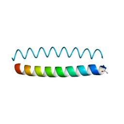
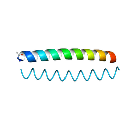
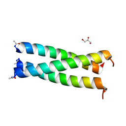 | | Coiled-coil Trimer with Glu:Trp:Lys Triad | | 分子名称: | GLYCEROL, coiled-coil trimer with Glu:Trp:Lys triad | | 著者 | Smith, M.S, Billings, W.M, Whitby, F.G, Miller, M.B, Price, J.L. | | 登録日 | 2017-02-23 | | 公開日 | 2017-08-09 | | 実験手法 | X-RAY DIFFRACTION (2.197 Å) | | 主引用文献 | Enhancing a long-range salt bridge with intermediate aromatic and nonpolar amino acids.
Org. Biomol. Chem., 15, 2017
|
|
3EV0
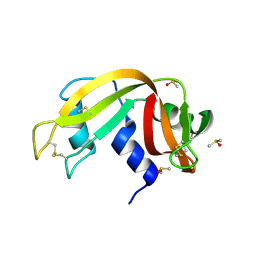 | | Crystal Structure of Ribonuclease A in 70% Dimethyl Sulfoxide | | 分子名称: | DIMETHYL SULFOXIDE, Ribonuclease pancreatic | | 著者 | Dechene, M, Wink, G, Smith, M, Swartz, P, Mattos, C. | | 登録日 | 2008-10-12 | | 公開日 | 2009-06-23 | | 最終更新日 | 2023-12-27 | | 実験手法 | X-RAY DIFFRACTION (1.76 Å) | | 主引用文献 | Multiple solvent crystal structures of ribonuclease A: An assessment of the method
Proteins, 76, 2009
|
|
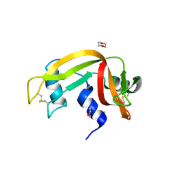
3EV3
 | | Crystal Structure of Ribonuclease A in 70% t-Butanol | | 分子名称: | Ribonuclease pancreatic, TERTIARY-BUTYL ALCOHOL | | 著者 | Dechene, M, Wink, G, Smith, M, Swartz, P, Mattos, C. | | 登録日 | 2008-10-12 | | 公開日 | 2009-06-23 | | 最終更新日 | 2023-12-27 | | 実験手法 | X-RAY DIFFRACTION (1.68 Å) | | 主引用文献 | Multiple solvent crystal structures of ribonuclease A: An assessment of the method
Proteins, 76, 2009
|
|
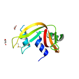
3EUY
 | | Crystal Structure of Ribonuclease A in 50% Dioxane | | 分子名称: | 1,4-DIETHYLENE DIOXIDE, Ribonuclease pancreatic | | 著者 | Dechene, M, Wink, G, Smith, M, Swartz, P, Mattos, C. | | 登録日 | 2008-10-12 | | 公開日 | 2009-06-23 | | 最終更新日 | 2023-12-27 | | 実験手法 | X-RAY DIFFRACTION (1.95 Å) | | 主引用文献 | Multiple solvent crystal structures of ribonuclease A: An assessment of the method
Proteins, 76, 2009
|
|
3EV6
 | | Crystal Structure of Ribonuclease A in 50% R,S,R-Bisfuranol | | 分子名称: | (3R,3aS,6aR)-hexahydrofuro[2,3-b]furan-3-ol, Ribonuclease pancreatic | | 著者 | Dechene, M, Wink, G, Smith, M, Swartz, P, Mattos, C. | | 登録日 | 2008-10-12 | | 公開日 | 2009-06-23 | | 最終更新日 | 2023-12-27 | | 実験手法 | X-RAY DIFFRACTION (1.76 Å) | | 主引用文献 | Multiple solvent crystal structures of ribonuclease A: An assessment of the method
Proteins, 76, 2009
|
|
3EUX
 | | Crystal Structure of Crosslinked Ribonuclease A | | 分子名称: | Ribonuclease pancreatic | | 著者 | Dechene, M, Wink, G, Smith, M, Swartz, P, Mattos, C. | | 登録日 | 2008-10-12 | | 公開日 | 2009-06-23 | | 最終更新日 | 2023-12-27 | | 実験手法 | X-RAY DIFFRACTION (1.65 Å) | | 主引用文献 | Multiple solvent crystal structures of ribonuclease A: An assessment of the method
Proteins, 76, 2009
|
|
3EV4
 | | Crystal Structure of Ribonuclease A in 50% Trifluoroethanol | | 分子名称: | Ribonuclease pancreatic, TRIFLUOROETHANOL | | 著者 | Dechene, M, Wink, G, Smith, M, Swartz, P, Mattos, C. | | 登録日 | 2008-10-12 | | 公開日 | 2009-06-23 | | 最終更新日 | 2023-12-27 | | 実験手法 | X-RAY DIFFRACTION (1.93 Å) | | 主引用文献 | Multiple solvent crystal structures of ribonuclease A: An assessment of the method
Proteins, 76, 2009
|
|
3EUZ
 | | Crystal Structure of Ribonuclease A in 50% Dimethylformamide | | 分子名称: | DIMETHYLFORMAMIDE, Ribonuclease pancreatic | | 著者 | Dechene, M, Wink, G, Smith, M, Swartz, P, Mattos, C. | | 登録日 | 2008-10-12 | | 公開日 | 2009-06-23 | | 最終更新日 | 2023-12-27 | | 実験手法 | X-RAY DIFFRACTION (1.84 Å) | | 主引用文献 | Multiple solvent crystal structures of ribonuclease A: An assessment of the method
Proteins, 76, 2009
|
|
3EV1
 | | Crystal Structure of Ribonuclease A in 70% Hexanediol | | 分子名称: | HEXANE-1,6-DIOL, Ribonuclease pancreatic | | 著者 | Dechene, M, Wink, G, Smith, M, Swartz, P, Mattos, C. | | 登録日 | 2008-10-12 | | 公開日 | 2009-06-23 | | 最終更新日 | 2023-12-27 | | 実験手法 | X-RAY DIFFRACTION (2 Å) | | 主引用文献 | Multiple solvent crystal structures of ribonuclease A: An assessment of the method
Proteins, 76, 2009
|
|
3EV2
 | | Crystal Structure of Ribonuclease A in 70% Isopropanol | | 分子名称: | ISOPROPYL ALCOHOL, Ribonuclease pancreatic | | 著者 | Dechene, M, Wink, G, Smith, M, Swartz, P, Mattos, C. | | 登録日 | 2008-10-12 | | 公開日 | 2009-06-23 | | 最終更新日 | 2023-12-27 | | 実験手法 | X-RAY DIFFRACTION (2.02 Å) | | 主引用文献 | Multiple solvent crystal structures of ribonuclease A: An assessment of the method
Proteins, 76, 2009
|
|
3EV5
 | | Crystal Structure of Ribonuclease A in 1M Trimethylamine N-Oxide | | 分子名称: | Ribonuclease pancreatic, trimethylamine oxide | | 著者 | Dechene, M, Wink, G, Smith, M, Swartz, P, Mattos, C. | | 登録日 | 2008-10-12 | | 公開日 | 2009-06-23 | | 最終更新日 | 2023-12-27 | | 実験手法 | X-RAY DIFFRACTION (1.68 Å) | | 主引用文献 | Multiple solvent crystal structures of ribonuclease A: An assessment of the method
Proteins, 76, 2009
|
|
4JXI
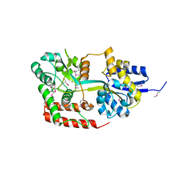 | | Directed evolution and rational design of a de novo designed esterase toward improved catalysis. Northeast Structural Genomics Consortium (NESG) Target OR184 | | 分子名称: | 3,6,9,12,15,18,21,24-OCTAOXAHEXACOSAN-1-OL, BROMIDE ION, GLYCEROL, ... | | 著者 | Kuzin, A, Smith, M.D, Richter, F, Lew, S, Seetharaman, R, Bryan, C, Lech, Z, Kiss, G, Moretti, R, Maglaqui, M, Xiao, R, Kohan, E, Smith, M, Everett, J.K, Nguyen, R, Pande, V, Hilvert, D, Kornhaber, G, Baker, D, Montelione, G.T, Hunt, J.F, Tong, L, Northeast Structural Genomics Consortium (NESG) | | 登録日 | 2013-03-28 | | 公開日 | 2013-05-22 | | 最終更新日 | 2023-12-06 | | 実験手法 | X-RAY DIFFRACTION (2.29 Å) | | 主引用文献 | Directed evolution and rational design of a de novo designed esterase toward improved catalysis. Northeast Structural Genomics Consortium (NESG) Target OR184
To be Published
|
|
8F5W
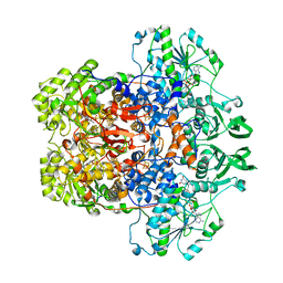
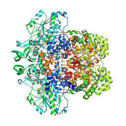 | | Dihydropyrimidine Dehydrogenase (DPD) C671S Mutant Soaked with Dihydrothymine and NADPH Quasi-Anaerobically | | 分子名称: | 1-DEOXY-1-(7,8-DIMETHYL-2,4-DIOXO-3,4-DIHYDRO-2H-BENZO[G]PTERIDIN-1-ID-10(5H)-YL)-5-O-PHOSPHONATO-D-RIBITOL, Dihydropyrimidine dehydrogenase [NADP(+)], FLAVIN-ADENINE DINUCLEOTIDE, ... | | 著者 | Kaley, N, Smith, M, Forouzesh, D, Liu, D, Moran, G. | | 登録日 | 2022-11-15 | | 公開日 | 2023-02-01 | | 最終更新日 | 2023-10-25 | | 実験手法 | X-RAY DIFFRACTION (1.97 Å) | | 主引用文献 | Mammalian dihydropyrimidine dehydrogenase: Added mechanistic details from transient-state analysis of charge transfer complexes.
Arch.Biochem.Biophys., 736, 2023
|
|
8F6N
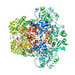
 | | Dihydropyrimidine Dehydrogenase (DPD) C671S Mutant Soaked with Thymine Quasi-Anaerobically | | 分子名称: | Dihydropyrimidine dehydrogenase [NADP(+)], FLAVIN MONONUCLEOTIDE, FLAVIN-ADENINE DINUCLEOTIDE, ... | | 著者 | Kaley, N, Smith, M, Forouzesh, D, Liu, D, Moran, G. | | 登録日 | 2022-11-16 | | 公開日 | 2023-02-01 | | 最終更新日 | 2023-10-25 | | 実験手法 | X-RAY DIFFRACTION (2.12 Å) | | 主引用文献 | Mammalian dihydropyrimidine dehydrogenase: Added mechanistic details from transient-state analysis of charge transfer complexes.
Arch.Biochem.Biophys., 736, 2023
|
|
8F61
 | | Dihydropyrimidine Dehydrogenase (DPD) C671S Mutant Soaked with Dihydrothymine Quasi-Anaerobically | | 分子名称: | (5S)-5-methyl-1,3-diazinane-2,4-dione, 1-DEOXY-1-(7,8-DIMETHYL-2,4-DIOXO-3,4-DIHYDRO-2H-BENZO[G]PTERIDIN-1-ID-10(5H)-YL)-5-O-PHOSPHONATO-D-RIBITOL, Dihydropyrimidine dehydrogenase [NADP(+)], ... | | 著者 | Kaley, N, Smith, M, Forouzesh, D, Liu, D, Moran, G. | | 登録日 | 2022-11-15 | | 公開日 | 2023-02-01 | | 最終更新日 | 2023-10-25 | | 実験手法 | X-RAY DIFFRACTION (2.14 Å) | | 主引用文献 | Mammalian dihydropyrimidine dehydrogenase: Added mechanistic details from transient-state analysis of charge transfer complexes.
Arch.Biochem.Biophys., 736, 2023
|
|
6O2E
 | |
6O2F
 | |
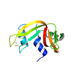
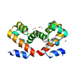
6URJ
 | | Barrier-to-autointegration factor soaked in Acetone: 1 of 14 in MSCS set | | 分子名称: | Barrier-to-autointegration factor, ETHANOL | | 著者 | Agarwal, S, Smith, M, De La Rosa, I, Kliment, A.V, Swartz, P, Segura-Totten, M, Mattos, C. | | 登録日 | 2019-10-23 | | 公開日 | 2020-10-07 | | 最終更新日 | 2024-03-13 | | 実験手法 | X-RAY DIFFRACTION (1.653 Å) | | 主引用文献 | Development of a structure-analysis pipeline using multiple-solvent crystal structures of barrier-to-autointegration factor.
Acta Crystallogr D Struct Biol, 76, 2020
|
|
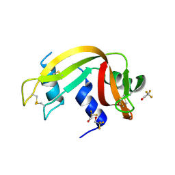
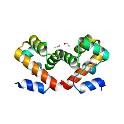
6USB
 | | Barrier-to-autointegration factor soaked in urea: 1 of 14 in MSCS set | | 分子名称: | Barrier-to-autointegration factor, ETHANOL | | 著者 | Agarwal, S, Smith, M, De La Rosa, I, Kliment, A.V, Swartz, P, Segura-Totten, M, Mattos, C. | | 登録日 | 2019-10-25 | | 公開日 | 2020-10-07 | | 最終更新日 | 2023-10-11 | | 実験手法 | X-RAY DIFFRACTION (1.68 Å) | | 主引用文献 | Development of a structure-analysis pipeline using multiple-solvent crystal structures of barrier-to-autointegration factor.
Acta Crystallogr D Struct Biol, 76, 2020
|
|
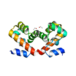
6US1
 | | Barrier-to-autointegration factor soaked in isobutanol: 1 of 14 in MSCS set | | 分子名称: | Barrier-to-autointegration factor, ETHANOL | | 著者 | Agarwal, S, Smith, M, De La Rosa, I, Kliment, A.V, Swartz, P, Segura-Totten, M, Mattos, C. | | 登録日 | 2019-10-24 | | 公開日 | 2020-10-07 | | 最終更新日 | 2024-03-13 | | 実験手法 | X-RAY DIFFRACTION (1.653 Å) | | 主引用文献 | Development of a structure-analysis pipeline using multiple-solvent crystal structures of barrier-to-autointegration factor.
Acta Crystallogr D Struct Biol, 76, 2020
|
|
6US7
 | | Barrier-to-autointegration factor soaked in TMAO: 1 of 14 in MSCS set | | 分子名称: | Barrier-to-autointegration factor, ETHANOL | | 著者 | Agarwal, S, Smith, M, De La Rosa, I, Kliment, A.V, Swartz, P, Segura-Totten, M, Mattos, C. | | 登録日 | 2019-10-24 | | 公開日 | 2020-10-07 | | 最終更新日 | 2024-03-13 | | 実験手法 | X-RAY DIFFRACTION (1.653 Å) | | 主引用文献 | Development of a structure-analysis pipeline using multiple-solvent crystal structures of barrier-to-autointegration factor.
Acta Crystallogr D Struct Biol, 76, 2020
|
|
6UNT
 | | Barrier-to-autointegration factor soaked in DMSO: 1 of 14 in MSCS set | | 分子名称: | Barrier-to-autointegration factor, ETHANOL | | 著者 | Agarwal, S, Smith, M, De La Rosa, I, Kliment, A.V, Swartz, P, Segura-Totten, M, Mattos, C. | | 登録日 | 2019-10-13 | | 公開日 | 2020-10-07 | | 最終更新日 | 2024-03-13 | | 実験手法 | X-RAY DIFFRACTION (1.75 Å) | | 主引用文献 | Development of a structure-analysis pipeline using multiple-solvent crystal structures of barrier-to-autointegration factor.
Acta Crystallogr D Struct Biol, 76, 2020
|
|
6URZ
 | | Barrier-to-autointegration factor soaked in methanol: 1 of 14 in MSCS set | | 分子名称: | Barrier-to-autointegration factor, ETHANOL, METHANOL | | 著者 | Agarwal, S, Smith, M, De La Rosa, I, Kliment, A.V, Swartz, P, Segura-Totten, M, Mattos, C. | | 登録日 | 2019-10-24 | | 公開日 | 2020-10-07 | | 最終更新日 | 2024-03-13 | | 実験手法 | X-RAY DIFFRACTION (1.653 Å) | | 主引用文献 | Development of a structure-analysis pipeline using multiple-solvent crystal structures of barrier-to-autointegration factor.
Acta Crystallogr D Struct Biol, 76, 2020
|
|
6URN
 | | Barrier-to-autointegration factor t-butanol: 1 of 14 in MSCS set | | 分子名称: | Barrier-to-autointegration factor, ETHANOL | | 著者 | Agarwal, S, Smith, M, De La Rosa, I, Kliment, A.V, Swartz, P, Segura-Totten, M, Mattos, C. | | 登録日 | 2019-10-23 | | 公開日 | 2020-10-07 | | 最終更新日 | 2024-03-13 | | 実験手法 | X-RAY DIFFRACTION (1.68 Å) | | 主引用文献 | Development of a structure-analysis pipeline using multiple-solvent crystal structures of barrier-to-autointegration factor.
Acta Crystallogr D Struct Biol, 76, 2020
|
|
6US0
 | | Barrier-to-autointegration factor soaked in R,S,R-bisfuranol (RSR): 1 of 14 in MSCS set | | 分子名称: | Barrier-to-autointegration factor, ETHANOL | | 著者 | Agarwal, S, Smith, M, De La Rosa, I, Kliment, A.V, Swartz, P, Segura-Totten, M, Mattos, C. | | 登録日 | 2019-10-24 | | 公開日 | 2020-10-07 | | 最終更新日 | 2024-03-13 | | 実験手法 | X-RAY DIFFRACTION (1.653 Å) | | 主引用文献 | Development of a structure-analysis pipeline using multiple-solvent crystal structures of barrier-to-autointegration factor.
Acta Crystallogr D Struct Biol, 76, 2020
|
|


