Loading
PDBj
メニューPDBj@FacebookPDBj@TwitterPDBj@YouTubewwPDB FoundationwwPDB
RCSB PDBPDBeBMRBAdv. SearchSearch help
PDB: 335 件

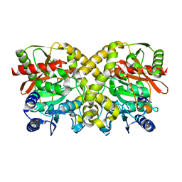
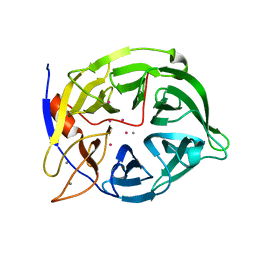
1CVM
DownloadVisualize
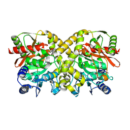
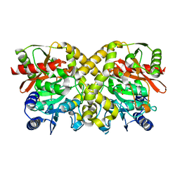
BU of 1cvm by Molmil
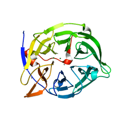
CADMIUM INHIBITED CRYSTAL STRUCTURE OF PHYTASE FROM BACILLUS AMYLOLIQUEFACIENS
分子名称: CADMIUM ION, CALCIUM ION, PHYTASE
著者Shin, S, Ha, N.-C, Oh, B.-H.
登録日1999-08-24
公開日2000-02-24
最終更新日2024-02-07
実験手法X-RAY DIFFRACTION (2.4 Å)
主引用文献Crystal structures of a novel, thermostable phytase in partially and fully calcium-loaded states.
Nat.Struct.Biol., 7, 2000
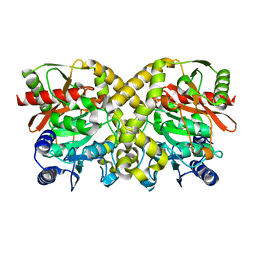
1GS3
DownloadVisualize
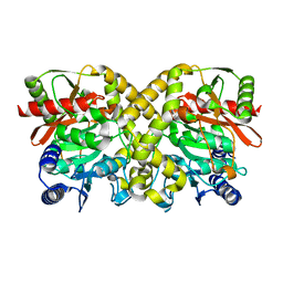
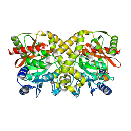
BU of 1gs3 by Molmil
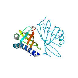
High resolution crystal structure of PI delta-5-3-Ketosteroid Isomerase mutants Y30F/Y55F/Y115F/D38N (Y32F/Y57F/Y119F/D40N, PI numbering)complexed with equilenin at 2.1 A resolution
分子名称: EQUILENIN, STEROID DELTA-ISOMERASE
著者Shin, S, Ha, N.-C, Oh, B.-H.
登録日2001-12-27
公開日2003-01-23
最終更新日2023-12-13
実験手法X-RAY DIFFRACTION (2.1 Å)
主引用文献Contribution of a Low-Barrier Hydrogen Bond to Catalysis by Delta-5-3-Ketosteroid Isomerase is not Extremely High Compared to that of an Ordinary Hydrogen Bond. Low-Barrier Hydrogen Bond of Pi Ksi
To be Published
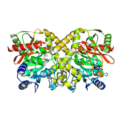
1H6L
DownloadVisualize
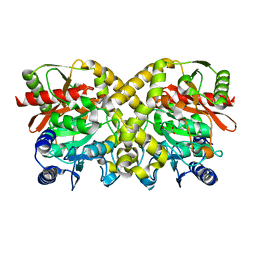
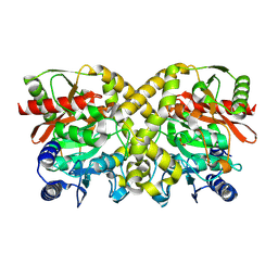
BU of 1h6l by Molmil
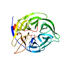
beta-propeller phytase in complex with phosphate and calcium ions
分子名称: 3-PHYTASE, CALCIUM ION, PHOSPHATE ION
著者Shin, S, Ha, N.C, Oh, B.H.
登録日2001-06-19
公開日2001-08-29
最終更新日2023-12-13
実験手法X-RAY DIFFRACTION (1.8 Å)
主引用文献Enzyme Mechanism and Catalytic Property of Beta Propeller Phytase
Structure, 9, 2001
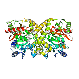
1QLG
DownloadVisualize
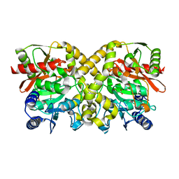
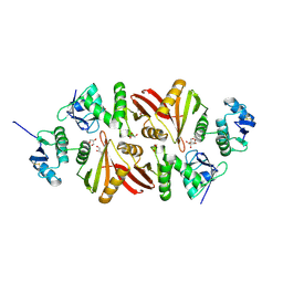
BU of 1qlg by Molmil
Crystal structure of phytase with magnesium from Bacillus amyloliquefaciens
分子名称: 3-PHYTASE, CALCIUM ION, MAGNESIUM ION
著者Shin, S, Ha, N.-C, Oh, B.-H.
登録日1999-08-31
公開日2000-02-03
最終更新日2023-12-13
実験手法X-RAY DIFFRACTION (2.2 Å)
主引用文献Crystal Structures of a Novel, Thermostable Phytase in Partially and Fully Calcium-Loaded States
Nat.Struct.Biol., 7, 2000
1O9N
DownloadVisualize
BU of 1o9n by Molmil
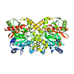
Crystal structure of the K62A mutant of Malonamidase E2 from Bradyrhizobium japonicum
分子名称: MALONAMIDASE E2
著者Shin, S, Oh, B.-H.
登録日2002-12-18
公開日2003-12-18
最終更新日2023-12-13
実験手法X-RAY DIFFRACTION (2 Å)
主引用文献Characterization of a Novel Ser-Cisser-Lys Catalytic Triad in Comparison with the Classical Ser-His-Asp Triad
J.Biol.Chem., 278, 2003
1OBI
DownloadVisualize
BU of 1obi by Molmil
Crystal structure of the G130A mutant of Malonamidase E2 from Bradyrhizobium japonicum
分子名称: MALONAMIDASE E2
著者Shin, S, Oh, B.-H.
登録日2003-01-31
公開日2003-12-18
最終更新日2023-12-13
実験手法X-RAY DIFFRACTION (2.2 Å)
主引用文献Characterization of a Novel Ser-Cisser-Lys Catalytic Triad in Comparison with the Classical Ser-His-Asp Triad
J.Biol.Chem., 278, 2003
1OBK
DownloadVisualize
BU of 1obk by Molmil
crystal structure of the R158Q mutant of Malonamidase E2 from Bradyrhizobium japonicum
分子名称: MALONAMIDASE E2
著者Shin, S, Oh, B.-H.
登録日2003-01-31
公開日2004-02-13
最終更新日2023-12-13
実験手法X-RAY DIFFRACTION (2.2 Å)
主引用文献Characterization of a Novel Ser-Cisser-Lys Catalytic Triad in Comparison with the Classical Ser-His-Asp Triad.
J.Biol.Chem., 278, 2003
1OCM
DownloadVisualize
BU of 1ocm by Molmil
THE CRYSTAL STRUCTURE OF MALONAMIDASE E2 COVALENTLY COMPLEXED WITH PYROPHOSPHATE FROM BRADYRHIZOBIUM JAPONICUM
分子名称: MALONAMIDASE E2, PYROPHOSPHATE 2-
著者Shin, S, Ha, N.-C, Lee, T.-H, Oh, B.-H.
登録日2003-02-08
公開日2003-02-25
最終更新日2024-10-09
実験手法X-RAY DIFFRACTION (1.9 Å)
主引用文献Characterization of a Novel Ser-Cisser-Lys Catalytic Triad in Comparison with the Classical Ser-His-Asp Triad.
J.Biol.Chem., 278, 2003
1O9O
DownloadVisualize
BU of 1o9o by Molmil
Crystal structure of the S131A mutant of Malonamidase E2 complexed with malonamate from Bradyrhizobium japonicum
分子名称: 3-AMINO-3-OXOPROPANOIC ACID, MALONAMIDASE E2
著者Shin, S, Oh, B.-H.
登録日2002-12-18
公開日2003-12-18
最終更新日2023-12-13
実験手法X-RAY DIFFRACTION (2.3 Å)
主引用文献Characterization of a Novel Ser-Cisser-Lys Catalytic Triad in Comparison with the Classical Ser-His-Asp Triad
J.Biol.Chem., 278, 2003
1O9Q
DownloadVisualize
BU of 1o9q by Molmil
Crystal structure of the S155C mutant of Malonamidase E2 from Bradyrhizobium japonicum
分子名称: MALONAMIDASE E2
著者Shin, S, Oh, B.-H.
登録日2002-12-18
公開日2003-12-18
最終更新日2023-12-13
実験手法X-RAY DIFFRACTION (1.8 Å)
主引用文献Characterization of a Novel Ser-Cisser-Lys Catalytic Triad in Comparison with the Classical Ser-His-Asp Triad
J.Biol.Chem., 278, 2003
1OCK
DownloadVisualize
BU of 1ock by Molmil
THE CRYSTAL STRUCTURE OF MALONAMIDASE E2 FROM BRADYRHIZOBIUM JAPONICUM
分子名称: MALONAMIDASE E2
著者Shin, S, Ha, N.-C, Lee, T.-H, Oh, B.-H.
登録日2003-02-08
公開日2003-03-03
最終更新日2024-05-08
実験手法X-RAY DIFFRACTION (1.8 Å)
主引用文献Characterization of a Novel Ser-Cisser-Lys Catalytic Triad in Comparison with the Classical Ser-His-Asp Triad.
J.Biol.Chem., 278, 2003
1O9P
DownloadVisualize
BU of 1o9p by Molmil
Crystal structure of the S131A mutant of Malonamidase E2 complexed with malonate from Bradyrhizobium japonicum
分子名称: MALONAMIDASE E2, MALONIC ACID
著者Shin, S, Oh, B.-H.
登録日2002-12-18
公開日2003-12-18
最終更新日2023-12-13
実験手法X-RAY DIFFRACTION (1.8 Å)
主引用文献Characterization of a Novel Ser-Cisser-Lys Catalytic Triad in Comparison with the Classical Ser-His-Asp Triad
J.Biol.Chem., 278, 2003
1OCL
DownloadVisualize
BU of 1ocl by Molmil
THE CRYSTAL STRUCTURE OF MALONAMIDASE E2 COMPLEXED WITH MALONATE FROM BRADYRHIZOBIUM JAPONICUM
分子名称: MALONAMIDASE E2, MALONIC ACID
著者Shin, S, Ha, N.-C, Lee, T.-H, Oh, B.-H.
登録日2003-02-08
公開日2003-02-25
最終更新日2023-12-13
実験手法X-RAY DIFFRACTION (2 Å)
主引用文献Characterization of a Novel Ser-Cisser-Lys Catalytic Triad in Comparison with the Classical Ser-His-Asp Triad.
J.Biol.Chem., 278, 2003
1OBL
DownloadVisualize
BU of 1obl by Molmil
crystal structure of the S133A mutant of Malonamidase E2 complexed with malonate from Bradyrhizobium japonicum
分子名称: MALONAMIDASE E2, MALONIC ACID
著者Shin, S, Oh, B.-H.
登録日2003-01-31
公開日2004-02-13
最終更新日2023-12-13
実験手法X-RAY DIFFRACTION (2 Å)
主引用文献Characterization of a Novel Ser-Cisser-Lys Catalytic Triad in Comparison with the Classical Ser-His-Asp Triad.
J.Biol.Chem., 278, 2003
1OBJ
DownloadVisualize
BU of 1obj by Molmil
Crystal structure of the T150A mutant of Malonamidase E2 from Bradyrhizobium japonicum
分子名称: MALONAMIDASE E2
著者Shin, S, Oh, B.-H.
登録日2003-01-31
公開日2003-12-18
最終更新日2023-12-13
実験手法X-RAY DIFFRACTION (1.9 Å)
主引用文献Characterization of a Novel Ser-Cisser-Lys Catalytic Triad in Comparison with the Classical Ser-His-Asp Triad
J.Biol.Chem., 278, 2003
1OCH
DownloadVisualize
BU of 1och by Molmil
Crystal structure of the S155C mutant of malonamidase E2 from Bradyrhizobium japonicum
分子名称: MALONAMIDASE E2
著者Shin, S, Oh, B.-H.
登録日2003-02-07
公開日2003-12-18
最終更新日2023-12-13
実験手法X-RAY DIFFRACTION (1.8 Å)
主引用文献Characterization of a Novel Ser-Cisser-Lys Catalytic Triad in Comparison with the Classical Ser-His-Asp Triad
J.Biol.Chem., 278, 2003
3AJV
DownloadVisualize
BU of 3ajv by Molmil
Splicing endonuclease from Aeropyrum pernix
分子名称: CHLORIDE ION, GLYCEROL, Putative uncharacterized protein, ...
著者Yoshinari, S, Watanabe, Y, Okuda, M, Shiba, T, Inaoka, K.D, Kurisu, G.
登録日2010-06-19
公開日2010-11-17
最終更新日2024-10-30
実験手法X-RAY DIFFRACTION (1.7 Å)
主引用文献A Conserved Lysine Residue in the Crenarchaea-Specific Loop is Important for the Crenarchaeal Splicing Endonuclease Activity.
J.Mol.Biol., 405, 2011
2DEP
DownloadVisualize
BU of 2dep by Molmil
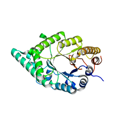
Crystal Structure of xylanase B from Clostridium stercorarium F9
分子名称: Thermostable celloxylanase
著者Fushinobu, S, Nishimoto, M, Miyanaga, A, Kitaoka, M, Hayashi, K.
登録日2006-02-16
公開日2007-01-16
最終更新日2023-10-25
実験手法X-RAY DIFFRACTION (1.8 Å)
主引用文献Molecular anatomy of the alkaliphilic xylanase from Bacillus halodurans C-125
To be Published
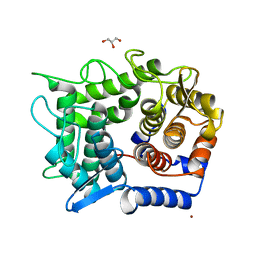
2DRR
DownloadVisualize
BU of 2drr by Molmil
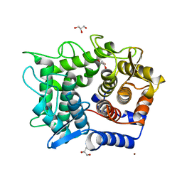
Crystal structure of reducing-end-xylose releasing exo-oligoxylanase D263N mutant
分子名称: GLYCEROL, NICKEL (II) ION, Xylanase Y
著者Fushinobu, S, Hidaka, M, Honda, Y, Wakagi, T, Shoun, H, Kitaoka, M.
登録日2006-06-12
公開日2006-06-27
最終更新日2023-10-25
実験手法X-RAY DIFFRACTION (1.6 Å)
主引用文献Structural explanation for the acquisition of glycosynthase activity
J.Biochem., 2009
2DRS
DownloadVisualize
BU of 2drs by Molmil
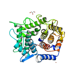
Crystal structure of reducing-end-xylose releasing exo-oligoxylanase D263S mutant
分子名称: GLYCEROL, NICKEL (II) ION, Xylanase Y
著者Fushinobu, S, Hidaka, M, Honda, Y, Wakagi, T, Shoun, H, Kitaoka, M.
登録日2006-06-12
公開日2006-06-27
最終更新日2023-10-25
実験手法X-RAY DIFFRACTION (2.1 Å)
主引用文献Structural explanation for the acquisition of glycosynthase activity
J.Biochem., 2009
2DRO
DownloadVisualize
BU of 2dro by Molmil
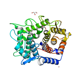
Crystal structure of reducing-end-xylose releasing exo-oligoxylanase D263C mutant
分子名称: GLYCEROL, NICKEL (II) ION, Xylanase Y
著者Fushinobu, S, Hidaka, M, Honda, Y, Wakagi, T, Shoun, H, Kitaoka, M.
登録日2006-06-12
公開日2006-06-27
最終更新日2023-10-25
実験手法X-RAY DIFFRACTION (1.7 Å)
主引用文献Structural explanation for the acquisition of glycosynthase activity
J.Biochem., 2009
2DRQ
DownloadVisualize
BU of 2drq by Molmil
Crystal structure of reducing-end-xylose releasing exo-oligoxylanase D263G mutant
分子名称: GLYCEROL, NICKEL (II) ION, Xylanase Y
著者Fushinobu, S, Hidaka, M, Honda, Y, Wakagi, T, Shoun, H, Kitaoka, M.
登録日2006-06-12
公開日2006-06-27
最終更新日2023-10-25
実験手法X-RAY DIFFRACTION (2.1 Å)
主引用文献Structural explanation for the acquisition of glycosynthase activity
J.Biochem., 2009
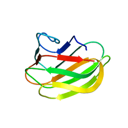
2RV9
DownloadVisualize
BU of 2rv9 by Molmil
Solution structure of chitosan-binding module 1 derived from chitosanase/glucanase from Paenibacillus sp. IK-5
分子名称: Glucanase
著者Shinya, S, Nishimura, S, Fukamizo, T.
登録日2015-05-12
公開日2016-04-06
最終更新日2024-05-15
実験手法SOLUTION NMR
主引用文献Mechanism of chitosan recognition by CBM32 carbohydrate-binding modules from a Paenibacillus sp. IK-5 chitosanase/glucanase.
Biochem.J., 473, 2016
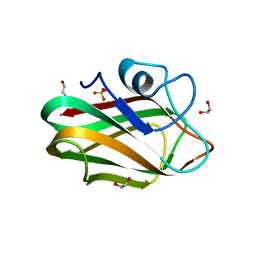
4ZXE
DownloadVisualize
BU of 4zxe by Molmil
X-ray crystal structure of chitosan-binding module 1 derived from chitosanase/glucanase from Paenibacillus sp. IK-5.
分子名称: 1,2-ETHANEDIOL, Glucanase/Chitosanase, SULFATE ION
著者Shinya, S, Oi, H, Kitaoku, Y, Ohnuma, T, Numata, T, Fukamizo, T.
登録日2015-05-20
公開日2016-04-13
最終更新日2024-03-20
実験手法X-RAY DIFFRACTION (1.4 Å)
主引用文献Mechanism of chitosan recognition by CBM32 carbohydrate-binding modules from a Paenibacillus sp. IK-5 chitosanase/glucanase
Biochem.J., 473, 2016
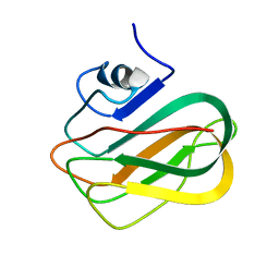
2RVA
DownloadVisualize
BU of 2rva by Molmil
Solution structure of chitosan-binding module 2 derived from chitosanase/glucanase from Paenibacillus sp. IK-5
分子名称: Glucanase
著者Shinya, S, Nishimura, S, Fukamizo, T.
登録日2015-05-13
公開日2016-04-06
最終更新日2024-05-15
実験手法SOLUTION NMR
主引用文献Mechanism of chitosan recognition by CBM32 carbohydrate-binding modules from a Paenibacillus sp. IK-5 chitosanase/glucanase.
Biochem.J., 473, 2016

227111

件を2024-11-06に公開中

PDB statisticsPDBj update infoContact PDBjnumon