1GMQ
 
 | | COMPLEX OF RIBONUCLEASE FROM STREPTOMYCES AUREOFACIENS WITH 2'-GMP AT 1.7 ANGSTROMS RESOLUTION | | 分子名称: | RIBONUCLEASE SA, SULFATE ION | | 著者 | Sevcik, J, Hill, C, Dauter, Z, Wilson, K. | | 登録日 | 1992-10-01 | | 公開日 | 1993-10-31 | | 最終更新日 | 2017-11-29 | | 実験手法 | X-RAY DIFFRACTION (1.8 Å) | | 主引用文献 | Complex of ribonuclease from Streptomyces aureofaciens with 2'-GMP at 1.7 A resolution.
Acta Crystallogr.,Sect.D, 49, 1993
|
|
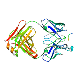
1AYX
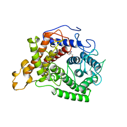 | | CRYSTAL STRUCTURE OF GLUCOAMYLASE FROM SACCHAROMYCOPSIS FIBULIGERA AT 1.7 ANGSTROMS | | 分子名称: | 2-AMINO-2-HYDROXYMETHYL-PROPANE-1,3-DIOL, GLUCOAMYLASE | | 著者 | Sevcik, J, Hostinova, E, Gasperik, J, Solovicova, A, Wilson, K.S, Dauter, Z. | | 登録日 | 1997-11-12 | | 公開日 | 1998-05-13 | | 最終更新日 | 2024-02-07 | | 実験手法 | X-RAY DIFFRACTION (1.7 Å) | | 主引用文献 | Structure of glucoamylase from Saccharomycopsis fibuligera at 1.7 A resolution.
Acta Crystallogr.,Sect.D, 54, 1998
|
|
4GHO
 | |
2F6D
 | | Structure of the complex of a glucoamylase from Saccharomycopsis fibuligera with acarbose | | 分子名称: | 4,6-dideoxy-4-{[(1S,4R,5S,6S)-4,5,6-trihydroxy-3-(hydroxymethyl)cyclohex-2-en-1-yl]amino}-alpha-D-glucopyranose-(1-4)-alpha-D-glucopyranose-(1-4)-alpha-D-glucopyranose, Glucoamylase GLU1, PHOSPHATE ION, ... | | 著者 | Sevcik, J, Hostinova, E, Solovicova, A, Gasperik, J, Dauter, Z, Wilson, K.S. | | 登録日 | 2005-11-29 | | 公開日 | 2006-05-23 | | 最終更新日 | 2023-08-23 | | 実験手法 | X-RAY DIFFRACTION (1.6 Å) | | 主引用文献 | Structure of the complex of a yeast glucoamylase with acarbose reveals the presence of a raw starch binding site on the catalytic domain.
Febs J., 273, 2006
|
|
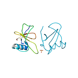
1AY7
 | | RIBONUCLEASE SA COMPLEX WITH BARSTAR | | 分子名称: | BARSTAR, GUANYL-SPECIFIC RIBONUCLEASE SA | | 著者 | Sevcik, J, Urbanikova, L, Dauter, Z, Wilson, K.S. | | 登録日 | 1997-11-14 | | 公開日 | 1999-03-02 | | 最終更新日 | 2023-08-02 | | 実験手法 | X-RAY DIFFRACTION (1.7 Å) | | 主引用文献 | Recognition of RNase Sa by the inhibitor barstar: structure of the complex at 1.7 A resolution.
Acta Crystallogr.,Sect.D, 54, 1998
|
|
2FBA
 | | Glucoamylase from Saccharomycopsis fibuligera at atomic resolution | | 分子名称: | 2-AMINO-2-HYDROXYMETHYL-PROPANE-1,3-DIOL, Glucoamylase GLU1 | | 著者 | Sevcik, J, Hostinova, E, Solovicova, A, Gasperik, J, Dauter, Z, Wilson, K.S. | | 登録日 | 2005-12-09 | | 公開日 | 2006-05-23 | | 最終更新日 | 2023-08-30 | | 実験手法 | X-RAY DIFFRACTION (1.1 Å) | | 主引用文献 | Structure of the complex of a yeast glucoamylase with acarbose reveals the presence of a raw starch binding site on the catalytic domain.
Febs J., 273, 2006
|
|
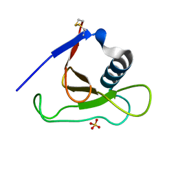
2SAR
 | | DETERMINATION AND RESTRAINED LEAST-SQUARES REFINEMENT OF THE CRYSTAL STRUCTURES OF RIBONUCLEASE SA AND ITS COMPLEX WITH 3'-GUANYLIC ACID AT 1.8 ANGSTROMS RESOLUTION | | 分子名称: | GUANOSINE-3'-MONOPHOSPHATE, RIBONUCLEASE SA, SULFATE ION | | 著者 | Sevcik, J, Dodson, E.J, Dodson, G.G. | | 登録日 | 1990-12-13 | | 公開日 | 1992-04-15 | | 最終更新日 | 2017-11-29 | | 実験手法 | X-RAY DIFFRACTION (1.8 Å) | | 主引用文献 | Determination and restrained least-squares refinement of the structures of ribonuclease Sa and its complex with 3'-guanylic acid at 1.8 A resolution.
Acta Crystallogr.,Sect.B, 47, 1991
|
|
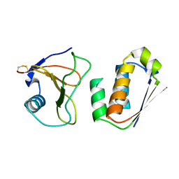
2V17
 | | Structure of the complex of antibody MN423 with a fragment of tau protein | | 分子名称: | MONOCLONAL ANTIBODY FAB FRAGMENT MN423, PEPTIDE FRAGMENT | | 著者 | Sevcik, J, Skrabana, R, Csokova, N, Dvorsky, R, Novak, M. | | 登録日 | 2007-05-22 | | 公開日 | 2007-12-25 | | 最終更新日 | 2023-12-13 | | 実験手法 | X-RAY DIFFRACTION (1.65 Å) | | 主引用文献 | X-Ray Structure of the Phf Core C-Terminus: Insight Into the Folding of the Intrinsically Disordered Protein Tau in Alzheimer'S Disease.
FEBS Lett., 581, 2007
|
|
1MGR
 | | Crystal structure of RNase Sa3,cytotoxic microbial ribonuclease | | 分子名称: | Guanyl-specific ribonuclease Sa3, SULFATE ION | | 著者 | Sevcik, J, Urbanikova, L, Leland, P.A, Raines, R.T. | | 登録日 | 2002-08-16 | | 公開日 | 2003-02-04 | | 最終更新日 | 2011-07-13 | | 実験手法 | X-RAY DIFFRACTION (1.7 Å) | | 主引用文献 | Links X-ray Structure of Two Crystalline Forms of a Streptomycete Ribonuclease with Cytotoxic Activity
J.Biol.Chem., 277, 2002
|
|
1MGW
 | | Crystal structure of RNase Sa3, cytotoxic microbial ribonuclease | | 分子名称: | Guanyl-specific ribonuclease Sa3, LITHIUM ION | | 著者 | Sevcik, J, Urbanikova, L, Leland, P.A, Raines, R.T. | | 登録日 | 2002-08-16 | | 公開日 | 2003-02-04 | | 最終更新日 | 2011-11-16 | | 実験手法 | X-RAY DIFFRACTION (2 Å) | | 主引用文献 | Links X-ray Structure of Two Crystalline Forms of a Streptomycete Ribonuclease with Cytotoxic Activity
J.Biol.Chem., 277, 2002
|
|
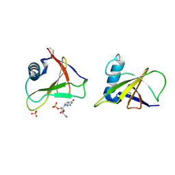
1GMP
 | | COMPLEX OF RIBONUCLEASE FROM STREPTOMYCES AUREOFACIENS WITH 2'-GMP AT 1.7 ANGSTROMS RESOLUTION | | 分子名称: | GUANOSINE-2'-MONOPHOSPHATE, RIBONUCLEASE SA, SULFATE ION | | 著者 | Sevcik, J, Hill, C, Dauter, Z, Wilson, K. | | 登録日 | 1992-10-01 | | 公開日 | 1993-10-31 | | 最終更新日 | 2017-11-29 | | 実験手法 | X-RAY DIFFRACTION (1.7 Å) | | 主引用文献 | Complex of ribonuclease from Streptomyces aureofaciens with 2'-GMP at 1.7 A resolution.
Acta Crystallogr.,Sect.D, 49, 1993
|
|
1GMR
 | | COMPLEX OF RIBONUCLEASE FROM STREPTOMYCES AUREOFACIENS WITH 2'-GMP AT 1.7 ANGSTROMS RESOLUTION | | 分子名称: | GUANOSINE-2'-MONOPHOSPHATE, RIBONUCLEASE SA, SULFATE ION | | 著者 | Sevcik, J, Hill, C, Dauter, Z, Wilson, K. | | 登録日 | 1992-10-01 | | 公開日 | 1993-10-31 | | 最終更新日 | 2017-11-29 | | 実験手法 | X-RAY DIFFRACTION (1.77 Å) | | 主引用文献 | Complex of ribonuclease from Streptomyces aureofaciens with 2'-GMP at 1.7 A resolution.
Acta Crystallogr.,Sect.D, 49, 1993
|
|
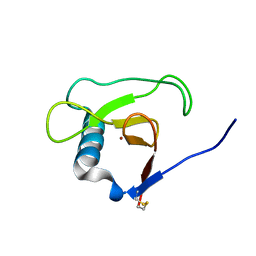
1RGE
 | | HYDROLASE, GUANYLORIBONUCLEASE | | 分子名称: | GUANOSINE-2'-MONOPHOSPHATE, RIBONUCLEASE, SULFATE ION | | 著者 | Sevcik, J, Dauter, Z, Lamzin, V.S, Wilson, K.S. | | 登録日 | 1995-06-05 | | 公開日 | 1996-10-14 | | 最終更新日 | 2017-11-29 | | 実験手法 | X-RAY DIFFRACTION (1.15 Å) | | 主引用文献 | Ribonuclease from Streptomyces aureofaciens at atomic resolution.
Acta Crystallogr.,Sect.D, 52, 1996
|
|
1RGF
 | | HYDROLASE, GUANYLORIBONUCLEASE | | 分子名称: | RIBONUCLEASE, SULFATE ION | | 著者 | Sevcik, J, Dauter, Z, Lamzin, V.S, Wilson, K.S. | | 登録日 | 1995-06-05 | | 公開日 | 1996-10-14 | | 最終更新日 | 2017-11-29 | | 実験手法 | X-RAY DIFFRACTION (1.2 Å) | | 主引用文献 | Ribonuclease from Streptomyces aureofaciens at atomic resolution.
Acta Crystallogr.,Sect.D, 52, 1996
|
|
1RGH
 | | HYDROLASE, GUANYLORIBONUCLEASE | | 分子名称: | RIBONUCLEASE, SULFATE ION | | 著者 | Sevcik, J, Dauter, Z, Lamzin, V.S, Wilson, K.S. | | 登録日 | 1995-06-05 | | 公開日 | 1996-10-14 | | 最終更新日 | 2011-07-13 | | 実験手法 | X-RAY DIFFRACTION (1.2 Å) | | 主引用文献 | Ribonuclease from Streptomyces aureofaciens at atomic resolution.
Acta Crystallogr.,Sect.D, 52, 1996
|
|
1RGG
 | | HYDROLASE, GUANYLORIBONUCLEASE | | 分子名称: | RIBONUCLEASE, SULFATE ION | | 著者 | Sevcik, J, Dauter, Z, Lamzin, V.S, Wilson, K.S. | | 登録日 | 1995-06-05 | | 公開日 | 1996-10-14 | | 最終更新日 | 2011-07-13 | | 実験手法 | X-RAY DIFFRACTION (1.2 Å) | | 主引用文献 | Ribonuclease from Streptomyces aureofaciens at atomic resolution.
Acta Crystallogr.,Sect.D, 52, 1996
|
|
1I70
 | |
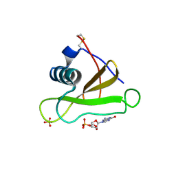
1RSN
 | | RIBONUCLEASE (RNASE SA) (E.C.3.1.4.8) COMPLEXED WITH EXO-2',3'-CYCLOPHOSPHOROTHIOATE | | 分子名称: | GUANOSINE-2',3'-CYCLOPHOSPHOROTHIOATE, RIBONUCLEASE SA, SULFATE ION | | 著者 | Sevcik, J, Dauter, Z, Lamzin, V.S, Wilson, K.S. | | 登録日 | 1995-09-01 | | 公開日 | 1995-12-07 | | 最終更新日 | 2011-07-13 | | 実験手法 | X-RAY DIFFRACTION (2 Å) | | 主引用文献 | Complex of ribonuclease Sa with a cyclic nucleotide and a proposed model for the reaction intermediate.
Eur.J.Biochem., 216, 1993
|
|
3DGY
 | |
3DH2
 | |
1LNI
 | | CRYSTAL STRUCTURE ANALYSIS OF A RIBONUCLEASE FROM STREPTOMYCES AUREOFACIENS AT ATOMIC RESOLUTION (1.0 A) | | 分子名称: | GLYCEROL, GUANYL-SPECIFIC RIBONUCLEASE SA, SULFATE ION | | 著者 | Sevcik, J, Lamzin, V.S, Dauter, Z, Wilson, K.S. | | 登録日 | 2002-05-03 | | 公開日 | 2002-07-31 | | 最終更新日 | 2023-08-16 | | 実験手法 | X-RAY DIFFRACTION (1 Å) | | 主引用文献 | Atomic resolution data reveal flexibility in the structure of RNase Sa.
Acta Crystallogr.,Sect.D, 58, 2002
|
|
1I8V
 | |
1SH6
 | |
1SH5
 | |
1PY3
 | |


