1IBI
 
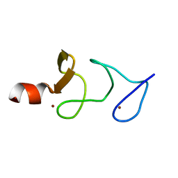 | | QUAIL CYSTEINE AND GLYCINE-RICH PROTEIN, NMR, 15 MINIMIZED MODEL STRUCTURES | | 分子名称: | CYSTEINE-RICH PROTEIN 2, ZINC ION | | 著者 | Schuler, W, Kloiber, K, Matt, T, Bister, K, Konrat, R. | | 登録日 | 2001-03-28 | | 公開日 | 2001-09-05 | | 最終更新日 | 2024-05-22 | | 実験手法 | SOLUTION NMR | | 主引用文献 | Application of cross-correlated NMR spin relaxation to the zinc-finger protein CRP2(LIM2): evidence for collective motions in LIM domains.
Biochemistry, 40, 2001
|
|
4CAU
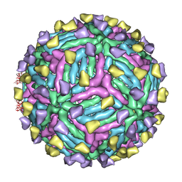 | | THREE-DIMENSIONAL STRUCTURE OF DENGUE VIRUS SEROTYPE 1 COMPLEXED WITH 2 HMAB 14C10 FAB | | 分子名称: | ENVELOPE PROTEIN E, FAB 14C10 | | 著者 | Teoh, E.P, Kukkaro, P, Teo, E.W, Lim, A.P, Tan, T.T, Yip, A, Schul, W, Aung, M, Kostyuchenko, V.A, Leo, Y.S, Chan, S.H, Smith, K.G, Chan, A.H, Zou, G, Ooi, E.E, Kemeny, D.M, Tan, G.K, Ng, J.K, Ng, M.L, Alonso, S, Fisher, D, Shi, P.Y, Hanson, B.J, Lok, S.M, Macary, P.A. | | 登録日 | 2013-10-09 | | 公開日 | 2013-10-16 | | 最終更新日 | 2024-05-08 | | 実験手法 | ELECTRON MICROSCOPY (7 Å) | | 主引用文献 | The Structural Basis for Serotype-Specific Neutralization of Dengue Virus by a Human Antibody.
Sci.Trans.Med, 4, 2012
|
|
3J05
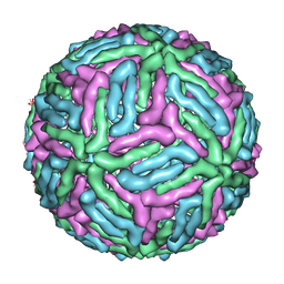 | | Three-dimensional structure of Dengue virus serotype 1 complexed with HMAb 14c10 Fab | | 分子名称: | envelope protein | | 著者 | Teoh, E.P, Kukkaro, P, Teo, E.W, Lim, A, Tan, T.T, Shi, P.Y, Yip, A, Schul, W, Leo, Y.S, Chan, S.H, Smith, K.G.C, Ooi, E.E, Kemeny, D.M, Ng, G, Ng, M.L, Alonso, S, Fisher, D, Hanson, B, Lok, S.M, MacAry, P.A. | | 登録日 | 2011-04-01 | | 公開日 | 2012-07-04 | | 最終更新日 | 2024-02-21 | | 実験手法 | ELECTRON MICROSCOPY (7 Å) | | 主引用文献 | The structural basis for serotype-specific neutralization of dengue virus by a human antibody.
Sci Transl Med, 4, 2012
|
|
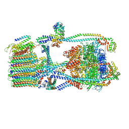
7UW9
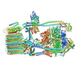 | | Citrus V-ATPase State 1, H in contact with subunit a | | 分子名称: | V-type proton ATPase catalytic subunit A, V-type proton ATPase subunit AP1 fragment, V-type proton ATPase subunit AP2 fragment, ... | | 著者 | Keon, K.A, Abdelaziz, R.A, Schulze, W.X, Schumacher, K, Rubinstein, J.L. | | 登録日 | 2022-05-03 | | 公開日 | 2022-07-06 | | 最終更新日 | 2024-06-12 | | 実験手法 | ELECTRON MICROSCOPY (4.2 Å) | | 主引用文献 | Structure of V-ATPase from citrus fruit.
Structure, 30, 2022
|
|
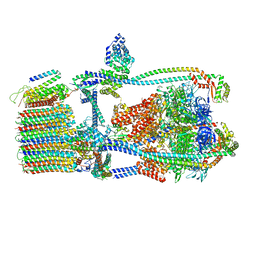
7UWB
 | | Citrus V-ATPase State 2, Highest-Resolution Class | | 分子名称: | V-type proton ATPase catalytic subunit A, V-type proton ATPase subunit AP1 fragment, V-type proton ATPase subunit AP2 fragment, ... | | 著者 | Keon, K.A, Abdelaziz, R.A, Schulze, W.X, Schumacher, K, Rubinstein, J.L. | | 登録日 | 2022-05-03 | | 公開日 | 2022-07-06 | | 最終更新日 | 2024-01-17 | | 実験手法 | ELECTRON MICROSCOPY (3.9 Å) | | 主引用文献 | Structure of V-ATPase from citrus fruit.
Structure, 30, 2022
|
|
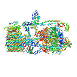
7UWA
 | | Citrus V-ATPase State 1, H in contact with subunits AB | | 分子名称: | V-type proton ATPase catalytic subunit A, V-type proton ATPase subunit AP1 fragment, V-type proton ATPase subunit AP2 fragment, ... | | 著者 | Abdelaziz, R.A, Keon, K.A, Schulze, W.X, Schumacher, K, Rubinstein, J.L. | | 登録日 | 2022-05-03 | | 公開日 | 2022-07-06 | | 最終更新日 | 2024-06-12 | | 実験手法 | ELECTRON MICROSCOPY (4.3 Å) | | 主引用文献 | Structure of V-ATPase from citrus fruit.
Structure, 30, 2022
|
|
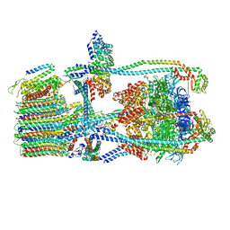
7UWD
 | | Citrus V-ATPase State 2, H in contact with subunits AB | | 分子名称: | V-type proton ATPase catalytic subunit A, V-type proton ATPase subunit, V-type proton ATPase subunit AP1 fragment, ... | | 著者 | Keon, K.A, Abdelaziz, R.A, Schulze, W.X, Schumacher, K, Rubinstein, J.L. | | 登録日 | 2022-05-03 | | 公開日 | 2022-07-06 | | 最終更新日 | 2024-01-17 | | 実験手法 | ELECTRON MICROSCOPY (4.1 Å) | | 主引用文献 | Structure of V-ATPase from citrus fruit.
Structure, 30, 2022
|
|
7UWC
 | | Citrus V-ATPase State 2, H in contact with subunit a | | 分子名称: | V-type proton ATPase catalytic subunit A, V-type proton ATPase subunit AP1 fragment, V-type proton ATPase subunit AP2 fragment, ... | | 著者 | Keon, K.A, Abdelaziz, R.A, Schulze, W.X, Schumacher, K, Rubinstein, J.L. | | 登録日 | 2022-05-03 | | 公開日 | 2022-07-06 | | 最終更新日 | 2024-01-17 | | 実験手法 | ELECTRON MICROSCOPY (4 Å) | | 主引用文献 | Structure of V-ATPase from citrus fruit.
Structure, 30, 2022
|
|
6F7S
 | |
6F7P
 | |
6F7J
 | |
6F8D
 | |
5OO6
 | |
5OOB
 | |


