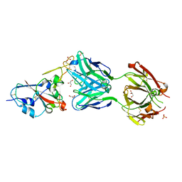
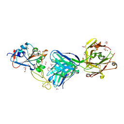
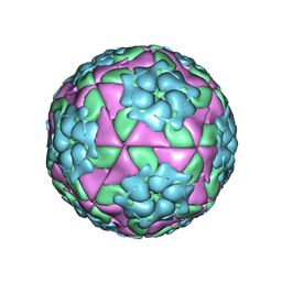
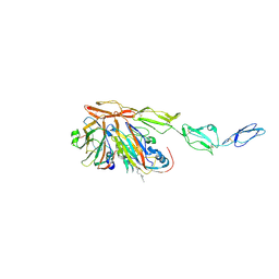
5MQW
 
 | | High-speed fixed-target serial virus crystallography | | 分子名称: | ADENOSINE-5'-TRIPHOSPHATE, CHLORIDE ION, GUANOSINE-5'-TRIPHOSPHATE, ... | | 著者 | Roedig, P, Ginn, H.M, Pakendorf, T, Sutton, G, Harlos, K, Walter, T.S, Meyer, J, Fischer, P, Duman, R, Vartiainen, I, Reime, B, Warmer, M, Brewster, A.S, Young, I.D, Michels-Clark, T, Sauter, N.K, Sikorsky, M, Nelson, S, Damiani, D.S, Alonso-Mori, R, Ren, J, Fry, E.E, David, C, Stuart, D.I, Wagner, A, Meents, A. | | 登録日 | 2016-12-21 | | 公開日 | 2017-06-21 | | 最終更新日 | 2024-01-17 | | 実験手法 | X-RAY DIFFRACTION (2.4 Å) | | 主引用文献 | High-speed fixed-target serial virus crystallography.
Nat. Methods, 14, 2017
|
|
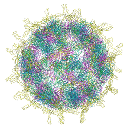
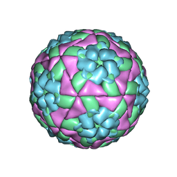
5GKA
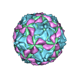 | | cryo-EM structure of human Aichi virus | | 分子名称: | Genome polyprotein, capsid protein VP0, capsid protein VP1 | | 著者 | Zhu, L, Wang, X.X, Ren, J.S, Tuthill, T.J, Fry, E.E, Rao, Z.H, Stuart, D.I. | | 登録日 | 2016-07-04 | | 公開日 | 2016-09-21 | | 最終更新日 | 2024-03-27 | | 実験手法 | ELECTRON MICROSCOPY (3.7 Å) | | 主引用文献 | Structure of human Aichi virus and implications for receptor binding
Nat Microbiol, 1, 2016
|
|
7ND4
 | | EM structure of SARS-CoV-2 Spike glycoprotein in complex with COVOX-88 Fab | | 分子名称: | 2-acetamido-2-deoxy-beta-D-glucopyranose, 2-acetamido-2-deoxy-beta-D-glucopyranose-(1-4)-2-acetamido-2-deoxy-beta-D-glucopyranose, COVOX-88 Fab heavy chain, ... | | 著者 | Duyvesteyn, H.M.E, Zhao, Y, Ren, J, Stuart, D. | | 登録日 | 2021-01-30 | | 公開日 | 2021-03-03 | | 最終更新日 | 2021-04-28 | | 実験手法 | ELECTRON MICROSCOPY (3.6 Å) | | 主引用文献 | The antigenic anatomy of SARS-CoV-2 receptor binding domain.
Cell, 184, 2021
|
|
7ND3
 | | EM structure of SARS-CoV-2 Spike glycoprotein in complex with COVOX-40 Fab | | 分子名称: | 2-acetamido-2-deoxy-beta-D-glucopyranose, 2-acetamido-2-deoxy-beta-D-glucopyranose-(1-4)-2-acetamido-2-deoxy-beta-D-glucopyranose, COVOX-40 heavy chain, ... | | 著者 | Duyvesteyn, H.M.E, Zhao, Y, Ren, J, Stuart, D. | | 登録日 | 2021-01-30 | | 公開日 | 2021-03-03 | | 最終更新日 | 2023-11-29 | | 実験手法 | ELECTRON MICROSCOPY (3.7 Å) | | 主引用文献 | The antigenic anatomy of SARS-CoV-2 receptor binding domain.
Cell, 184, 2021
|
|
7ND8
 | | EM structure of SARS-CoV-2 Spike glycoprotein in complex with COVOX-384 Fab | | 分子名称: | 2-acetamido-2-deoxy-beta-D-glucopyranose, 2-acetamido-2-deoxy-beta-D-glucopyranose-(1-4)-2-acetamido-2-deoxy-beta-D-glucopyranose, COVOX-384 Fab heavy chain, ... | | 著者 | Duyvesteyn, H.M.E, Zhao, Y, Ren, J, Stuart, D. | | 登録日 | 2021-01-30 | | 公開日 | 2021-03-03 | | 最終更新日 | 2021-04-28 | | 実験手法 | ELECTRON MICROSCOPY (3.5 Å) | | 主引用文献 | The antigenic anatomy of SARS-CoV-2 receptor binding domain.
Cell, 184, 2021
|
|
7NDC
 | | EM structure of SARS-CoV-2 Spike glycoprotein (all RBD down) in complex with COVOX-159 | | 分子名称: | 2-acetamido-2-deoxy-beta-D-glucopyranose, 2-acetamido-2-deoxy-beta-D-glucopyranose-(1-4)-2-acetamido-2-deoxy-beta-D-glucopyranose, COVOX-159 Fab light chain, ... | | 著者 | Duyvesteyn, H.M.E, Zhao, Y, Ren, J, Stuart, D. | | 登録日 | 2021-01-30 | | 公開日 | 2021-03-03 | | 最終更新日 | 2021-04-28 | | 実験手法 | ELECTRON MICROSCOPY (4.1 Å) | | 主引用文献 | The antigenic anatomy of SARS-CoV-2 receptor binding domain.
Cell, 184, 2021
|
|
7ND7
 | | EM structure of SARS-CoV-2 Spike glycoprotein in complex with COVOX-316 Fab | | 分子名称: | 2-acetamido-2-deoxy-beta-D-glucopyranose, 2-acetamido-2-deoxy-beta-D-glucopyranose-(1-4)-2-acetamido-2-deoxy-beta-D-glucopyranose, 2-acetamido-2-deoxy-beta-D-glucopyranose-(1-4)-[alpha-L-fucopyranose-(1-6)]2-acetamido-2-deoxy-beta-D-glucopyranose, ... | | 著者 | Duyvesteyn, H.M.E, Zhao, Y, Ren, J, Stuart, D. | | 登録日 | 2021-01-30 | | 公開日 | 2021-03-03 | | 最終更新日 | 2021-04-28 | | 実験手法 | ELECTRON MICROSCOPY (3.6 Å) | | 主引用文献 | The antigenic anatomy of SARS-CoV-2 receptor binding domain.
Cell, 184, 2021
|
|
7ND9
 | | EM structure of SARS-CoV-2 Spike glycoprotein (one RBD up) in complex with COVOX-253H55L Fab | | 分子名称: | 2-acetamido-2-deoxy-beta-D-glucopyranose, 2-acetamido-2-deoxy-beta-D-glucopyranose-(1-4)-2-acetamido-2-deoxy-beta-D-glucopyranose, 2-acetamido-2-deoxy-beta-D-glucopyranose-(1-4)-[alpha-L-fucopyranose-(1-6)]2-acetamido-2-deoxy-beta-D-glucopyranose, ... | | 著者 | Duyvesteyn, H.M.E, Zhao, Y, Ren, J, Stuart, D. | | 登録日 | 2021-01-30 | | 公開日 | 2021-03-03 | | 最終更新日 | 2021-04-28 | | 実験手法 | ELECTRON MICROSCOPY (2.8 Å) | | 主引用文献 | The antigenic anatomy of SARS-CoV-2 receptor binding domain.
Cell, 184, 2021
|
|
7NDD
 | | EM structure of SARS-CoV-2 Spike glycoprotein (one RBD up) in complex with COVOX-159 | | 分子名称: | 2-acetamido-2-deoxy-beta-D-glucopyranose, 2-acetamido-2-deoxy-beta-D-glucopyranose-(1-4)-2-acetamido-2-deoxy-beta-D-glucopyranose, COVOX-159 Fab heavy chain, ... | | 著者 | Duyvesteyn, H.M.E, Zhao, Y, Ren, J, Stuart, D. | | 登録日 | 2021-01-30 | | 公開日 | 2021-03-03 | | 最終更新日 | 2021-04-28 | | 実験手法 | ELECTRON MICROSCOPY (4.2 Å) | | 主引用文献 | The antigenic anatomy of SARS-CoV-2 receptor binding domain.
Cell, 184, 2021
|
|
7NDA
 | | EM structure of SARS-CoV-2 Spike glycoprotein (all RBD down) in complex with COVOX-253H55L Fab | | 分子名称: | 2-acetamido-2-deoxy-beta-D-glucopyranose, 2-acetamido-2-deoxy-beta-D-glucopyranose-(1-4)-2-acetamido-2-deoxy-beta-D-glucopyranose, 2-acetamido-2-deoxy-beta-D-glucopyranose-(1-4)-[alpha-L-fucopyranose-(1-6)]2-acetamido-2-deoxy-beta-D-glucopyranose, ... | | 著者 | Duyvesteyn, H.M.E, Zhao, Y, Ren, J, Stuart, D. | | 登録日 | 2021-01-30 | | 公開日 | 2021-03-03 | | 最終更新日 | 2021-04-28 | | 実験手法 | ELECTRON MICROSCOPY (3.3 Å) | | 主引用文献 | The antigenic anatomy of SARS-CoV-2 receptor binding domain.
Cell, 184, 2021
|
|
7ND5
 | | EM structure of SARS-CoV-2 Spike glycoprotein in complex with COVOX-150 Fab | | 分子名称: | 2-acetamido-2-deoxy-beta-D-glucopyranose, 2-acetamido-2-deoxy-beta-D-glucopyranose-(1-4)-2-acetamido-2-deoxy-beta-D-glucopyranose, COVOX-150 Fab heavy chain, ... | | 著者 | Duyvesteyn, H.M.E, Zhao, Y, Ren, J, Stuart, D. | | 登録日 | 2021-01-30 | | 公開日 | 2021-03-03 | | 最終更新日 | 2021-04-28 | | 実験手法 | ELECTRON MICROSCOPY (3.4 Å) | | 主引用文献 | The antigenic anatomy of SARS-CoV-2 receptor binding domain.
Cell, 184, 2021
|
|
7ND6
 | | EM structure of SARS-CoV-2 Spike glycoprotein in complex with COVOX-40 Fab | | 分子名称: | 2-acetamido-2-deoxy-beta-D-glucopyranose, 2-acetamido-2-deoxy-beta-D-glucopyranose-(1-4)-2-acetamido-2-deoxy-beta-D-glucopyranose, COVOX-158 Fab heavy chain, ... | | 著者 | Duyvesteyn, H.M.E, Zhao, Y, Ren, J, Stuart, D. | | 登録日 | 2021-01-30 | | 公開日 | 2021-03-03 | | 最終更新日 | 2021-04-28 | | 実験手法 | ELECTRON MICROSCOPY (7.3 Å) | | 主引用文献 | The antigenic anatomy of SARS-CoV-2 receptor binding domain.
Cell, 184, 2021
|
|
7NDB
 | | EM structure of SARS-CoV-2 Spike glycoprotein in complex with COVOX-253H165L Fab | | 分子名称: | 2-acetamido-2-deoxy-beta-D-glucopyranose, 2-acetamido-2-deoxy-beta-D-glucopyranose-(1-4)-2-acetamido-2-deoxy-beta-D-glucopyranose, 2-acetamido-2-deoxy-beta-D-glucopyranose-(1-4)-[alpha-L-fucopyranose-(1-6)]2-acetamido-2-deoxy-beta-D-glucopyranose, ... | | 著者 | Duyvesteyn, H.M.E, Zhao, Y, Ren, J, Stuart, D. | | 登録日 | 2021-01-30 | | 公開日 | 2021-03-03 | | 最終更新日 | 2021-04-28 | | 実験手法 | ELECTRON MICROSCOPY (4.6 Å) | | 主引用文献 | The antigenic anatomy of SARS-CoV-2 receptor binding domain.
Cell, 184, 2021
|
|
7NEG
 | | Crystal structure of the N501Y mutant receptor binding domain of SARS-CoV-2 Spike glycoprotein in complex with COVOX-269 Fab | | 分子名称: | 2-acetamido-2-deoxy-beta-D-glucopyranose-(1-4)-[alpha-L-fucopyranose-(1-6)]2-acetamido-2-deoxy-beta-D-glucopyranose, Antibody COVOX-269 Fab heavy chain, Antibody COVOX-269 Fab light chain, ... | | 著者 | Zhou, D, Ren, J, Stuart, D. | | 登録日 | 2021-02-04 | | 公開日 | 2021-03-03 | | 最終更新日 | 2024-01-31 | | 実験手法 | X-RAY DIFFRACTION (2.19 Å) | | 主引用文献 | Reduced neutralization of SARS-CoV-2 B.1.1.7 variant by convalescent and vaccine sera.
Cell, 184, 2021
|
|
7NEH
 | | Crystal structure of the receptor binding domain of SARS-CoV-2 Spike glycoprotein in complex with COVOX-269 Fab | | 分子名称: | 1,2-ETHANEDIOL, 2-acetamido-2-deoxy-beta-D-glucopyranose-(1-4)-[alpha-L-fucopyranose-(1-6)]2-acetamido-2-deoxy-beta-D-glucopyranose, CHLORIDE ION, ... | | 著者 | Zhou, D, Ren, J, Stuart, D. | | 登録日 | 2021-02-04 | | 公開日 | 2021-03-03 | | 最終更新日 | 2024-01-31 | | 実験手法 | X-RAY DIFFRACTION (1.77 Å) | | 主引用文献 | Reduced neutralization of SARS-CoV-2 B.1.1.7 variant by convalescent and vaccine sera.
Cell, 184, 2021
|
|
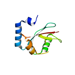
5E6O
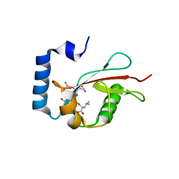 | | Crystal structure of C. elegans LGG-2 bound to an AIM/LIR motif | | 分子名称: | Protein lgg-2, TRP-GLU-GLU-LEU | | 著者 | Qi, X, Ren, J.Q, Wu, F, Zhang, H, Feng, W. | | 登録日 | 2015-10-10 | | 公開日 | 2016-01-06 | | 最終更新日 | 2024-03-20 | | 実験手法 | X-RAY DIFFRACTION (1.8 Å) | | 主引用文献 | Structural Basis of the Differential Function of the Two C. elegans Atg8 Homologs, LGG-1 and LGG-2, in Autophagy
Mol.Cell, 60, 2015
|
|
5D8A
 | | Crystal structure of recombinant foot-and-mouth-disease virus A22-H2093F empty capsid | | 分子名称: | VP1, VP2, VP3, ... | | 著者 | Kotecha, A, Seago, J, Scott, K, Burman, A, Loureiro, S, Ren, J, Porta, C, Ginn, H.M, Jackson, T, Perez-Martin, E, Siebert, C.A, Paul, G, Huiskonen, J.T, Jones, I.M, Esnouf, R.M, Fry, E.E, Maree, F.F, Charleston, B, Stuart, D.I. | | 登録日 | 2015-08-16 | | 公開日 | 2015-09-23 | | 最終更新日 | 2024-01-10 | | 実験手法 | X-RAY DIFFRACTION (2.4 Å) | | 主引用文献 | Structure-based energetics of protein interfaces guides foot-and-mouth disease virus vaccine design.
Nat.Struct.Mol.Biol., 22, 2015
|
|
5ED9
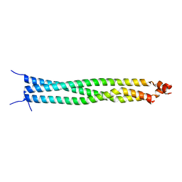 | | Crystal structure of CC1 of mouse SUN2 | | 分子名称: | SUN domain-containing protein 2 | | 著者 | Nie, S, Ke, H.M, Gao, F, Ren, J.Q, Wang, M.Z, Huo, L, Gong, W.M, Feng, W. | | 登録日 | 2015-10-21 | | 公開日 | 2016-01-13 | | 最終更新日 | 2024-03-20 | | 実験手法 | X-RAY DIFFRACTION (2.009 Å) | | 主引用文献 | Coiled-Coil Domains of SUN Proteins as Intrinsic Dynamic Regulators
Structure, 24, 2016
|
|
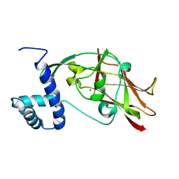
5E6N
 | | Crystal structure of C. elegans LGG-2 | | 分子名称: | Protein lgg-2 | | 著者 | Qi, X, Ren, J.Q, Wu, F, Zhang, H, Feng, W. | | 登録日 | 2015-10-10 | | 公開日 | 2016-01-06 | | 最終更新日 | 2024-03-20 | | 実験手法 | X-RAY DIFFRACTION (2.102 Å) | | 主引用文献 | Structural Basis of the Differential Function of the Two C. elegans Atg8 Homologs, LGG-1 and LGG-2, in Autophagy
Mol.Cell, 60, 2015
|
|
5ED8
 | | Crystal structure of CC2-SUN of mouse SUN2 | | 分子名称: | MAGNESIUM ION, MKIAA0668 protein | | 著者 | Nie, S, Ke, H.M, Gao, F, Ren, J.Q, Wang, M.Z, Huo, L, Gong, W.M, Feng, W. | | 登録日 | 2015-10-21 | | 公開日 | 2016-01-13 | | 最終更新日 | 2024-03-20 | | 実験手法 | X-RAY DIFFRACTION (2.5 Å) | | 主引用文献 | Coiled-Coil Domains of SUN Proteins as Intrinsic Dynamic Regulators
Structure, 24, 2016
|
|
8B9F
 | | Structure of Echovirus 11 complexed with DAF (CD55) calculated from symmetry expansion | | 分子名称: | Complement decay-accelerating factor, Genome polyprotein, SPHINGOSINE | | 著者 | Stuart, D.I, Ren, J, Qin, L, Zhou, D. | | 登録日 | 2022-10-05 | | 公開日 | 2022-12-07 | | 最終更新日 | 2023-01-04 | | 実験手法 | ELECTRON MICROSCOPY (3.93 Å) | | 主引用文献 | Switching of Receptor Binding Poses between Closely Related Enteroviruses.
Viruses, 14, 2022
|
|
8B8R
 | | Complex of Echovirus 11 with its attaching receptor decay-accelerating factor (CD55) | | 分子名称: | DECAY ACCELERATING FACTOR (CD55), SPHINGOSINE, VP1, ... | | 著者 | Stuart, D.I, Ren, J, Zhou, D, Qin, L. | | 登録日 | 2022-10-04 | | 公開日 | 2022-12-07 | | 最終更新日 | 2023-01-04 | | 実験手法 | ELECTRON MICROSCOPY (3.1 Å) | | 主引用文献 | Switching of Receptor Binding Poses between Closely Related Enteroviruses.
Viruses, 14, 2022
|
|
5DDJ
 | | Crystal structure of recombinant foot-and-mouth-disease virus O1M-S2093Y empty capsid | | 分子名称: | Foot and mouth disease virus, VP1, VP2, ... | | 著者 | Kotecha, A, Seago, J, Scott, K, Burman, A, Loureiro, S, Ren, J, Porta, C, Ginn, H.M, Jackson, T, Perez-Martin, E, Siebert, C.A, Paul, G, Huiskonen, J.T, Jones, I.M, Esnouf, R.M, Fry, E.E, Maree, F.F, Charleston, B, Stuart, D.I. | | 登録日 | 2015-08-25 | | 公開日 | 2015-09-23 | | 最終更新日 | 2024-01-10 | | 実験手法 | X-RAY DIFFRACTION (3.5 Å) | | 主引用文献 | Structure-based energetics of protein interfaces guides foot-and-mouth disease virus vaccine design.
Nat.Struct.Mol.Biol., 22, 2015
|
|
8CII
 | | Delta-RBD complex with BA.2-07 fab, SARS1-34 fab and C1 nanobody | | 分子名称: | BA.2-07 fab Heavy Chain, BA.2-07 fab Light Chain, C1 nanobody, ... | | 著者 | Duyvesteyn, H.M.E, Ren, J, Stuart, D.I, Fry, E.E. | | 登録日 | 2023-02-09 | | 公開日 | 2024-02-21 | | 実験手法 | ELECTRON MICROSCOPY (2.7 Å) | | 主引用文献 | Potent cross-reactive mAbs from BA.4/5 breakthrough infection
To Be Published
|
|
5JQ3
 | | Crystal structure of Ebola glycoprotein | | 分子名称: | 2-acetamido-2-deoxy-beta-D-glucopyranose, Envelope glycoprotein 1,Envelope glycoprotein 1,Envelope glycoprotein 1, Envelope glycoprotein 2, ... | | 著者 | Zhao, Y, Ren, J, Stuart, D.I. | | 登録日 | 2016-05-04 | | 公開日 | 2016-06-29 | | 最終更新日 | 2024-01-10 | | 実験手法 | X-RAY DIFFRACTION (2.23 Å) | | 主引用文献 | Toremifene interacts with and destabilizes the Ebola virus glycoprotein.
Nature, 535, 2016
|
|


