1V6N
 
 | | Peanut lectin with 9mer peptide (PVIWSSATG) | | 分子名称: | CALCIUM ION, Galactose-binding lectin, MANGANESE (II) ION | | 著者 | Kundhavai Natchiar, S, Arockia Jeyaprakash, A, Ramya, T.N.C, Thomas, C.J, Suguna, K, Surolia, A, Vijayan, M. | | 登録日 | 2003-12-02 | | 公開日 | 2004-02-10 | | 最終更新日 | 2023-10-25 | | 実験手法 | X-RAY DIFFRACTION (3.5 Å) | | 主引用文献 | Structural plasticity of peanut lectin: an X-ray analysis involving variation in pH, ligand binding and crystal structure.
Acta Crystallogr.,Sect.D, 60, 2004
|
|
1V6M
 | | Peanut Lectin with 9mer peptide (IWSSAGNVA) | | 分子名称: | CALCIUM ION, Galactose-binding lectin, MANGANESE (II) ION | | 著者 | Kundhavai Natchiar, S, Arockia Jeyaprakash, A, Ramya, T.N.C, Thomas, C.J, Suguna, K, Surolia, A, Vijayan, M. | | 登録日 | 2003-12-02 | | 公開日 | 2004-02-10 | | 最終更新日 | 2023-10-25 | | 実験手法 | X-RAY DIFFRACTION (2.7 Å) | | 主引用文献 | Structural plasticity of peanut lectin: an X-ray analysis involving variation in pH, ligand binding and crystal structure.
Acta Crystallogr.,Sect.D, 60, 2004
|
|
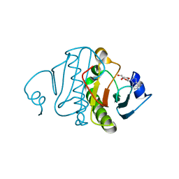
3O1Z
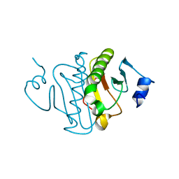 | | High resolution crystal structure of histidine triad nucleotide-binding protein 1 (Hint1) double cysteine mutant from rabbit | | 分子名称: | Histidine triad nucleotide-binding protein 1 | | 著者 | Dolot, R.M, Ozga, M, Krakowiak, A.K, Nawrot, B, Stec, W.J. | | 登録日 | 2010-07-22 | | 公開日 | 2010-08-04 | | 最終更新日 | 2023-09-06 | | 実験手法 | X-RAY DIFFRACTION (1.3 Å) | | 主引用文献 | Histidine Triad Nucleotide-binding Protein 1 (HINT-1) Phosphoramidase Transforms Nucleoside 5'-O-Phosphorothioates to Nucleoside 5'-O-Phosphates.
J.Biol.Chem., 285, 2010
|
|
1U9B
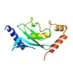 | | MURINE/HUMAN UBIQUITIN-CONJUGATING ENZYME UBC9 | | 分子名称: | UBIQUITIN-CONJUGATING ENZYME E9 | | 著者 | Tong, H, Hateboer, G, Perrakis, A, Bernards, R, Sixma, T.K. | | 登録日 | 1997-05-20 | | 公開日 | 1997-07-07 | | 最終更新日 | 2024-05-22 | | 実験手法 | X-RAY DIFFRACTION (2 Å) | | 主引用文献 | Crystal structure of murine/human Ubc9 provides insight into the variability of the ubiquitin-conjugating system.
J.Biol.Chem., 272, 1997
|
|
1U9A
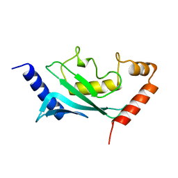 | | HUMAN UBIQUITIN-CONJUGATING ENZYME UBC9 | | 分子名称: | UBIQUITIN-CONJUGATING ENZYME | | 著者 | Tong, H, Hateboer, G, Perrakis, A, Bernards, R, Sixma, T.K. | | 登録日 | 1997-02-11 | | 公開日 | 1997-05-15 | | 最終更新日 | 2024-05-22 | | 実験手法 | X-RAY DIFFRACTION (2 Å) | | 主引用文献 | Crystal structure of murine/human Ubc9 provides insight into the variability of the ubiquitin-conjugating system.
J.Biol.Chem., 272, 1997
|
|
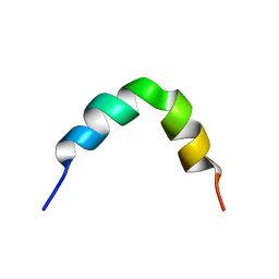
2L96
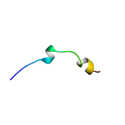 | | Solution structure of LAK160-P7 | | 分子名称: | LAK160-P7 | | 著者 | Vermeer, L.S, Bui, T.T, Lan, Y, Jumagulova, E, Kozlowska, J, McIntyre, C, Drake, A.F, Mason, J.A. | | 登録日 | 2011-02-01 | | 公開日 | 2012-02-01 | | 最終更新日 | 2024-05-01 | | 実験手法 | SOLUTION NMR | | 主引用文献 | The role of proline induced conformational flexibility in determining the antibacterial potency of linear cationic alpha-helical peptides
To be Published
|
|
3QGZ
 | | Re-investigated high resolution crystal structure of histidine triad nucleotide-binding protein 1 (HINT1) from rabbit complexed with adenosine | | 分子名称: | ADENOSINE, Histidine triad nucleotide-binding protein 1 | | 著者 | Dolot, R.M, Ozga, M, Krakowiak, A, Nawrot, B, Stec, W.J. | | 登録日 | 2011-01-25 | | 公開日 | 2011-02-16 | | 最終更新日 | 2023-09-13 | | 実験手法 | X-RAY DIFFRACTION (1.1 Å) | | 主引用文献 | High-resolution X-ray crystal structure of rabbit histidine triad nucleotide-binding protein 1 (rHINT1) - adenosine complex at 1.10A resolution
Acta Crystallogr.,Sect.D, 67, 2011
|
|
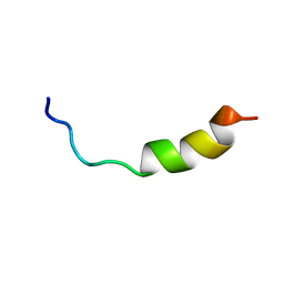
2L99
 | | Solution structure of LAK160-P10 | | 分子名称: | LAK160-P10 | | 著者 | Vermeer, L.S, Bui, T.T, Lan, Y, Jumagulova, E, Kozlowska, J, McIntyre, C, Drake, A.F, Mason, J.A. | | 登録日 | 2011-02-03 | | 公開日 | 2012-02-01 | | 最終更新日 | 2024-05-01 | | 実験手法 | SOLUTION NMR | | 主引用文献 | The role of proline induced conformational flexibility in determining the antibacterial potency of linear cationic alpha-helical peptides
To be Published
|
|
2L9A
 | | Solution structure of LAK160-P12 | | 分子名称: | LAK160-P12 | | 著者 | Vermeer, L.S, Bui, T.T, Lan, Y, Jumagulova, E, Kozlowska, J, McIntyre, C, Drake, A.F, Mason, J.A. | | 登録日 | 2011-02-03 | | 公開日 | 2012-02-01 | | 最終更新日 | 2024-05-01 | | 実験手法 | SOLUTION NMR | | 主引用文献 | The role of proline induced conformational flexibility in determining the antibacterial potency of linear cationic alpha-helical peptides
To be Published
|
|
2MPH
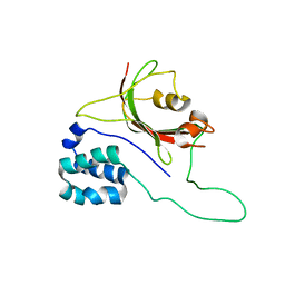 | |
2CKL
 | | Ring1b-Bmi1 E3 catalytic domain structure | | 分子名称: | IODIDE ION, POLYCOMB GROUP RING FINGER PROTEIN 4, UBIQUITIN LIGASE PROTEIN RING2, ... | | 著者 | Buchwald, G, van der Stoop, P, Weichenrieder, O, Perrakis, A, van Lohuizen, M, Sixma, T.K. | | 登録日 | 2006-04-20 | | 公開日 | 2006-05-11 | | 最終更新日 | 2024-05-08 | | 実験手法 | X-RAY DIFFRACTION (2 Å) | | 主引用文献 | Structure and E3-Ligase Activity of the Ring-Ring Complex of Polycomb Proteins Bmi1 and Ring1B.
Embo J., 25, 2006
|
|


