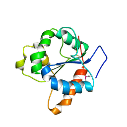
1K0D
 
 | | Ure2p in Complex with Glutathione | | 分子名称: | GLUTATHIONE, URE2 PROTEIN | | 著者 | Bousset, L, Belrhali, H, Melki, R, Morera, S. | | 登録日 | 2001-09-19 | | 公開日 | 2001-12-21 | | 最終更新日 | 2024-02-07 | | 実験手法 | X-RAY DIFFRACTION (2.2 Å) | | 主引用文献 | Crystal structures of the yeast prion Ure2p functional region in complex with glutathione and related compounds.
Biochemistry, 40, 2001
|
|
4WO0
 | | Crystal structure of transthyretin in complex with apigenin | | 分子名称: | 5,7-dihydroxy-2-(4-hydroxyphenyl)-4H-chromen-4-one, Transthyretin | | 著者 | Zanotti, G, Cianci, M, Folli, C, Berni, R. | | 登録日 | 2014-10-15 | | 公開日 | 2015-08-05 | | 最終更新日 | 2024-01-10 | | 実験手法 | X-RAY DIFFRACTION (1.339 Å) | | 主引用文献 | Structural evidence for asymmetric ligand binding to transthyretin.
Acta Crystallogr.,Sect.D, 71, 2015
|
|
1FZU
 | | RNAse T1 V78A mutant | | 分子名称: | CALCIUM ION, GUANOSINE-2'-MONOPHOSPHATE, GUANYL-SPECIFIC RIBONUCLEASE T1 | | 著者 | De Vos, S, Loris, R, Steyaert, J. | | 登録日 | 2000-10-04 | | 公開日 | 2000-10-25 | | 最終更新日 | 2021-11-03 | | 実験手法 | X-RAY DIFFRACTION (1.8 Å) | | 主引用文献 | Hydrophobic core manipulations in ribonuclease T1.
Biochemistry, 40, 2001
|
|
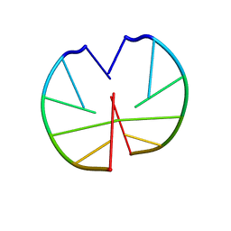
1G02
 | | Ribonuclease T1 V16S mutant | | 分子名称: | CALCIUM ION, GUANOSINE-2'-MONOPHOSPHATE, GUANYL-SPECIFIC RIBONUCLEASE T1 | | 著者 | De Vos, S, Loris, R, Steyaert, J. | | 登録日 | 2000-10-05 | | 公開日 | 2000-10-25 | | 最終更新日 | 2011-07-13 | | 実験手法 | X-RAY DIFFRACTION (1.86 Å) | | 主引用文献 | Hydrophobic core manipulations in ribonuclease T1.
Biochemistry, 40, 2001
|
|
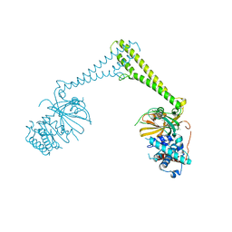
1FXK
 | |
8P19
 | |
8P14
 | |
1FYD
 | | CRYSTAL STRUCTURE OF NH3-DEPENDENT NAD+ SYNTHETASE FROM BACILLUS SUBTILIS COMPLEXED WITH ONE MOLECULE AMP, ONE PYROPHOSPHATE ION AND ONE MG2+ ION | | 分子名称: | ADENOSINE MONOPHOSPHATE, MAGNESIUM ION, NH(3)-DEPENDENT NAD(+) SYNTHETASE, ... | | 著者 | Devedjiev, Y, Symersky, J, Singh, R, Brouillette, W, Muccio, D, Jedrzejas, M, Brouillette, C, DeLucas, L. | | 登録日 | 2000-09-28 | | 公開日 | 2001-06-06 | | 最終更新日 | 2024-02-07 | | 実験手法 | X-RAY DIFFRACTION (2.25 Å) | | 主引用文献 | Stabilization of active-site loops in NH3-dependent NAD+ synthetase from Bacillus subtilis.
Acta Crystallogr.,Sect.D, 57, 2001
|
|
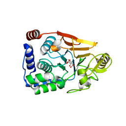
1JC1
 | | CRYSTAL STRUCTURE ANALYSIS OF A REDOX-SENSITIVE GREEN FLUORESCENT PROTEIN VARIANT IN A OXIDIZED FORM | | 分子名称: | GREEN FLUORESCENT PROTEIN | | 著者 | Hanson, G.T, Aggeler, R, Oglesbee, D, Cannon, M, Capaldi, R.A, Tsien, R.Y, Remington, S.J. | | 登録日 | 2001-06-07 | | 公開日 | 2003-09-09 | | 最終更新日 | 2023-11-15 | | 実験手法 | X-RAY DIFFRACTION (1.9 Å) | | 主引用文献 | Investigating mitochondrial redox potential with redox-sensitive green fluorescent protein indicators.
J.Biol.Chem., 279, 2004
|
|
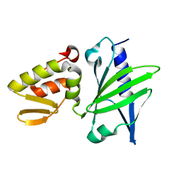
1FYV
 | | CRYSTAL STRUCTURE OF THE TIR DOMAIN OF HUMAN TLR1 | | 分子名称: | TOLL-LIKE RECEPTOR 1 | | 著者 | Xu, Y, Tao, X, Shen, B, Horng, T, Medzhitov, R, Manley, J.L, Tong, L. | | 登録日 | 2000-10-03 | | 公開日 | 2000-11-22 | | 最終更新日 | 2018-01-31 | | 実験手法 | X-RAY DIFFRACTION (2.9 Å) | | 主引用文献 | Structural basis for signal transduction by the Toll/interleukin-1 receptor domains.
Nature, 408, 2000
|
|
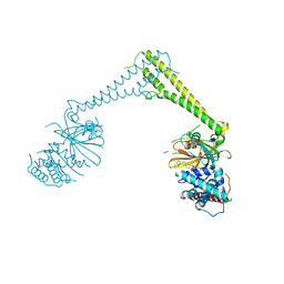
8P1P
 | | USP28 in complex with AZ1 | | 分子名称: | 2-[[5-bromanyl-2-[[4-fluoranyl-3-(trifluoromethyl)phenyl]methoxy]phenyl]methylamino]ethanol, CHLORIDE ION, DIMETHYL SULFOXIDE, ... | | 著者 | Sauer, F, Karal-Nair, R, Kisker, C. | | 登録日 | 2023-05-12 | | 公開日 | 2024-05-22 | | 実験手法 | X-RAY DIFFRACTION (2.76 Å) | | 主引用文献 | USP28 in complex with AZ1
To Be Published
|
|
1JAJ
 | |
8P1Q
 | | USP28 in complex with FT206 | | 分子名称: | 3-azanyl-N-[(2S)-6-[(1S,5R)-3,8-diazabicyclo[3.2.1]octan-3-yl]-1,2,3,4-tetrahydronaphthalen-2-yl]-6-methyl-thieno[2,3-b]pyridine-2-carboxamide, DIMETHYL SULFOXIDE, Ubiquitin carboxyl-terminal hydrolase 28 | | 著者 | Sauer, F, Karal Nair, R, Kisker, C. | | 登録日 | 2023-05-12 | | 公開日 | 2024-05-22 | | 実験手法 | X-RAY DIFFRACTION (2.79 Å) | | 主引用文献 | USP28 in complex with FT206
To Be Published
|
|
8P1Y
 | |
4WW0
 | | Truncated FtsH from A. aeolicus | | 分子名称: | ADENOSINE-5'-DIPHOSPHATE, ATP-dependent zinc metalloprotease FtsH, ZINC ION | | 著者 | Vostrukhina, M, Baumann, U, Schacherl, M, Bieniossek, C, Lanz, M, Baumgartner, R. | | 登録日 | 2014-11-09 | | 公開日 | 2015-05-06 | | 最終更新日 | 2024-01-10 | | 実験手法 | X-RAY DIFFRACTION (2.96 Å) | | 主引用文献 | The structure of Aquifex aeolicus FtsH in the ADP-bound state reveals a C2-symmetric hexamer.
Acta Crystallogr.,Sect.D, 71, 2015
|
|
5JPE
 | | Yeast-specific serine/threonine protein phosphatase (PPZ1) of Candida albicans | | 分子名称: | CITRATE ANION, GLYCEROL, Serine/threonine-protein phosphatase | | 著者 | Choy, M.S, Chen, E.H, Peti, W, Page, R. | | 登録日 | 2016-05-03 | | 公開日 | 2016-08-31 | | 最終更新日 | 2023-09-27 | | 実験手法 | X-RAY DIFFRACTION (2.611 Å) | | 主引用文献 | Molecular Insights into the Fungus-Specific Serine/Threonine Protein Phosphatase Z1 in Candida albicans.
Mbio, 7, 2016
|
|
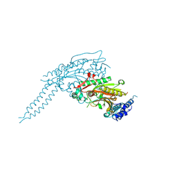
8P4T
 | | The spike complex of the Lujo Virus | | 分子名称: | 2-acetamido-2-deoxy-beta-D-glucopyranose, 2-acetamido-2-deoxy-beta-D-glucopyranose-(1-4)-2-acetamido-2-deoxy-beta-D-glucopyranose, Glycoprotein, ... | | 著者 | Eilon-Ashkenazy, M, Diskin, R. | | 登録日 | 2023-05-23 | | 公開日 | 2024-06-12 | | 実験手法 | ELECTRON MICROSCOPY (2.96 Å) | | 主引用文献 | The spike complex of the Lujo Virus
To Be Published
|
|
7B0B
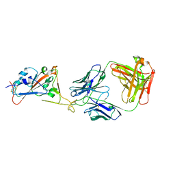 | | Fab HbnC3t1p1_C6 bound to SARS-CoV-2 RBD | | 分子名称: | 2-acetamido-2-deoxy-beta-D-glucopyranose-(1-4)-2-acetamido-2-deoxy-beta-D-glucopyranose, Fab heavy chain, IGK@ protein, ... | | 著者 | Borenstein-Katz, A, Diskin, R. | | 登録日 | 2020-11-19 | | 公開日 | 2021-12-01 | | 最終更新日 | 2024-01-31 | | 実験手法 | X-RAY DIFFRACTION (2.98 Å) | | 主引用文献 | Somatic hypermutation introduces bystander mutations that prepare SARS-CoV-2 antibodies for emerging variants.
Immunity, 56, 2023
|
|
1G84
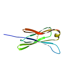 | | THE SOLUTION STRUCTURE OF THE C EPSILON2 DOMAIN FROM IGE | | 分子名称: | IMMUNOGLOBULIN E | | 著者 | McDonnell, J.M, Cowburn, D, Gould, H.J, Sutton, B.J, Calvert, R, Beavil, R.E, Beavil, A.J, Henry, A.J. | | 登録日 | 2000-11-16 | | 公開日 | 2001-05-16 | | 最終更新日 | 2021-10-27 | | 実験手法 | SOLUTION NMR | | 主引用文献 | The structure of the IgE Cepsilon2 domain and its role in stabilizing the complex with its high-affinity receptor FcepsilonRIalpha.
Nat.Struct.Biol., 8, 2001
|
|
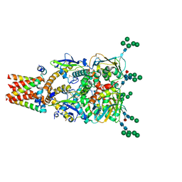
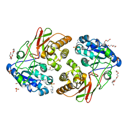
5JXJ
 | | Structure of the proprotein convertase furin complexed to meta-guanidinomethyl-Phac-RVR-Amba in presence of EDTA | | 分子名称: | 2UC-ARG-VAL-ARG-00S, CALCIUM ION, CHLORIDE ION, ... | | 著者 | Dahms, S.O, Arciniega, M, Steinmetzer, T, Huber, R, Than, M.E. | | 登録日 | 2016-05-13 | | 公開日 | 2016-10-05 | | 最終更新日 | 2024-07-10 | | 実験手法 | X-RAY DIFFRACTION (2 Å) | | 主引用文献 | Structure of the unliganded form of the proprotein convertase furin suggests activation by a substrate-induced mechanism.
Proc.Natl.Acad.Sci.USA, 113, 2016
|
|
1G9K
 | |
5JYB
 | | Crystal structure of 3 mutant of Ba3275 (S116A, E243A, H313A), the member of S66 family of serine peptidases | | 分子名称: | 1,2-ETHANEDIOL, 2-BUTANOL, 4-(2-HYDROXYETHYL)-1-PIPERAZINE ETHANESULFONIC ACID, ... | | 著者 | Nocek, B, Jedrzejczak, R, Joachimiak, A, CSGID, Center for Structural Genomics of Infectious Diseases (CSGID) | | 登録日 | 2016-05-13 | | 公開日 | 2016-06-15 | | 最終更新日 | 2023-09-27 | | 実験手法 | X-RAY DIFFRACTION (1.647 Å) | | 主引用文献 | Crystal structure of 3 mutant of Ba3275 (S116A, E243A, H313A), the member of S66 family of serine peptidases
To Be Published
|
|
1G3A
 | |
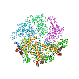
8P5D
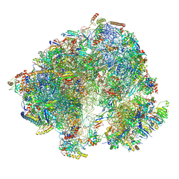 | | Spraguea lophii ribosome in the closed conformation by cryo sub tomogram averaging | | 分子名称: | 40S Ribosomal protein S19, 40S ribosomal protein S0, 40S ribosomal protein S10, ... | | 著者 | Gil Diez, P, McLaren, M, Isupov, M.N, Daum, B, Conners, R, Williams, B. | | 登録日 | 2023-05-23 | | 公開日 | 2023-06-21 | | 最終更新日 | 2023-12-20 | | 実験手法 | ELECTRON MICROSCOPY (10.8 Å) | | 主引用文献 | CryoEM reveals that ribosomes in microsporidian spores are locked in a dimeric hibernating state.
Nat Microbiol, 8, 2023
|
|
7B0W
 | | Crystal structure of the E. coli type 1 pilus assembly inhibitor FimI bound to FimC | | 分子名称: | 1,2-ETHANEDIOL, Chaperone protein FimC, FORMIC ACID, ... | | 著者 | Scharer, M.A, Zigova, Z, Giese, C, Puorger, C, Ignatov, O, Capitani, G, Glockshuber, R. | | 登録日 | 2020-11-23 | | 公開日 | 2021-12-08 | | 最終更新日 | 2024-01-31 | | 実験手法 | X-RAY DIFFRACTION (1.75 Å) | | 主引用文献 | Comprehensive kinetic characterization of bacterial pilus rod assembly and assembly termination
To Be Published
|
|


