3EPV
 
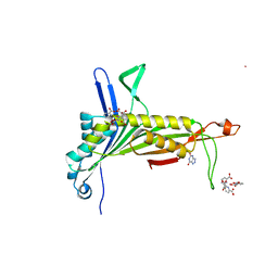
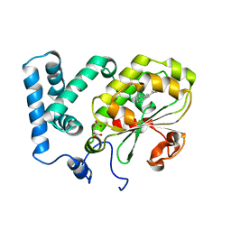
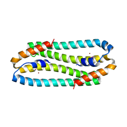 | | X-ray Structure of the Metal-sensor CnrX in both the Apo- and Copper-bound Forms | | 分子名称: | COPPER (II) ION, Nickel and cobalt resistance protein cnrR | | 著者 | Pompidor, G, Maillard, A.P, Girard, E, Gambarelli, S, Kahn, R, Coves, J. | | 登録日 | 2008-09-30 | | 公開日 | 2008-11-25 | | 最終更新日 | 2011-07-13 | | 実験手法 | X-RAY DIFFRACTION (1.742 Å) | | 主引用文献 | X-ray structure of the metal-sensor CnrX in both the apo- and copper-bound forms.
Febs Lett., 2008
|
|
3RWL
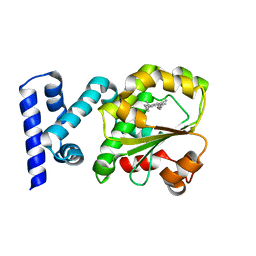
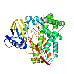 | | Structure of P450pyr hydroxylase | | 分子名称: | Cytochrome P450 alkane hydroxylase 1 CYP153A7, PROTOPORPHYRIN IX CONTAINING FE | | 著者 | Pompidor, G. | | 登録日 | 2011-05-09 | | 公開日 | 2012-04-18 | | 最終更新日 | 2024-02-28 | | 実験手法 | X-RAY DIFFRACTION (2 Å) | | 主引用文献 | Evolving P450pyr hydroxylase for highly enantioselective hydroxylation at non-activated carbon atom.
Chem.Commun.(Camb.), 48, 2012
|
|
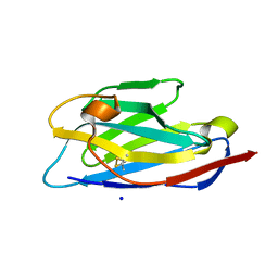
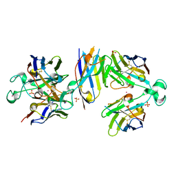
3LGR
 | | Xylanase II from Trichoderma reesei cocrystallized with tris-dipicolinate europium | | 分子名称: | EUROPIUM ION, Endo-1,4-beta-xylanase 2, PYRIDINE-2,6-DICARBOXYLIC ACID | | 著者 | Pompidor, G, Kahn, R, Maury, O. | | 登録日 | 2010-01-21 | | 公開日 | 2011-01-19 | | 最終更新日 | 2019-12-25 | | 実験手法 | X-RAY DIFFRACTION (1.64 Å) | | 主引用文献 | A dipicolinate lanthanide complex for solving protein structures using anomalous diffraction.
Acta Crystallogr.,Sect.D, 66, 2010
|
|
2PC2
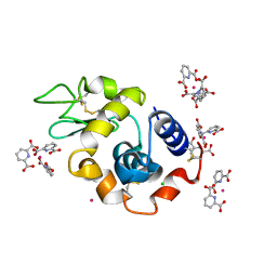 | | Lysozyme Cocrystallized with Tris-dipicolinate Eu complex | | 分子名称: | CHLORIDE ION, EUROPIUM ION, Lysozyme C, ... | | 著者 | Pompidor, G, Vicat, J, Kahn, R. | | 登録日 | 2007-03-29 | | 公開日 | 2008-04-01 | | 最終更新日 | 2017-10-18 | | 実験手法 | X-RAY DIFFRACTION (1.538 Å) | | 主引用文献 | A dipicolinate lanthanide complex for solving protein structures using anomalous diffraction
Acta Crystallogr.,Sect.D, 66, 2010
|
|
2PE7
 | | Thaumatin from Thaumatococcus Danielli in complex with tris-dipicolinate Europium | | 分子名称: | EUROPIUM ION, L(+)-TARTARIC ACID, PYRIDINE-2,6-DICARBOXYLIC ACID, ... | | 著者 | Pompidor, G, Vicat, J, Kahn, R. | | 登録日 | 2007-04-02 | | 公開日 | 2008-04-22 | | 最終更新日 | 2023-08-30 | | 実験手法 | X-RAY DIFFRACTION (1.46 Å) | | 主引用文献 | A dipicolinate lanthanide complex for solving protein structures using anomalous diffraction.
Acta Crystallogr.,Sect.D, 66, 2010
|
|
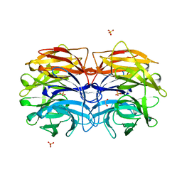
2PES
 | | Urate Oxidase in complex with tris-dipicolinate Lutetium | | 分子名称: | 8-AZAXANTHINE, LUTETIUM (III) ION, PYRIDINE-2,6-DICARBOXYLIC ACID, ... | | 著者 | Pompidor, G, Vicat, J, Kahn, R. | | 登録日 | 2007-04-03 | | 公開日 | 2008-04-22 | | 最終更新日 | 2024-02-21 | | 実験手法 | X-RAY DIFFRACTION (1.6 Å) | | 主引用文献 | A dipicolinate lanthanide complex for solving protein structures using anomalous diffraction.
Acta Crystallogr.,Sect.D, 66, 2010
|
|
6XZF
 | |
6XYF
 | |
6XYM
 | |
6Y0E
 | |
6Y1R
 | | Nb22-LBT | | 分子名称: | Nb22-LBT, SULFATE ION, TERBIUM(III) ION | | 著者 | Pompidor, G, Zimmermann, S, Loew, C, Schneider, T. | | 登録日 | 2020-02-13 | | 公開日 | 2021-02-24 | | 実験手法 | X-RAY DIFFRACTION (1.85 Å) | | 主引用文献 | Engineered nanobodies with a lanthanide binding motif for crystallographic phasing
To Be Published
|
|
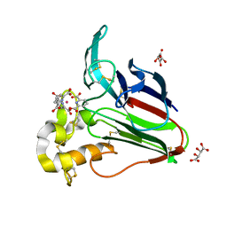
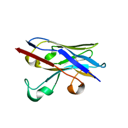
6GXR
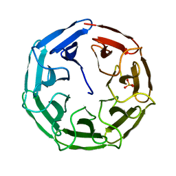 | | Crystal structure of BP39L lectin from Burkholderia pseudomallei at 1.7 A resolution | | 分子名称: | BP39L lectin, SULFATE ION | | 著者 | Sykorova, P, Novotna, J, Demo, G, Pompidor, G, Dubska, E, Komarek, J, Fujdiarova, E, Haronikova, L, Varrot, A, Imberty, A, Shilova, N, Bovin, N, Pokorna, M, Wimmerova, M. | | 登録日 | 2018-06-27 | | 公開日 | 2019-12-04 | | 最終更新日 | 2024-05-15 | | 実験手法 | X-RAY DIFFRACTION (1.7 Å) | | 主引用文献 | Characterization of novel lectins from Burkholderia pseudomallei and Chromobacterium violaceum with seven-bladed beta-propeller fold.
Int.J.Biol.Macromol., 152, 2020
|
|
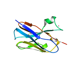
6GXS
 | | Crystal structure of CV39L lectin from Chromobacterium violaceum at 1.8 A resolution | | 分子名称: | 1,2-ETHANEDIOL, CV39L lectin, DI(HYDROXYETHYL)ETHER, ... | | 著者 | Sykorova, P, Novotna, J, Demo, G, Pompidor, G, Dubska, E, Komarek, J, Fujdiarova, E, Haronikova, L, Varrot, A, Imberty, A, Shilova, N, Bovin, N, Pokorna, M, Wimmerova, M. | | 登録日 | 2018-06-27 | | 公開日 | 2019-12-04 | | 最終更新日 | 2024-05-15 | | 実験手法 | X-RAY DIFFRACTION (1.8 Å) | | 主引用文献 | Characterization of novel lectins from Burkholderia pseudomallei and Chromobacterium violaceum with seven-bladed beta-propeller fold.
Int.J.Biol.Macromol., 152, 2020
|
|
4CJ6
 | | Crystal structure of the complex of the Cellular Retinal Binding Protein Mutant R234W with 9-cis-retinal | | 分子名称: | RETINAL, RETINALDEHYDE-BINDING PROTEIN 1 | | 著者 | Bolze, C.S, Helbling, R.E, Owen, R.L, Pearson, A.R, Pompidor, G, Dworkowski, F, Fuchs, M.R, Furrer, J, Golczak, M, Palczewski, K, Cascella, M, Stocker, A. | | 登録日 | 2013-12-19 | | 公開日 | 2014-01-08 | | 最終更新日 | 2023-12-20 | | 実験手法 | X-RAY DIFFRACTION (1.896 Å) | | 主引用文献 | Human Cellular Retinaldehyde-Binding Protein Has Secondary Thermal 9-Cis-Retinal Isomerase Activity.
J.Am.Chem.Soc., 136, 2014
|
|
4CIZ
 | | Crystal structure of the complex of the Cellular Retinal Binding Protein with 9-cis-retinal | | 分子名称: | L(+)-TARTARIC ACID, RETINAL, RETINALDEHYDE-BINDING PROTEIN 1 | | 著者 | Bolze, C.S, Helbling, R.E, Owen, R.L, Pearson, A.R, Pompidor, G, Dworkowski, F, Fuchs, M.R, Furrer, J, Golczak, M, Palczewski, K, Cascella, M, Stocker, A. | | 登録日 | 2013-12-18 | | 公開日 | 2014-01-08 | | 最終更新日 | 2023-12-20 | | 実験手法 | X-RAY DIFFRACTION (3.403 Å) | | 主引用文献 | Human Cellular Retinaldehyde-Binding Protein Has Secondary Thermal 9-Cis-Retinal Isomerase Activity.
J.Am.Chem.Soc., 136, 2014
|
|


