Loading
PDBj
MenuPDBj@FacebookPDBj@TwitterPDBj@YouTubewwPDB FoundationwwPDB
RCSB PDBPDBeBMRBAdv. SearchSearch help
PDB: 98 results

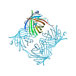
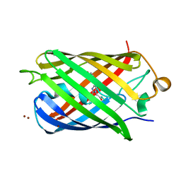
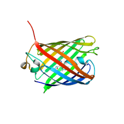
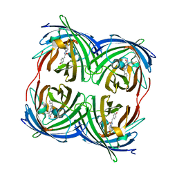
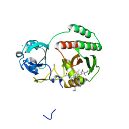
2PXW
DownloadVisualize
BU of 2pxw by Molmil
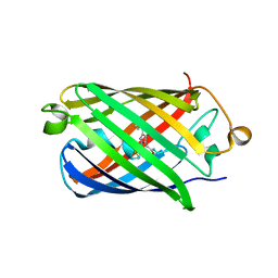
Crystal Structure of N66D Mutant of Green Fluorescent Protein from Zoanthus sp. at 2.4 A Resolution (Transition State)
Descriptor: GFP-like fluorescent chromoprotein FP506
Authors:Pletnev, S.V, Pletneva, N.V, Tikhonova, T.V, Pletnev, V.Z.
Deposit date:2007-05-14
Release date:2007-09-25
Last modified:2024-10-30
Method:X-RAY DIFFRACTION (2.4 Å)
Cite:Refined crystal structures of red and green fluorescent proteins from the button polyp Zoanthus.
Acta Crystallogr.,Sect.D, 63, 2007
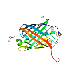
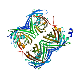
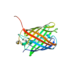
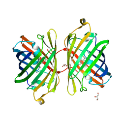
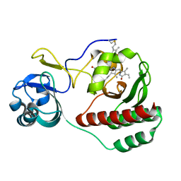
2PXS
DownloadVisualize
BU of 2pxs by Molmil
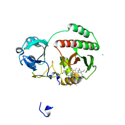
Crystal Structure of N66D Mutant of Green Fluorescent Protein from Zoanthus sp. at 2.2 A Resolution (Mature State)
Descriptor: GFP-like fluorescent chromoprotein FP506
Authors:Pletnev, S.V, Pletneva, N.V, Tikhonova, T.V, Pletnev, V.Z.
Deposit date:2007-05-14
Release date:2007-09-25
Last modified:2024-04-03
Method:X-RAY DIFFRACTION (2.2 Å)
Cite:Refined crystal structures of red and green fluorescent proteins from the button polyp Zoanthus.
Acta Crystallogr.,Sect.D, 63, 2007
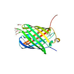
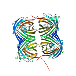
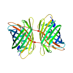
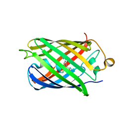
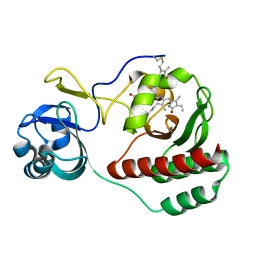
8SXC
DownloadVisualize
BU of 8sxc by Molmil
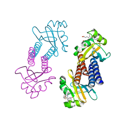
Crystal Structure of Fluorescent Protein Fusion Red 2
Descriptor: Far-red Fluorescent Protein Fusion Red 2, GLYCEROL, HEXAETHYLENE GLYCOL, ...
Authors:Pletnev, S, Pletneva, N, Pletnev, V.
Deposit date:2023-05-21
Release date:2024-02-07
Method:X-RAY DIFFRACTION (1.5 Å)
Cite:Crystal Structure of Fluorescent Protein Fusion Red 2
To Be Published
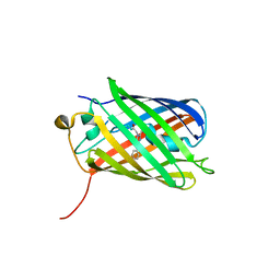
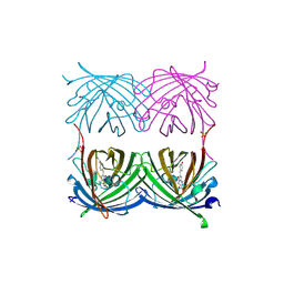
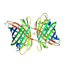
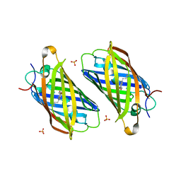
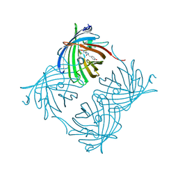
3BX9
DownloadVisualize
BU of 3bx9 by Molmil
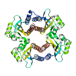
Monomeric Far-red Fluorescent Protein mKate Crystallized at pH 2.0
Descriptor: CITRIC ACID, Far-red fluorescent protein mKate, GLYCEROL
Authors:Pletnev, S, Pletneva, N, Pletnev, V.
Deposit date:2008-01-11
Release date:2008-07-22
Last modified:2023-11-15
Method:X-RAY DIFFRACTION (1.8 Å)
Cite:A Crystallographic Study of Bright Far-Red Fluorescent Protein mKate Reveals pH-induced cis-trans Isomerization of the Chromophore.
J.Biol.Chem., 283, 2008
3BXA
DownloadVisualize
BU of 3bxa by Molmil
Monomeric Far-red Fluorescent Protein mKate Crystallized at pH 4.2
Descriptor: CITRIC ACID, Far-red fluorescent protein mKate
Authors:Pletnev, S, Pletneva, N, Pletnev, V.
Deposit date:2008-01-12
Release date:2008-07-22
Last modified:2017-10-25
Method:X-RAY DIFFRACTION (1.75 Å)
Cite:A Crystallographic Study of Bright Far-Red Fluorescent Protein mKate Reveals pH-induced cis-trans Isomerization of the Chromophore.
J.Biol.Chem., 283, 2008
6U1A
DownloadVisualize
BU of 6u1a by Molmil
Crystal Structure of Fluorescent Protein FusionRed
Descriptor: CALCIUM ION, NICKEL (II) ION, Red fluorescent protein
Authors:Pletnev, S, Muslinkina, L, Pletneva, N, Pletnev, V.Z.
Deposit date:2019-08-15
Release date:2020-04-22
Last modified:2023-11-15
Method:X-RAY DIFFRACTION (1.09 Å)
Cite:Two independent routes of post-translational chemistry in fluorescent protein FusionRed.
Int.J.Biol.Macromol., 155, 2020
7SF9
DownloadVisualize
BU of 7sf9 by Molmil
Branchiostoma Floridae Violet Fluorescent Protein
Descriptor: Violet Fluorescent Protein
Authors:Pletnev, S, Pletneva, N, Pletnev, V.Z, Muslinkina, L.
Deposit date:2021-10-03
Release date:2022-03-30
Last modified:2023-10-18
Method:X-RAY DIFFRACTION (1.8 Å)
Cite:A novel violet fluorescent protein contains a unique oxidized tyrosine as the simplest chromophore ever reported in fluorescent proteins.
Protein Sci., 31, 2022
7SFA
DownloadVisualize
BU of 7sfa by Molmil
Branchiostoma floridae fluorescent protein LanFP10A2
Descriptor: Fluorescent protein lanFP10A2
Authors:Pletnev, S, Pletneva, N, Pletnev, V.Z, Muslinkina, L.
Deposit date:2021-10-03
Release date:2022-03-30
Last modified:2023-11-15
Method:X-RAY DIFFRACTION (1.65 Å)
Cite:A novel violet fluorescent protein contains a unique oxidized tyrosine as the simplest chromophore ever reported in fluorescent proteins.
Protein Sci., 31, 2022
2ICR
DownloadVisualize
BU of 2icr by Molmil
Red fluorescent protein zRFP574 from Zoanthus sp.
Descriptor: Red fluorescent protein zoanRFP, SULFATE ION
Authors:Pletnev, S, Pletneva, N, Tikhonova, T, Pletnev, V.
Deposit date:2006-09-13
Release date:2007-10-02
Last modified:2023-11-15
Method:X-RAY DIFFRACTION (1.51 Å)
Cite:Refined crystal structures of red and green fluorescent proteins from the button polyp Zoanthus.
Acta Crystallogr.,Sect.D, 63, 2007
3BXC
DownloadVisualize
BU of 3bxc by Molmil
Monomeric Far-red Fluorescent Protein mKate Crystallized at pH 9.0
Descriptor: Far-red fluorescent protein mKate
Authors:Pletnev, S, Pletneva, N, Pletnev, V.
Deposit date:2008-01-12
Release date:2008-07-22
Last modified:2019-07-24
Method:X-RAY DIFFRACTION (2.6 Å)
Cite:A Crystallographic Study of Bright Far-Red Fluorescent Protein mKate Reveals pH-induced cis-trans Isomerization of the Chromophore.
J.Biol.Chem., 283, 2008
3BXB
DownloadVisualize
BU of 3bxb by Molmil
Monomeric Far-red Fluorescent Protein mKate Crystallized at pH 7.0
Descriptor: Far-red fluorescent protein mKate
Authors:Pletnev, S, Pletneva, N, Pletnev, V.
Deposit date:2008-01-12
Release date:2008-07-22
Last modified:2019-07-24
Method:X-RAY DIFFRACTION (2.6 Å)
Cite:A Crystallographic Study of Bright Far-Red Fluorescent Protein mKate Reveals pH-induced cis-trans Isomerization of the Chromophore.
J.Biol.Chem., 283, 2008
3GL4
DownloadVisualize
BU of 3gl4 by Molmil
X-ray structure of photobleached killerred
Descriptor: KillerRed
Authors:Pletnev, S, Pletneva, N.V, Pletnev, V.Z.
Deposit date:2009-03-11
Release date:2009-09-08
Last modified:2023-11-15
Method:X-RAY DIFFRACTION (2.15 Å)
Cite:Structural basis for phototoxicity of the genetically encoded photosensitizer KillerRed.
J.Biol.Chem., 284, 2009
3GB3
DownloadVisualize
BU of 3gb3 by Molmil
X-ray structure of genetically encoded photosensitizer KillerRed in native form
Descriptor: KillerRed, SULFATE ION
Authors:Pletnev, S, Pletneva, N.V, Pletnev, V.Z.
Deposit date:2009-02-18
Release date:2009-09-08
Last modified:2024-10-09
Method:X-RAY DIFFRACTION (1.75 Å)
Cite:Structural basis for phototoxicity of the genetically encoded photosensitizer KillerRed.
J.Biol.Chem., 284, 2009
3PIB
DownloadVisualize
BU of 3pib by Molmil
Crystal structure of red fluorescent protein eqFP578 crystallized at pH 5.5
Descriptor: GLYCEROL, eqFP578 fluorescent protein
Authors:Pletnev, S, Pletneva, N.V, Pletnev, V.Z.
Deposit date:2010-11-05
Release date:2011-05-25
Last modified:2012-02-22
Method:X-RAY DIFFRACTION (1.154 Å)
Cite:Crystallographic study of red fluorescent protein eqFP578 and its far-red variant Katushka reveals opposite pH-induced isomerization of chromophore.
Protein Sci., 20, 2011
3PJ7
DownloadVisualize
BU of 3pj7 by Molmil
Crystal structure of far-red fluorescent protein Katushka crystallized at pH 8.5
Descriptor: Red fluorescent protein eqFP578
Authors:Pletnev, S, Pletneva, N.V, Pletnev, V.Z.
Deposit date:2010-11-08
Release date:2011-05-25
Last modified:2012-02-22
Method:X-RAY DIFFRACTION (1.85 Å)
Cite:Crystallographic study of red fluorescent protein eqFP578 and its far-red variant Katushka reveals opposite pH-induced isomerization of chromophore.
Protein Sci., 20, 2011
3PJB
DownloadVisualize
BU of 3pjb by Molmil
Crystal structure of red fluorescent protein eqFP578 crystallized at pH 4.0
Descriptor: GLYCEROL, Red fluorescent protein eqFP578
Authors:Pletnev, S, Pletneva, N.V, Pletnev, V.Z.
Deposit date:2010-11-09
Release date:2011-05-25
Last modified:2018-01-24
Method:X-RAY DIFFRACTION (1.75 Å)
Cite:Crystallographic study of red fluorescent protein eqFP578 and its far-red variant Katushka reveals opposite pH-induced isomerization of chromophore.
Protein Sci., 20, 2011
4EDO
DownloadVisualize
BU of 4edo by Molmil
Crystal structure of far-red fluorescent protein eqFP650
Descriptor: far-red fluorescent protein eqFP650
Authors:Pletnev, S, Pletnev, V.Z, Pletneva, N.V.
Deposit date:2012-03-27
Release date:2012-09-05
Last modified:2013-03-27
Method:X-RAY DIFFRACTION (1.8 Å)
Cite:Structural basis for bathochromic shift of fluorescence in far-red fluorescent proteins eqFP650 and eqFP670.
Acta Crystallogr.,Sect.D, 68, 2012
3PJ5
DownloadVisualize
BU of 3pj5 by Molmil
Crystal structure of far-red fluorescent protein Katushka crystallized at pH 5.0
Descriptor: Red fluorescent protein eqFP578, SULFATE ION
Authors:Pletnev, S, Pletneva, N.V, Pletnev, V.Z.
Deposit date:2010-11-08
Release date:2011-05-25
Last modified:2012-02-22
Method:X-RAY DIFFRACTION (1.6 Å)
Cite:Crystallographic study of red fluorescent protein eqFP578 and its far-red variant Katushka reveals opposite pH-induced isomerization of chromophore.
Protein Sci., 20, 2011
4EDS
DownloadVisualize
BU of 4eds by Molmil
Crystal structure of far-red fluorescent protein eqFP670
Descriptor: far-red fluorescent protein eqFP650
Authors:Pletnev, S, Pletnev, V.Z, Pletneva, N.V.
Deposit date:2012-03-27
Release date:2012-09-05
Last modified:2013-03-27
Method:X-RAY DIFFRACTION (1.6 Å)
Cite:Structural basis for bathochromic shift of fluorescence in far-red fluorescent proteins eqFP650 and eqFP670.
Acta Crystallogr.,Sect.D, 68, 2012
4XTQ
DownloadVisualize
BU of 4xtq by Molmil
Crystal structure of a mutant (C20S) of a near-infrared fluorescent protein BphP1-FP
Descriptor: 3-[2-[(Z)-[5-[(Z)-[(3R,4R)-3-ethenyl-4-methyl-5-oxidanylidene-pyrrolidin-2-ylidene]methyl]-3-(3-hydroxy-3-oxopropyl)-4-methyl-pyrrol-2-ylidene]methyl]-5-[(Z)-(4-ethenyl-3-methyl-5-oxidanylidene-pyrrol-2-ylidene)methyl]-4-methyl-1H-pyrrol-3-yl]propanoic acid, BphP1-FP/C20S, CHLORIDE ION
Authors:Pletnev, S, Malashkevich, V.N.
Deposit date:2015-01-23
Release date:2015-12-09
Last modified:2019-12-04
Method:X-RAY DIFFRACTION (1.64 Å)
Cite:Molecular Basis of Spectral Diversity in Near-Infrared Phytochrome-Based Fluorescent Proteins.
Chem.Biol., 22, 2015
7LSD
DownloadVisualize
BU of 7lsd by Molmil
Crystal structure of near-infrared fluorescent protein miRFP718nano
Descriptor: BILIVERDINE IX ALPHA, miRFP718nano
Authors:Pletnev, S.
Deposit date:2021-02-18
Release date:2022-02-23
Last modified:2024-10-16
Method:X-RAY DIFFRACTION (1.7 Å)
Cite:Rational design of a small near-infrared fluorescent protein from CBCR suitable for deep-tissue SWIR imaging.
To Be Published
7LSC
DownloadVisualize
BU of 7lsc by Molmil
Crystal structure of near-infrared fluorescent protein miRFP670nano3
Descriptor: 3-[2-[(~{Z})-[5-[(~{Z})-[(3~{S},4~{R})-3-ethenyl-4-methyl-5-oxidanylidene-pyrrolidin-2-ylidene]methyl]-3-(3-hydroxy-3-oxopropyl)-4-methyl-pyrrol-2-ylidene]methyl]-5-[(~{Z})-(4-ethenyl-3-methyl-5-oxidanylidene-pyrrol-2-ylidene)methyl]-4-methyl-1~{H}-pyrrol-3-yl]propanoic acid, miRFP670nano3
Authors:Pletnev, S.
Deposit date:2021-02-18
Release date:2022-02-23
Last modified:2023-10-18
Method:X-RAY DIFFRACTION (1.8 Å)
Cite:Near-infrared fluorescent protein with enhanced brightness.
To Be Published
5VIV
DownloadVisualize
BU of 5viv by Molmil
Crystal structure of monomeric near-infrared fluorescent protein miRFP670
Descriptor: 3-[2-[[5-[(4-ethenyl-3-methyl-5-oxidanylidene-pyrrol-2-yl)methyl]-3-(3-hydroxy-3-oxopropyl)-4-methyl-1~{H}-pyrrol-2-yl] methyl]-5-[[(3~{R},4~{R})-3-ethyl-4-methyl-5-oxidanylidene-3,4-dihydropyrrol-2-yl]methyl]-4-methyl-1~{H}-pyrrol-3-yl]pro panoic acid, 3-[5-[[(3~{R},4~{R})-3-ethenyl-4-methyl-5-oxidanylidene-3,4-dihydropyrrol-2-yl]methyl]-2-[[5-[(4-ethenyl-3-methyl-5-oxidanylidene-pyrrol-2-yl)methyl]-3-(3-hydroxy-3-oxopropyl)-4-methyl-1~{H}-pyrrol-2-yl]methyl]-4-methyl-1~{H}-pyrrol-3-yl]propanoic acid, CHLORIDE ION, ...
Authors:Pletnev, S.
Deposit date:2017-04-17
Release date:2017-06-21
Last modified:2024-10-16
Method:X-RAY DIFFRACTION (1.33 Å)
Cite:Designing brighter near-infrared fluorescent proteins: insights from structural and biochemical studies.
Chem Sci, 8, 2017
5VIK
DownloadVisualize
BU of 5vik by Molmil
Crystal structure of monomeric near-infrared fluorescent protein miRFP703
Descriptor: BILIVERDINE IX ALPHA, near-infrared fluorescent protein miRFP703
Authors:Pletnev, S.
Deposit date:2017-04-17
Release date:2017-06-07
Last modified:2024-10-23
Method:X-RAY DIFFRACTION (1.35 Å)
Cite:Designing brighter near-infrared fluorescent proteins: insights from structural and biochemical studies.
Chem Sci, 8, 2017
5VIQ
DownloadVisualize
BU of 5viq by Molmil
Crystal structure of monomeric near-infrared fluorescent protein miRFP709
Descriptor: BILIVERDINE IX ALPHA, monomeric near-infrared fluorescent protein miRFP709
Authors:Pletnev, S.
Deposit date:2017-04-17
Release date:2017-06-07
Last modified:2024-10-16
Method:X-RAY DIFFRACTION (1.34 Å)
Cite:Designing brighter near-infrared fluorescent proteins: insights from structural and biochemical studies.
Chem Sci, 8, 2017


1234>

226707

數據於2024-10-30公開中

PDB statisticsPDBj update infoContact PDBjnumon