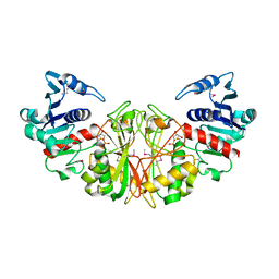
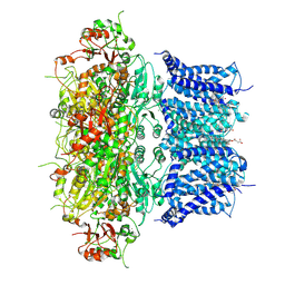
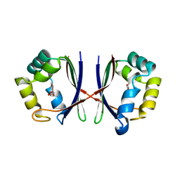
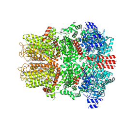
1F9T
 
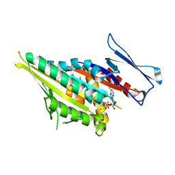 | | CRYSTAL STRUCTURES OF KINESIN MUTANTS REVEAL A SIGNALLING PATHWAY FOR ACTIVATION OF THE MOTOR ATPASE | | 分子名称: | ADENOSINE-5'-DIPHOSPHATE, KINESIN-LIKE PROTEIN KAR3, MAGNESIUM ION | | 著者 | Yun, M, Zhang, X, Park, C.-G, Park, H.-W, Endow, S.A. | | 登録日 | 2000-07-11 | | 公開日 | 2001-06-13 | | 最終更新日 | 2024-02-07 | | 実験手法 | X-RAY DIFFRACTION (1.5 Å) | | 主引用文献 | A structural pathway for activation of the kinesin motor ATPase.
EMBO J., 20, 2001
|
|
9JO4
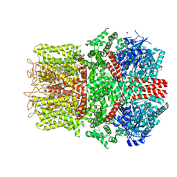
 | | Cryo-EM structure of human BKca channel-compound 51b complex | | 分子名称: | 5-(5-morpholin-4-ylpentylamino)-2-[2,3,5,6-tetrakis(fluoranyl)-4-(trifluoromethyl)phenoxy]phenol, CALCIUM ION, CHOLESTEROL HEMISUCCINATE, ... | | 著者 | Kim, S, Park, S, Lee, N.Y, Lee, E.Y, Lee, N, Roh, E.C, Kim, Y.G, Kim, H.J, Jin, M.S, Park, C.S, Kim, Y.C. | | 登録日 | 2024-09-24 | | 公開日 | 2025-02-26 | | 最終更新日 | 2025-03-12 | | 実験手法 | ELECTRON MICROSCOPY (3.4 Å) | | 主引用文献 | Discovery of Diphenyl Ether Derivatives as Novel BK Ca Channel Activators: Structure-Activity Relationship, Cryo-EM Complex Structures, and In Vivo Animal Studies.
J.Med.Chem., 68, 2025
|
|
9JO3
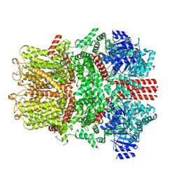
 | | Cryo-EM structure of human BKca channel-compound 10b complex | | 分子名称: | 5-azanyl-2-[2,3,5,6-tetrakis(fluoranyl)-4-(trifluoromethyl)phenoxy]phenol, CALCIUM ION, CHOLESTEROL HEMISUCCINATE, ... | | 著者 | Kim, S, Park, S, Lee, N.Y, Lee, E.Y, Lee, N, Roh, E.C, Kim, Y.G, Kim, H.J, Jin, M.S, Park, C.S, Kim, Y.C. | | 登録日 | 2024-09-24 | | 公開日 | 2025-02-26 | | 最終更新日 | 2025-03-12 | | 実験手法 | ELECTRON MICROSCOPY (2.8 Å) | | 主引用文献 | Discovery of Diphenyl Ether Derivatives as Novel BK Ca Channel Activators: Structure-Activity Relationship, Cryo-EM Complex Structures, and In Vivo Animal Studies.
J.Med.Chem., 68, 2025
|
|
1DC3
 | |
1DC4
 | | STRUCTURAL ANALYSIS OF GLYCERALDEHYDE 3-PHOSPHATE DEHYDROGENASE FROM ESCHERICHIA COLI: DIRECT EVIDENCE FOR SUBSTRATE BINDING AND COFACTOR-INDUCED CONFORMATIONAL CHANGES | | 分子名称: | GLYCERALDEHYDE 3-PHOSPHATE DEHYDROGENASE, SN-GLYCEROL-3-PHOSPHATE | | 著者 | Yun, M, Park, C.-G, Kim, J.-Y, Park, H.-W. | | 登録日 | 1999-11-04 | | 公開日 | 2000-08-23 | | 最終更新日 | 2024-10-30 | | 実験手法 | X-RAY DIFFRACTION (2.5 Å) | | 主引用文献 | Structural analysis of glyceraldehyde 3-phosphate dehydrogenase from Escherichia coli: direct evidence of substrate binding and cofactor-induced conformational changes.
Biochemistry, 39, 2000
|
|
1DC6
 | | STRUCTURAL ANALYSIS OF GLYCERALDEHYDE 3-PHOSPHATE DEHYDROGENASE FROM ESCHERICHIA COLI: DIRECT EVIDENCE FOR SUBSTRATE BINDING AND COFACTOR-INDUCED CONFORMATIONAL CHANGES. | | 分子名称: | GLYCERALDEHYDE-3-PHOSPHATE DEHYDROGENASE, NICOTINAMIDE-ADENINE-DINUCLEOTIDE | | 著者 | Yun, M, Park, C.-G, Kim, J.-Y, Park, H.-W. | | 登録日 | 1999-11-04 | | 公開日 | 2000-08-23 | | 最終更新日 | 2024-02-07 | | 実験手法 | X-RAY DIFFRACTION (2 Å) | | 主引用文献 | Structural analysis of glyceraldehyde 3-phosphate dehydrogenase from Escherichia coli: direct evidence of substrate binding and cofactor-induced conformational changes.
Biochemistry, 39, 2000
|
|
1DC5
 | |
8JB9
 | |
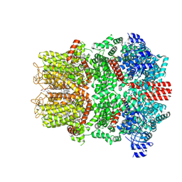
4ZZ7
 | | Crystal structure of methylmalonate-semialdehyde dehydrogenase (DddC) from Oceanimonas doudoroffii | | 分子名称: | Methylmalonate-semialdehyde dehydrogenase, NICOTINAMIDE-ADENINE-DINUCLEOTIDE | | 著者 | Do, H, Lee, C.W, Lee, S.G, Kang, H, Park, C.M, Kim, H.J, Park, H, Park, H, Lee, J.H. | | 登録日 | 2015-05-22 | | 公開日 | 2016-04-06 | | 最終更新日 | 2024-03-20 | | 実験手法 | X-RAY DIFFRACTION (2.9 Å) | | 主引用文献 | Crystal structure and modeling of the tetrahedral intermediate state of methylmalonate-semialdehyde dehydrogenase (MMSDH) from Oceanimonas doudoroffii.
J. Microbiol., 54, 2016
|
|
8Z3S
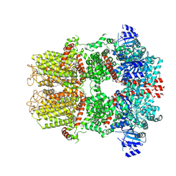
 | | Activation mechanism and novel binding site of the BKCa channel activator CTIBD | | 分子名称: | 4-[4-(4-chlorophenyl)-3-(trifluoromethyl)-1,2-oxazol-5-yl]benzene-1,3-diol, CALCIUM ION, CHOLESTEROL HEMISUCCINATE, ... | | 著者 | Lee, N, Kim, S, Jo, H, Lee, N.Y, Jin, M.S, Park, C.S. | | 登録日 | 2024-04-16 | | 公開日 | 2024-07-31 | | 最終更新日 | 2024-09-11 | | 実験手法 | ELECTRON MICROSCOPY (3.9 Å) | | 主引用文献 | Activation mechanism and novel binding sites of the BK Ca channel activator CTIBD.
Life Sci Alliance, 7, 2024
|
|
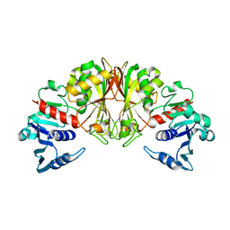
1ESM
 | | STRUCTURAL BASIS FOR THE FEEDBACK REGULATION OF ESCHERICHIA COLI PANTOTHENATE KINASE BY COENZYME A | | 分子名称: | COENZYME A, PANTOTHENATE KINASE | | 著者 | Yun, M, Park, C.G, Kim, J.Y, Rock, C.O, Jackowski, S, Park, H.W. | | 登録日 | 2000-04-10 | | 公開日 | 2000-09-20 | | 最終更新日 | 2024-11-06 | | 実験手法 | X-RAY DIFFRACTION (2.5 Å) | | 主引用文献 | Structural basis for the feedback regulation of Escherichia coli pantothenate kinase by coenzyme A.
J.Biol.Chem., 275, 2000
|
|
1ESN
 | | STRUCTURAL BASIS FOR THE FEEDBACK REGULATION OF ESCHERICHIA COLI PANTOTHENATE KINASE BY COENZYME A | | 分子名称: | MAGNESIUM ION, PANTOTHENATE KINASE, PHOSPHOAMINOPHOSPHONIC ACID-ADENYLATE ESTER | | 著者 | Yun, M, Park, C.G, Kim, J.Y, Rock, C.O, Jackowski, S, Park, H.W. | | 登録日 | 2000-04-10 | | 公開日 | 2000-11-17 | | 最終更新日 | 2024-11-06 | | 実験手法 | X-RAY DIFFRACTION (2.6 Å) | | 主引用文献 | Structural basis for the feedback regulation of Escherichia coli pantothenate kinase by coenzyme A.
J.Biol.Chem., 275, 2000
|
|
7C0J
 | |
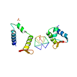
1X8D
 | | Crystal structure of E. coli YiiL protein containing L-rhamnose | | 分子名称: | Hypothetical protein yiiL, L-RHAMNOSE | | 著者 | Ryu, K.S, Kim, J.I, Cho, S.J, Park, D, Park, C, Lee, J.O, Choi, B.S. | | 登録日 | 2004-08-18 | | 公開日 | 2005-05-17 | | 最終更新日 | 2024-03-13 | | 実験手法 | X-RAY DIFFRACTION (1.8 Å) | | 主引用文献 | Structural Insights into the Monosaccharide Specificity of Escherichia coli Rhamnose Mutarotase
J.Mol.Biol., 349, 2005
|
|
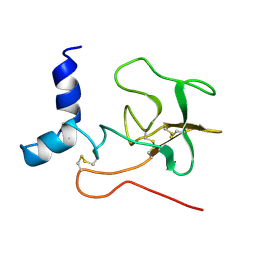
7C0I
 | | Crystal structure of chimeric mutant of E3L in complex with Z-DNA | | 分子名称: | DNA (5'-D(*TP*CP*GP*CP*GP*CP*G)-3'), Double-stranded RNA-binding protein,Double-stranded RNA-specific adenosine deaminase, SULFATE ION | | 著者 | Choi, H.J, Park, C.H, Kim, J.S. | | 登録日 | 2020-05-01 | | 公開日 | 2020-12-16 | | 最終更新日 | 2023-11-29 | | 実験手法 | X-RAY DIFFRACTION (2.4 Å) | | 主引用文献 | Dual conformational recognition by Z-DNA binding protein is important for the B-Z transition process.
Nucleic Acids Res., 48, 2020
|
|
1GUB
 | | Hinge-bending motion of D-allose binding protein from Escherichia coli: three open conformations | | 分子名称: | D-ALLOSE-BINDING PERIPLASMIC PROTEIN, NICKEL (II) ION | | 著者 | Magnusson, U, Chaudhuri, B.N, Ko, J, Park, C, Jones, T.A, Mowbray, S.L. | | 登録日 | 2002-01-24 | | 公開日 | 2003-03-06 | | 最終更新日 | 2023-12-13 | | 実験手法 | X-RAY DIFFRACTION (3.1 Å) | | 主引用文献 | Structure of D-Allose Binding Protein from Escherichia Coli Bound to D-Allose at 1.8 A Resolution
J.Mol.Biol., 286, 1999
|
|
1GUD
 | | Hinge-bending motion of D-allose binding protein from Escherichia coli: three open conformations | | 分子名称: | D-ALLOSE-BINDING PERIPLASMIC PROTEIN, ZINC ION | | 著者 | Magnusson, U, Chaudhuri, B.N, Ko, J, Park, C, Jones, T.A, Mowbray, S.L. | | 登録日 | 2002-01-24 | | 公開日 | 2003-03-06 | | 最終更新日 | 2023-12-13 | | 実験手法 | X-RAY DIFFRACTION (1.71 Å) | | 主引用文献 | Structure of D-Allose Binding Protein from Escherichia Coli Bound to D-Allose at 1.8 A Resolution
J.Mol.Biol., 286, 1999
|
|
2PF1
 | |
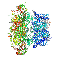
9B6G
 | | Cryo-EM structure of the mouse TRPM8 channel in complex with the antagonist AMTB | | 分子名称: | 2-acetamido-2-deoxy-beta-D-glucopyranose, CHOLESTEROL HEMISUCCINATE, N-(3-aminopropyl)-2-[(3-methylphenyl)methoxy]-N-[(thiophen-2-yl)methyl]benzamide, ... | | 著者 | Yin, Y, Park, C.-G, Zhang, F, Fedor, J, Feng, S, Suo, Y, Im, W, Lee, S.-Y. | | 登録日 | 2024-03-25 | | 公開日 | 2024-08-21 | | 最終更新日 | 2024-11-13 | | 実験手法 | ELECTRON MICROSCOPY (2.81 Å) | | 主引用文献 | Mechanisms of sensory adaptation and inhibition of the cold and menthol receptor TRPM8.
Sci Adv, 10, 2024
|
|
9B6I
 | | Cryo-EM structure of the avian great tit TRPM8 channel in complex with the antagonist TC-I 2014 | | 分子名称: | 3-{7-(trifluoromethyl)-5-[2-(trifluoromethyl)phenyl]-1H-benzimidazol-2-yl}-1-oxa-2-azaspiro[4.5]dec-2-ene, CALCIUM ION, CHOLESTEROL HEMISUCCINATE, ... | | 著者 | Yin, Y, Park, C.-G, Zhang, F, Fedor, J, Feng, S, Suo, Y, Im, W, Lee, S.-Y. | | 登録日 | 2024-03-25 | | 公開日 | 2024-08-21 | | 最終更新日 | 2024-10-09 | | 実験手法 | ELECTRON MICROSCOPY (3.26 Å) | | 主引用文献 | Mechanisms of sensory adaptation and inhibition of the cold and menthol receptor TRPM8.
Sci Adv, 10, 2024
|
|
9B6K
 | | Cryo-EM structure of the mouse TRPM8 channel in complex with Ca2+ in the absence of PI(4,5)P2 | | 分子名称: | CALCIUM ION, Transient receptor potential cation channel subfamily M member 8 | | 著者 | Yin, Y, Park, C.-G, Zhang, F, Fedor, J, Feng, S, Suo, Y, Im, W, Lee, S.-Y. | | 登録日 | 2024-03-25 | | 公開日 | 2024-08-21 | | 最終更新日 | 2024-11-06 | | 実験手法 | ELECTRON MICROSCOPY (4.13 Å) | | 主引用文献 | Mechanisms of sensory adaptation and inhibition of the cold and menthol receptor TRPM8.
Sci Adv, 10, 2024
|
|
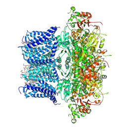
9B6F
 | | Cryo-EM structure of the mouse TRPM8 channel in complex with the antagonist AMG2850 | | 分子名称: | (8R)-8-[4-(trifluoromethyl)phenyl]-N-[(2S)-1,1,1-trifluoropropan-2-yl]-5,8-dihydro-1,7-naphthyridine-7(6H)-carboxamide, CHOLESTEROL HEMISUCCINATE, Transient receptor potential cation channel subfamily M member 8 | | 著者 | Yin, Y, Park, C.-G, Zhang, F, Fedor, J, Feng, S, Suo, Y, Im, W, Lee, S.-Y. | | 登録日 | 2024-03-25 | | 公開日 | 2024-08-21 | | 最終更新日 | 2024-10-23 | | 実験手法 | ELECTRON MICROSCOPY (3.42 Å) | | 主引用文献 | Mechanisms of sensory adaptation and inhibition of the cold and menthol receptor TRPM8.
Sci Adv, 10, 2024
|
|
9B6D
 | | Cryo-EM structure of the mouse TRPM8 channel in the ligand-free desensitized state | | 分子名称: | CHOLESTEROL HEMISUCCINATE, Transient receptor potential cation channel subfamily M member 8 | | 著者 | Yin, Y, Park, C.-G, Zhang, F, Fedor, J, Feng, S, Suo, Y, Im, W, Lee, S.-Y. | | 登録日 | 2024-03-25 | | 公開日 | 2024-08-21 | | 最終更新日 | 2024-11-20 | | 実験手法 | ELECTRON MICROSCOPY (3.3 Å) | | 主引用文献 | Mechanisms of sensory adaptation and inhibition of the cold and menthol receptor TRPM8.
Sci Adv, 10, 2024
|
|
9B6E
 | | Cryo-EM structure of the mouse TRPM8 channel in complex with the antagonist TC-I 2014 | | 分子名称: | 3-{7-(trifluoromethyl)-5-[2-(trifluoromethyl)phenyl]-1H-benzimidazol-2-yl}-1-oxa-2-azaspiro[4.5]dec-2-ene, CHOLESTEROL HEMISUCCINATE, Transient receptor potential cation channel subfamily M member 8 | | 著者 | Yin, Y, Park, C.-G, Zhang, F, Fedor, J, Feng, S, Suo, Y, Im, W, Lee, S.-Y. | | 登録日 | 2024-03-25 | | 公開日 | 2024-08-21 | | 最終更新日 | 2024-11-13 | | 実験手法 | ELECTRON MICROSCOPY (2.91 Å) | | 主引用文献 | Mechanisms of sensory adaptation and inhibition of the cold and menthol receptor TRPM8.
Sci Adv, 10, 2024
|
|
9B6J
 | | Cryo-EM structure of the mouse TRPM8 channel in complex with PI(4,5)P2 and Ca2+ | | 分子名称: | CALCIUM ION, Transient receptor potential cation channel subfamily M member 8, [(2R)-2-octanoyloxy-3-[oxidanyl-[(1R,2R,3S,4R,5R,6S)-2,3,6-tris(oxidanyl)-4,5-diphosphonooxy-cyclohexyl]oxy-phosphoryl]oxy-propyl] octanoate | | 著者 | Yin, Y, Park, C.-G, Zhang, F, Fedor, J, Feng, S, Suo, Y, Im, W, Lee, S.-Y. | | 登録日 | 2024-03-25 | | 公開日 | 2024-08-21 | | 実験手法 | ELECTRON MICROSCOPY (3.53 Å) | | 主引用文献 | Mechanisms of sensory adaptation and inhibition of the cold and menthol receptor TRPM8.
Sci Adv, 10, 2024
|
|


