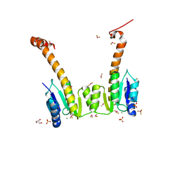
1PJE
 
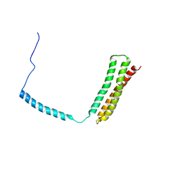
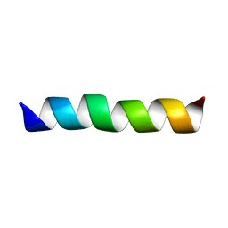
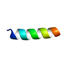 | | Structure of the channel-forming trans-membrane domain of Virus protein "u"(Vpu) from HIV-1 | | 分子名称: | VPU protein | | 著者 | Park, S.H, Mrse, A.A, Nevzorov, A.A, Mesleh, M.F, Oblatt-Montal, M, Montal, M, Opella, S.J. | | 登録日 | 2003-06-02 | | 公開日 | 2003-10-14 | | 最終更新日 | 2024-05-22 | | 実験手法 | SOLID-STATE NMR | | 主引用文献 | Three-dimensional structure of the channel-forming trans-membrane domain of Virus protein "u" (Vpu) from HIV-1
J.Mol.Biol., 333, 2003
|
|
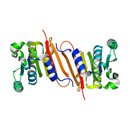
5Y8T
 | | Crystal structure of Bacillus subtilis PadR in complex with p-coumaric acid | | 分子名称: | 4'-HYDROXYCINNAMIC ACID, Transcriptional regulator | | 著者 | Park, S.C, Kwak, Y.M, Song, W.S, Hong, M, Yoon, S.I. | | 登録日 | 2017-08-21 | | 公開日 | 2017-11-29 | | 最終更新日 | 2023-11-22 | | 実験手法 | X-RAY DIFFRACTION (2 Å) | | 主引用文献 | Structural basis of effector and operator recognition by the phenolic acid-responsive transcriptional regulator PadR.
Nucleic Acids Res., 45, 2017
|
|
1ROM
 | |
4RDH
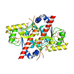
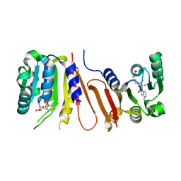
 | | Crystal structure of E. coli tRNA N6-threonylcarbamoyladenosine dehydratase, TcdA | | 分子名称: | ADENOSINE MONOPHOSPHATE, GLYCEROL, SULFATE ION, ... | | 著者 | Park, S.Y, Kim, S, Lee, H. | | 登録日 | 2014-09-19 | | 公開日 | 2015-08-05 | | 最終更新日 | 2015-10-07 | | 実験手法 | X-RAY DIFFRACTION (2.1 Å) | | 主引用文献 | The Structure of Escherichia coli TcdA (Also Known As CsdL) Reveals a Novel Topology and Provides Insight into the tRNA Binding Surface Required for N(6)-Threonylcarbamoyladenosine Dehydratase Activity.
J.Mol.Biol., 427, 2015
|
|
2DN1
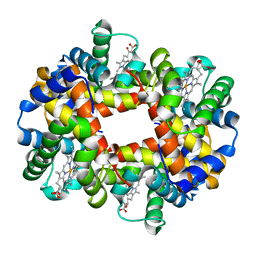
 | | 1.25A resolution crystal structure of human hemoglobin in the oxy form | | 分子名称: | Hemoglobin alpha subunit, Hemoglobin beta subunit, OXYGEN MOLECULE, ... | | 著者 | Park, S.-Y, Yokoyama, T, Shibayama, N, Shiro, Y, Tame, J.R. | | 登録日 | 2006-04-25 | | 公開日 | 2006-05-09 | | 最終更新日 | 2024-03-13 | | 実験手法 | X-RAY DIFFRACTION (1.25 Å) | | 主引用文献 | 1.25 a resolution crystal structures of human haemoglobin in the oxy, deoxy and carbonmonoxy forms.
J.Mol.Biol., 360, 2006
|
|
1OWA
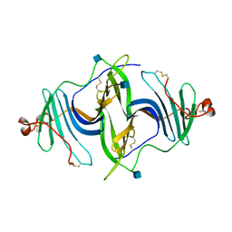
 | | Solution Structural Studies on Human Erythrocyte Alpha Spectrin N Terminal Tetramerization Domain | | 分子名称: | Spectrin alpha chain, erythrocyte | | 著者 | Park, S, Caffrey, M.S, Johnson, M.E, Fung, L.W. | | 登録日 | 2003-03-28 | | 公開日 | 2004-03-30 | | 最終更新日 | 2024-05-22 | | 実験手法 | SOLUTION NMR | | 主引用文献 | Solution structural studies on human erythrocyte alpha-spectrin tetramerization site.
J.Biol.Chem., 278, 2003
|
|
1XKO
 | | Structure of Thermotoga maritima CheX | | 分子名称: | CHEMOTAXIS protein cheX | | 著者 | Park, S.Y, Chao, X, Gonzalez-Bonet, G, Beel, B.D, Bilwes, A.M, Crane, B.R. | | 登録日 | 2004-09-29 | | 公開日 | 2004-12-07 | | 最終更新日 | 2023-08-23 | | 実験手法 | X-RAY DIFFRACTION (2.48 Å) | | 主引用文献 | Structure and Function of an Unusual Family of Protein Phosphatases; The Bacterial Chemotaxis Proteins CheC and CheX.
Mol.Cell, 16, 2004
|
|
5EEP
 | | Crystal structure of E. coli CsdE | | 分子名称: | CsdA-binding activator | | 著者 | Park, S.Y. | | 登録日 | 2015-10-23 | | 公開日 | 2016-06-08 | | 最終更新日 | 2024-03-20 | | 実験手法 | X-RAY DIFFRACTION (2.4 Å) | | 主引用文献 | The crystal structure of Escherichia coli CsdE
Int.J.Biol.Macromol., 87, 2016
|
|
5I0Z
 | |
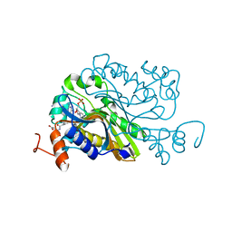
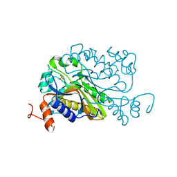
4RDI
 | | Crystal structure of E. coli tRNA N6-threonylcarbamoyladenosine dehydratase, TcdA | | 分子名称: | ADENOSINE-5'-TRIPHOSPHATE, GLYCEROL, tRNA threonylcarbamoyladenosine dehydratase | | 著者 | Park, S.Y, Kim, S, Lee, H. | | 登録日 | 2014-09-19 | | 公開日 | 2015-08-05 | | 最終更新日 | 2015-10-07 | | 実験手法 | X-RAY DIFFRACTION (1.95 Å) | | 主引用文献 | The Structure of Escherichia coli TcdA (Also Known As CsdL) Reveals a Novel Topology and Provides Insight into the tRNA Binding Surface Required for N(6)-Threonylcarbamoyladenosine Dehydratase Activity.
J.Mol.Biol., 427, 2015
|
|
2DN3
 | | 1.25A resolution crystal structure of human hemoglobin in the carbonmonoxy form | | 分子名称: | CARBON MONOXIDE, Hemoglobin alpha subunit, Hemoglobin beta subunit, ... | | 著者 | Park, S.-Y, Yokoyama, T, Shibayama, N, Shiro, Y, Tame, J.R. | | 登録日 | 2006-04-25 | | 公開日 | 2006-05-09 | | 最終更新日 | 2024-03-13 | | 実験手法 | X-RAY DIFFRACTION (1.25 Å) | | 主引用文献 | 1.25 a resolution crystal structures of human haemoglobin in the oxy, deoxy and carbonmonoxy forms.
J.Mol.Biol., 360, 2006
|
|
2DN2
 | | 1.25A resolution crystal structure of human hemoglobin in the deoxy form | | 分子名称: | Hemoglobin alpha subunit, Hemoglobin beta subunit, PROTOPORPHYRIN IX CONTAINING FE | | 著者 | Park, S.-Y, Yokoyama, T, Shibayama, N, Shiro, Y, Tame, J.R. | | 登録日 | 2006-04-25 | | 公開日 | 2006-05-09 | | 最終更新日 | 2023-10-25 | | 実験手法 | X-RAY DIFFRACTION (1.25 Å) | | 主引用文献 | 1.25 a resolution crystal structures of human haemoglobin in the oxy, deoxy and carbonmonoxy forms.
J.Mol.Biol., 360, 2006
|
|
6JLA
 | | Crystal structure of a mouse ependymin related protein | | 分子名称: | 2-acetamido-2-deoxy-beta-D-glucopyranose, 2-acetamido-2-deoxy-beta-D-glucopyranose-(1-4)-[alpha-L-fucopyranose-(1-6)]2-acetamido-2-deoxy-beta-D-glucopyranose, Mammalian ependymin-related protein 1 | | 著者 | Park, S. | | 登録日 | 2019-03-04 | | 公開日 | 2020-03-04 | | 最終更新日 | 2020-09-16 | | 実験手法 | X-RAY DIFFRACTION (2.4 Å) | | 主引用文献 | Structures of three ependymin-related proteins suggest their function as a hydrophobic molecule binder.
Iucrj, 6, 2019
|
|
2GOH
 | | Three-dimensional Structure of the Trans-membrane Domain of Vpu from HIV-1 in Aligned Phospholipid Bicelles | | 分子名称: | VPU protein | | 著者 | Park, S.H, De Angelis, A.A, Nevzorov, A.A, Wu, C.H, Opella, S.J. | | 登録日 | 2006-04-12 | | 公開日 | 2006-08-08 | | 最終更新日 | 2024-05-29 | | 実験手法 | SOLUTION NMR | | 主引用文献 | Three-Dimensional Structure of the Transmembrane Domain of Vpu from HIV-1 in Aligned Phospholipid Bicelles.
Biophys.J., 91, 2006
|
|
4POO
 | | The crystal structure of Bacillus subtilis YtqB in complex with SAM | | 分子名称: | Putative RNA methylase, S-ADENOSYLMETHIONINE | | 著者 | Park, S.C, Song, W.S, Yoon, S.I. | | 登録日 | 2014-02-26 | | 公開日 | 2014-04-02 | | 最終更新日 | 2023-11-08 | | 実験手法 | X-RAY DIFFRACTION (2.2 Å) | | 主引用文献 | Structural analysis of a putative SAM-dependent methyltransferase, YtqB, from Bacillus subtilis
Biochem.Biophys.Res.Commun., 446, 2014
|
|
6A9A
 | | Ternary complex crystal structure of dCH with dCMP and THF | | 分子名称: | (6S)-5,6,7,8-TETRAHYDROFOLATE, 2'-DEOXYCYTIDINE-5'-MONOPHOSPHATE, Deoxycytidylate 5-hydroxymethyltransferase, ... | | 著者 | Park, S.H, Song, H.K. | | 登録日 | 2018-07-12 | | 公開日 | 2019-01-02 | | 最終更新日 | 2023-11-22 | | 実験手法 | X-RAY DIFFRACTION (1.9 Å) | | 主引用文献 | A cytosine modification mechanism revealed by the structure of a ternary complex of deoxycytidylate hydroxymethylase from bacteriophage T4 with its cofactor and substrate.
Iucrj, 6, 2019
|
|
6A9B
 | |
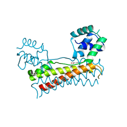
1L5Y
 | | CRYSTAL STRUCTURE OF MG2+ / BEF3-BOUND RECEIVER DOMAIN OF SINORHIZOBIUM MELILOTI DCTD | | 分子名称: | BERYLLIUM DIFLUORIDE, BERYLLIUM TETRAFLUORIDE ION, BERYLLIUM TRIFLUORIDE ION, ... | | 著者 | Park, S, Meyer, M, Jones, A.D, Yennawar, H.P, Yennawar, N.H, Nixon, B.T. | | 登録日 | 2002-03-08 | | 公開日 | 2002-10-23 | | 最終更新日 | 2023-08-16 | | 実験手法 | X-RAY DIFFRACTION (2.1 Å) | | 主引用文献 | Two-component signaling in the AAA + ATPase DctD: binding Mg2+ and BeF3- selects between alternate dimeric states of the receiver domain
FASEB J., 16, 2002
|
|
2GOF
 | | Three-dimensional structure of the trans-membrane domain of Vpu from HIV-1 in aligned phospholipid bicelles | | 分子名称: | VPU protein | | 著者 | Park, S.H, De Angelis, A.A, Nevzorov, A.A, Wu, C.H, Opella, S.J. | | 登録日 | 2006-04-12 | | 公開日 | 2006-08-08 | | 最終更新日 | 2024-05-29 | | 実験手法 | SOLUTION NMR | | 主引用文献 | Three-Dimensional Structure of the Transmembrane Domain of Vpu from HIV-1 in Aligned Phospholipid Bicelles.
Biophys.J., 91, 2006
|
|
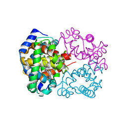
5H2B
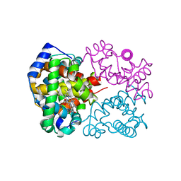
 | | Structure of a novel antibody G196 | | 分子名称: | G196 antibody Heavy chain, G196 antibody Light chain | | 著者 | Park, S.Y, Sugiyama, K. | | 登録日 | 2016-10-14 | | 公開日 | 2017-03-22 | | 最終更新日 | 2020-02-26 | | 実験手法 | X-RAY DIFFRACTION (2.001 Å) | | 主引用文献 | G196 epitope tag system: a novel monoclonal antibody, G196, recognizes the small, soluble peptide DLVPR with high affinity.
Sci Rep, 7, 2017
|
|
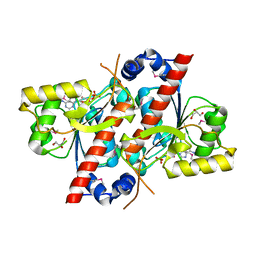
4R27
 | | Crystal structure of beta-glycosidase BGL167 | | 分子名称: | Glycoside hydrolase | | 著者 | Park, S.J, Choi, J.M, Kyeong, H.H, Kim, S.G, Kim, H.S. | | 登録日 | 2014-08-09 | | 公開日 | 2015-05-27 | | 最終更新日 | 2023-11-08 | | 実験手法 | X-RAY DIFFRACTION (2.03 Å) | | 主引用文献 | Rational design of a beta-glycosidase with high regiospecificity for triterpenoid tailoring
Chembiochem, 16, 2015
|
|
4PON
 | | The crystal structure of a putative SAM-dependent methyltransferase, YtqB, from Bacillus subtilis | | 分子名称: | Putative RNA methylase | | 著者 | Park, S.C, Song, W.S, Yoon, S.I. | | 登録日 | 2014-02-26 | | 公開日 | 2014-04-02 | | 最終更新日 | 2023-11-08 | | 実験手法 | X-RAY DIFFRACTION (1.9 Å) | | 主引用文献 | Structural analysis of a putative SAM-dependent methyltransferase, YtqB, from Bacillus subtilis
Biochem.Biophys.Res.Commun., 446, 2014
|
|
5X11
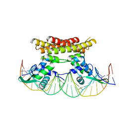 | | Crystal structure of Bacillus subtilis PadR in complex with operator DNA | | 分子名称: | DNA (28-MER), Transcriptional regulator | | 著者 | Park, S.C, Kwak, Y.M, Song, W.S, Hong, M, Yoon, S.I. | | 登録日 | 2017-01-24 | | 公開日 | 2017-11-22 | | 最終更新日 | 2023-11-22 | | 実験手法 | X-RAY DIFFRACTION (2.65 Å) | | 主引用文献 | Structural basis of effector and operator recognition by the phenolic acid-responsive transcriptional regulator PadR
Nucleic Acids Res., 45, 2017
|
|
2E46
 | |
4OKV
 | |


