Loading
PDBj
メニューPDBj@FacebookPDBj@TwitterPDBj@YouTubewwPDB FoundationwwPDB
RCSB PDBPDBeBMRBAdv. SearchSearch help
PDB: 8 件

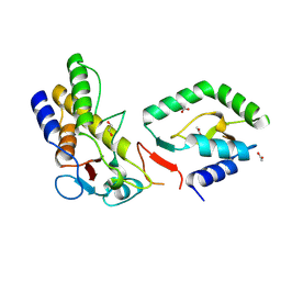
7YXV
DownloadVisualize
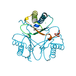
BU of 7yxv by Molmil
Crystal structure of a tautomerase superfamily member form Acinetobacter baumanii
分子名称: Anthranilate synthase, CHLORIDE ION, NITRATE ION
著者Pankov, G, Hunter, W.N.
登録日2022-02-16
公開日2022-05-18
最終更新日2024-02-07
実験手法X-RAY DIFFRACTION (1.7 Å)
主引用文献The structure of a tautomerase superfamily member linked to the type VI secretion system of Acinetobacter baumannii.
Acta Crystallogr.,Sect.F, 79, 2023
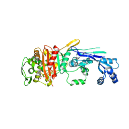
6XV5
DownloadVisualize
BU of 6xv5 by Molmil
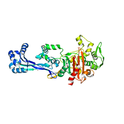
Crystal structure of penicillin-binding protein 2 from Yersinia pestis in complex with ertapenem
分子名称: L(+)-TARTARIC ACID, Peptidoglycan D,D-transpeptidase MrdA
著者Pankov, G, Hunter, W.N, Dawson, A.
登録日2020-01-21
公開日2021-02-03
最終更新日2024-01-24
実験手法X-RAY DIFFRACTION (2.673 Å)
主引用文献The structure of penicillin-binding protein 2 from Yersinia pestis
To Be Published
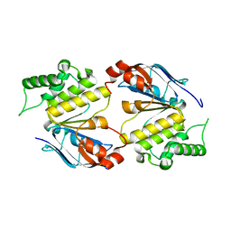
6HS3
DownloadVisualize
BU of 6hs3 by Molmil
Crystal structure of an ABC transporter related protein from Burkholderia pseudomallei
分子名称: ABC transporter family protein, CHLORIDE ION
著者Pankov, G, Hunter, W.N, Dawson, A.
登録日2018-09-28
公開日2018-11-07
最終更新日2024-01-24
実験手法X-RAY DIFFRACTION (2.4 Å)
主引用文献The structure of lipopolysaccharide transport protein B (LptB) from Burkholderia pseudomallei.
Acta Crystallogr.,Sect.F, 75, 2019
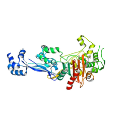
6SYN
DownloadVisualize
BU of 6syn by Molmil
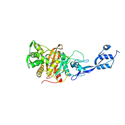
Crystal structure of Y. pestis penicillin-binding protein 3
分子名称: (2R,4S)-2-[(1R)-1-{[(2S)-2-carboxy-2-phenylacetyl]amino}-2-oxoethyl]-5,5-dimethyl-1,3-thiazolidine-4-carboxylic acid, ACETATE ION, Peptidoglycan D,D-transpeptidase FtsI
著者Pankov, G, Hunter, W.N, Dawson, A.
登録日2019-09-30
公開日2020-10-14
最終更新日2024-01-24
実験手法X-RAY DIFFRACTION (2.63 Å)
主引用文献The structure of penicillin-binding protein 3 from Yersinia pestis
To Be Published
6TIX
DownloadVisualize
BU of 6tix by Molmil
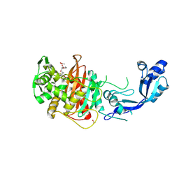
Crystal structure of penicillin-binding protein 2 from Yersinia pestis in complex with mecillinam
分子名称: 2-[(1R)-1-{[(E)-azepan-1-ylmethylidene]amino}-2-oxoethyl]-5,5-dimethyl-1,3-thiazolidine-4-carboxylic acid, Peptidoglycan D,D-transpeptidase MrdA
著者Pankov, G, Hunter, W.N, Dawson, A.
登録日2019-11-22
公開日2020-12-02
最終更新日2024-01-24
実験手法X-RAY DIFFRACTION (2.8 Å)
主引用文献The structure of penicillin-binding protein 2 from Yersinia pestis in complex with mecillinam
To Be Published
6TII
DownloadVisualize
BU of 6tii by Molmil
Crystal structure of penicillin-binding protein 2 from Yersinia pestis
分子名称: BROMIDE ION, Peptidoglycan D,D-transpeptidase MrdA
著者Pankov, G, Hunter, W.N, Dawson, A.
登録日2019-11-22
公開日2020-12-02
最終更新日2024-01-24
実験手法X-RAY DIFFRACTION (2.26 Å)
主引用文献The structure of penicillin-binding protein 2 from Yersinia pestis
To Be Published
6TUD
DownloadVisualize
BU of 6tud by Molmil
Crystal structure of Y. pestis penicillin-binding protein 3
分子名称: Peptidoglycan D,D-transpeptidase FtsI
著者Pankov, G, Hunter, W.N, Dawson, A.
登録日2020-01-06
公開日2021-01-27
最終更新日2024-01-24
実験手法X-RAY DIFFRACTION (3 Å)
主引用文献The structure of penicillin-binding protein 3 from Yersinia pestis
To Be Published
5G1S
DownloadVisualize
BU of 5g1s by Molmil
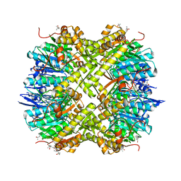
Open conformation of Francisella tularensis ClpP at 1.7 A
分子名称: (4R)-2-METHYLPENTANE-2,4-DIOL, (4S)-2-METHYL-2,4-PENTANEDIOL, ACETATE ION, ...
著者Diaz-Saez, L, Pankov, G, Hunter, W.N.
登録日2016-03-30
公開日2016-10-19
最終更新日2024-05-01
実験手法X-RAY DIFFRACTION (1.7 Å)
主引用文献Open and compressed conformations of Francisella tularensis ClpP.
Proteins, 85, 2017

219869

件を2024-05-15に公開中

PDB statisticsPDBj update infoContact PDBjnumon