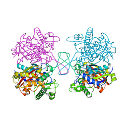
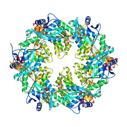
2WK4
 
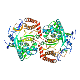 | | Dimeric structure of D347G D348G mutant of the sapporovirus RNA dependent RNA polymerase | | 分子名称: | GLYCEROL, PROTEASE-POLYMERASE P70 | | 著者 | Fullerton, S.W.B, Robel, I, Schuldt, L, Gebhardt, J, Tucker, P.A, Rohayem, J. | | 登録日 | 2009-06-05 | | 公開日 | 2010-09-01 | | 最終更新日 | 2023-12-13 | | 実験手法 | X-RAY DIFFRACTION (2.98 Å) | | 主引用文献 | Dimeric Structure of D347G D348G Mutant of the Sapporovirus Sapporovirus RNA Dependent RNA Polymerase
To be Published
|
|
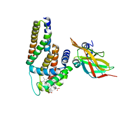
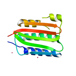
2WMX
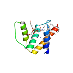
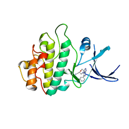 | | Crystal structure of checkpoint kinase 1 (Chk1) in complex with inhibitors | | 分子名称: | 1-[(2S)-4-(5-phenyl-1H-pyrazolo[3,4-b]pyridin-4-yl)morpholin-2-yl]methanamine, SERINE/THREONINE-PROTEIN KINASE CHK1 | | 著者 | Matthews, T.P, Klair, S, Burns, S, Boxall, K, Cherry, M, Fisher, M, Westwood, I.M, Walton, M.I, McHardy, T, Cheung, K.-M.J, Van Montfort, R, Williams, D, Aherne, G.W, Garrett, M.D, Reader, J, Collins, I. | | 登録日 | 2009-07-03 | | 公開日 | 2009-07-28 | | 最終更新日 | 2023-12-13 | | 実験手法 | X-RAY DIFFRACTION (2.45 Å) | | 主引用文献 | Identification of Inhibitors of Checkpoint Kinase 1 Through Template Screening.
J.Med.Chem., 52, 2009
|
|
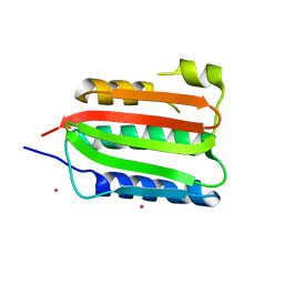
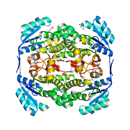
2WHE
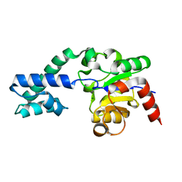 | | Structure of native Beta-Phosphoglucomutase in an open conformation without bound ligands. | | 分子名称: | BETA-PHOSPHOGLUCOMUTASE, MAGNESIUM ION | | 著者 | Bowler, M.W, Baxter, N.J, Webster, C.E, Pollard, S, Alizadeh, T, Hounslow, A.M, Cliff, M.J, Bermel, W, Williams, N.H, Hollfelder, F, Blackburn, G.M, Waltho, J.P. | | 登録日 | 2009-05-04 | | 公開日 | 2009-09-15 | | 最終更新日 | 2023-12-13 | | 実験手法 | X-RAY DIFFRACTION (1.55 Å) | | 主引用文献 | Atomic Details of Near-Transition State Conformers for Enzyme Phosphoryl Transfer Revealed by Mgf-3 Rather Than by Phosphoranes.
Proc.Natl.Acad.Sci.USA, 107, 2010
|
|
5K4T
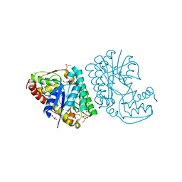 | |
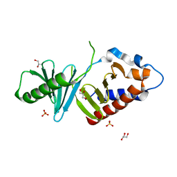
2WH5
 | | Crystal structure of human acyl-CoA binding domain 4 complexed with stearoyl-CoA | | 分子名称: | ACYL-COA-BINDING DOMAIN-CONTAINING PROTEIN 4, COENZYME A, STEARIC ACID, ... | | 著者 | Yue, W.W, Shafqat, N, Ugochukwu, E, Savitsky, P, Johansson, C, Salah, E, Roos, A.K, Chaikuad, A, von Delft, F, Arrowsmith, C, Weigelt, J, Edwards, A, Bountra, C, Oppermann, U. | | 登録日 | 2009-04-30 | | 公開日 | 2009-07-28 | | 最終更新日 | 2023-12-13 | | 実験手法 | X-RAY DIFFRACTION (2.6 Å) | | 主引用文献 | Crystal Structure of Human Acyl-Coa Binding Domain 4
To be Published
|
|
2WMQ
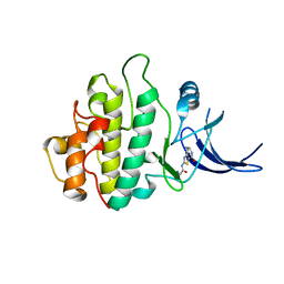 | | Crystal structure of checkpoint kinase 1 (Chk1) in complex with inhibitors | | 分子名称: | N-(4-OXO-5,6,7,8-TETRAHYDRO-4H-[1,3]THIAZOLO[5,4-C]AZEPIN-2-YL)ACETAMIDE, SERINE/THREONINE-PROTEIN KINASE CHK1 | | 著者 | Matthews, T.P, Klair, S, Burns, S, Boxall, K, Cherry, M, Fisher, M, Westwood, I.M, Walton, M.I, McHardy, T, Cheung, K.-M.J, Van Montfort, R, Williams, D, Aherne, G.W, Garrett, M.D, Reader, J, Collins, I. | | 登録日 | 2009-07-03 | | 公開日 | 2009-07-28 | | 最終更新日 | 2023-12-13 | | 実験手法 | X-RAY DIFFRACTION (2.48 Å) | | 主引用文献 | Identification of Inhibitors of Checkpoint Kinase 1 Through Template Screening.
J.Med.Chem., 52, 2009
|
|
2WKX
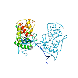 | | Crystal structure of the native E. coli zinc amidase AmiD | | 分子名称: | CHLORIDE ION, GLYCEROL, N-ACETYLMURAMOYL-L-ALANINE AMIDASE AMID, ... | | 著者 | Petrella, S, Kerff, F, Herman, R, Genereux, C, Pennartz, A, Sauvage, E, Joris, B, Charlier, P. | | 登録日 | 2009-06-18 | | 公開日 | 2010-01-12 | | 最終更新日 | 2023-12-13 | | 実験手法 | X-RAY DIFFRACTION (1.8 Å) | | 主引用文献 | Specific Structural Features of the N-Acetylmuramoyl-L-Alanine Amidase Amid from Escherichia Coli and Mechanistic Implications for Enzymes of This Family.
J.Mol.Biol., 397, 2010
|
|
2WMU
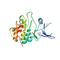 | | Crystal structure of checkpoint kinase 1 (Chk1) in complex with inhibitors | | 分子名称: | 6-MORPHOLIN-4-YL-9H-PURINE, SERINE/THREONINE-PROTEIN KINASE CHK1 | | 著者 | Matthews, T.P, Klair, S, Burns, S, Boxall, K, Cherry, M, Fisher, M, Westwood, I.M, Walton, M.I, McHardy, T, Cheung, K.-M.J, Van Montfort, R, Williams, D, Aherne, G.W, Garrett, M.D, Reader, J, Collins, I. | | 登録日 | 2009-07-03 | | 公開日 | 2009-07-28 | | 最終更新日 | 2023-12-13 | | 実験手法 | X-RAY DIFFRACTION (2.6 Å) | | 主引用文献 | Identification of Inhibitors of Checkpoint Kinase 1 Through Template Screening.
J.Med.Chem., 52, 2009
|
|
5XZ5
 | |
3OVU
 | | Crystal Structure of Human Alpha-Haemoglobin Complexed with AHSP and the First NEAT Domain of IsdH from Staphylococcus aureus | | 分子名称: | Alpha-hemoglobin-stabilizing protein, Hemoglobin subunit alpha, Iron-regulated surface determinant protein H, ... | | 著者 | Jacques, D.A, Krishna Kumar, K, Caradoc-Davies, T.T, Langley, D.B, Mackay, J.P, Guss, J.M, Gell, D.A. | | 登録日 | 2010-09-17 | | 公開日 | 2011-09-21 | | 最終更新日 | 2024-03-20 | | 実験手法 | X-RAY DIFFRACTION (2.83 Å) | | 主引用文献 | A new haem pocket structure in alpha-haemoglobin
To be Published
|
|
8K35
 | | Structure of the bacteriophage lambda tail tip complex | | 分子名称: | IRON/SULFUR CLUSTER, Tail tip assembly protein I, Tail tip protein L, ... | | 著者 | Xiao, H, Tan, L, Cheng, L.P, Liu, H.R. | | 登録日 | 2023-07-14 | | 公開日 | 2023-11-15 | | 最終更新日 | 2024-01-17 | | 実験手法 | ELECTRON MICROSCOPY (3.44 Å) | | 主引用文献 | Structure of the siphophage neck-Tail complex suggests that conserved tail tip proteins facilitate receptor binding and tail assembly.
Plos Biol., 21, 2023
|
|
8K38
 | | The structure of bacteriophage lambda portal-adaptor | | 分子名称: | Head completion protein, Portal protein B | | 著者 | Xiao, H, Tan, L, Cheng, L.P, Liu, H.R. | | 登録日 | 2023-07-14 | | 公開日 | 2023-11-15 | | 最終更新日 | 2024-01-17 | | 実験手法 | ELECTRON MICROSCOPY (3.2 Å) | | 主引用文献 | Structure of the siphophage neck-Tail complex suggests that conserved tail tip proteins facilitate receptor binding and tail assembly.
Plos Biol., 21, 2023
|
|
8DVQ
 | | CA domain of VanSA histidine kinase | | 分子名称: | CADMIUM ION, Sensor protein VanS | | 著者 | Loll, P.J. | | 登録日 | 2022-07-29 | | 公開日 | 2023-03-22 | | 最終更新日 | 2024-05-22 | | 実験手法 | X-RAY DIFFRACTION (2.19 Å) | | 主引用文献 | Structure of VanS from vancomycin-resistant enterococci: A sensor kinase with weak ATP binding.
J.Biol.Chem., 299, 2023
|
|
3OZI
 | | Crystal structure of the TIR domain from the flax disease resistance protein L6 | | 分子名称: | COBALT (II) ION, L6tr | | 著者 | Ve, T, Bernoux, M, Williams, S, Valkov, E, Warren, C, Hatters, D, Ellis, J.G, Dodds, P.N, Kobe, B. | | 登録日 | 2010-09-25 | | 公開日 | 2011-04-27 | | 最終更新日 | 2023-11-01 | | 実験手法 | X-RAY DIFFRACTION (2.3 Å) | | 主引用文献 | Structural and Functional Analysis of a Plant Resistance Protein TIR Domain Reveals Interfaces for Self-Association, Signaling, and Autoregulation.
Cell Host Microbe, 9, 2011
|
|
6SIQ
 | | Fragment AZ-012 binding at the p53pT387/14-3-3 sigma interface | | 分子名称: | 14-3-3 protein sigma, 4-chloranyl-7-propan-2-yloxy-1-benzothiophene-2-carboximidamide, CHLORIDE ION, ... | | 著者 | Leysen, S, Wolter, M, Guillory, X, Genet, S, Somsen, B, Patel, J, Castaldi, P, Ottmann, C. | | 登録日 | 2019-08-10 | | 公開日 | 2020-06-17 | | 最終更新日 | 2024-01-24 | | 実験手法 | X-RAY DIFFRACTION (1.60121 Å) | | 主引用文献 | Fragment-based Differential Targeting of PPI Stabilizer Interfaces.
J.Med.Chem., 63, 2020
|
|
8DX0
 | | VanSC CA domain | | 分子名称: | Histidine kinase, MAGNESIUM ION | | 著者 | Loll, P.J. | | 登録日 | 2022-08-02 | | 公開日 | 2023-03-22 | | 最終更新日 | 2024-04-03 | | 実験手法 | X-RAY DIFFRACTION (1.45 Å) | | 主引用文献 | Structure of VanS from vancomycin-resistant enterococci: A sensor kinase with weak ATP binding.
J.Biol.Chem., 299, 2023
|
|
6OJX
 | |
2WUW
 | | Crystallographic analysis of counter-ion effects on subtilisin enzymatic action in acetonitrile (native data) | | 分子名称: | ACETONITRILE, CALCIUM ION, SODIUM ION, ... | | 著者 | Cianci, M, Tomaszewki, B, Helliwell, J.R, Halling, P.J. | | 登録日 | 2009-10-09 | | 公開日 | 2010-12-08 | | 最終更新日 | 2023-12-20 | | 実験手法 | X-RAY DIFFRACTION (2.23 Å) | | 主引用文献 | Crystallographic Analysis of Counterion Effects on Subtilisin Enzymatic Action in Acetonitrile.
J.Am.Chem.Soc., 132, 2010
|
|
6OKV
 | |
8DWZ
 | | CA domain of VanSA histidine kinase, 7 keV data | | 分子名称: | CADMIUM ION, Sensor protein VanS | | 著者 | Loll, P.J. | | 登録日 | 2022-08-02 | | 公開日 | 2023-03-22 | | 最終更新日 | 2024-05-22 | | 実験手法 | X-RAY DIFFRACTION (2.21 Å) | | 主引用文献 | Structure of VanS from vancomycin-resistant enterococci: A sensor kinase with weak ATP binding.
J.Biol.Chem., 299, 2023
|
|
2WYU
 | | High resolution structure of Thermus thermophilus enoyl-acyl carrier protein reductase apo-form | | 分子名称: | ENOYL-[ACYL CARRIER PROTEIN] REDUCTASE, GLYCEROL, SODIUM ION | | 著者 | Otero, J.M, Noel, A.J, Guardado-Calvo, P, Llamas-Saiz, A.L, van Raaij, M.J. | | 登録日 | 2009-11-20 | | 公開日 | 2010-11-24 | | 最終更新日 | 2023-12-20 | | 実験手法 | X-RAY DIFFRACTION (1.5 Å) | | 主引用文献 | High-resolution structures of Thermus thermophilus enoyl-acyl carrier protein reductase in the apo form, in complex with NAD+ and in complex with NAD+ and triclosan.
Acta Crystallogr. Sect. F Struct. Biol. Cryst. Commun., 68, 2012
|
|
3P2W
 | |
6SA4
 | | SALSA / DMBT1 / GP340 SRCR domain 1 | | 分子名称: | CHLORIDE ION, Deleted in malignant brain tumors 1 protein, GLYCEROL, ... | | 著者 | Reichhardt, M.P, Lea, S.M, Johnson, S. | | 登録日 | 2019-07-16 | | 公開日 | 2020-03-18 | | 最終更新日 | 2024-01-24 | | 実験手法 | X-RAY DIFFRACTION (1.77 Å) | | 主引用文献 | Structures of SALSA/DMBT1 SRCR domains reveal the conserved ligand-binding mechanism of the ancient SRCR fold.
Life Sci Alliance, 3, 2020
|
|
8U7F
 | | Crystal structure of CIB_12 beta-galactosidase from Cuniculiplasma divulgatum | | 分子名称: | CIB_12 Beta-galactosidase, GLYCEROL | | 著者 | Stogios, P.J, Skarina, T, Di Leo, R, Yakunin, A, Golyshin, P, Savchenko, A. | | 登録日 | 2023-09-15 | | 公開日 | 2024-07-24 | | 実験手法 | X-RAY DIFFRACTION (2.55 Å) | | 主引用文献 | Crystal structure of CIB_12 beta-galactosidase from Cuniculiplasma divulgatum
To Be Published
|
|
5Y84
 | | Hapalindole U and DMSPP Bound AmbP3 | | 分子名称: | AmbP3, DIMETHYLALLYL S-THIOLODIPHOSPHATE, Hapalindole U | | 著者 | Wong, C.P, Awakawa, T, Nakashima, Y. | | 登録日 | 2017-08-18 | | 公開日 | 2018-07-18 | | 最終更新日 | 2023-11-22 | | 実験手法 | X-RAY DIFFRACTION (2 Å) | | 主引用文献 | Two Distinct Substrate Binding Modes for the Normal and Reverse Prenylation of Hapalindoles by the Prenyltransferase AmbP3
Angew. Chem. Int. Ed. Engl., 57, 2018
|
|


