Loading
PDBj
メニューPDBj@FacebookPDBj@TwitterPDBj@YouTubewwPDB FoundationwwPDB
RCSB PDBPDBeBMRBAdv. SearchSearch help
PDB: 2 件

3V3D
DownloadVisualize
BU of 3v3d by Molmil
Crystal Structure of an eYFP single mutant
分子名称: SULFATE ION, Yellow fluorescent protein
著者Ngan, N.B, Van Hecke, K, Van Meervelt, L.
登録日2011-12-13
公開日2012-12-26
最終更新日2024-11-06
実験手法X-RAY DIFFRACTION (1.95 Å)
主引用文献Improving the second-order nonlinear optical response of fluorescent proteins: the symmetry argument.
J.Am.Chem.Soc., 135, 2013
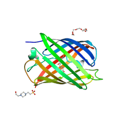
4EMQ
DownloadVisualize
BU of 4emq by Molmil
Crystal structure of a single mutant of Dronpa, the green-on-state PDM1-4
分子名称: 4-(2-HYDROXYETHYL)-1-PIPERAZINE ETHANESULFONIC ACID, DI(HYDROXYETHYL)ETHER, Fluorescent protein Dronpa, ...
著者Ngan, N.B, Van Hecke, K, Van Meervelt, L.
登録日2012-04-12
公開日2012-11-21
最終更新日2024-10-09
実験手法X-RAY DIFFRACTION (1.95 Å)
主引用文献Structural basis for the influence of a single mutation K145N on the oligomerization and photoswitching rate of Dronpa.
Acta Crystallogr.,Sect.D, 68, 2012

227111

件を2024-11-06に公開中

PDB statisticsPDBj update infoContact PDBjnumon