8XPC
 
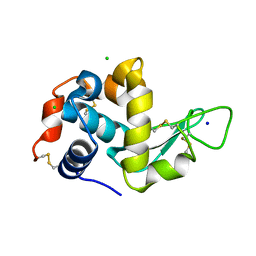
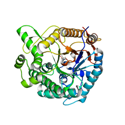
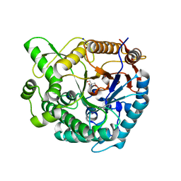 | | Crystal structure of Tris-bound TsaBgl (DATA I) | | 分子名称: | 2-AMINO-2-HYDROXYMETHYL-PROPANE-1,3-DIOL, SODIUM ION, beta-glucosidase | | 著者 | Nam, K.H. | | 登録日 | 2024-01-03 | | 公開日 | 2024-01-31 | | 最終更新日 | 2024-04-17 | | 実験手法 | X-RAY DIFFRACTION (1.55 Å) | | 主引用文献 | Structural analysis of Tris binding in beta-glucosidases.
Biochem.Biophys.Res.Commun., 700, 2024
|
|
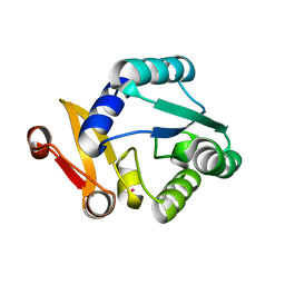
8ZM5
 | |
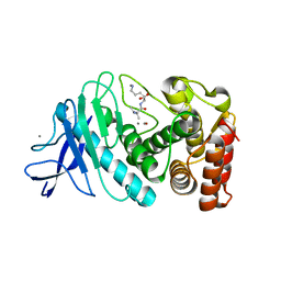
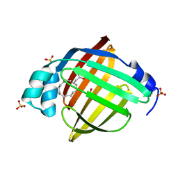
8XPD
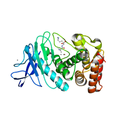
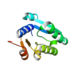
 | | Crystal structure of Tris-bound TsaBgl (DATA II) | | 分子名称: | 2-AMINO-2-HYDROXYMETHYL-PROPANE-1,3-DIOL, SODIUM ION, beta-glucosidase | | 著者 | Nam, K.H. | | 登録日 | 2024-01-03 | | 公開日 | 2024-01-31 | | 最終更新日 | 2024-04-17 | | 実験手法 | X-RAY DIFFRACTION (1.7 Å) | | 主引用文献 | Structural analysis of Tris binding in beta-glucosidases.
Biochem.Biophys.Res.Commun., 700, 2024
|
|
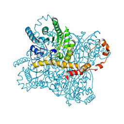
8YEA
 | |
8YBG
 | |
8YYO
 | |
8WGK
 | |
8WDG
 | |
8YPX
 | |
8WDI
 | |
8WFW
 | |
8WGL
 | |
8WFV
 | |
8ZM6
 | |
8WDH
 | |
8ZM4
 | |
6LL3
 | |
3L1J
 | |
3L1H
 | |
3L1I
 | |
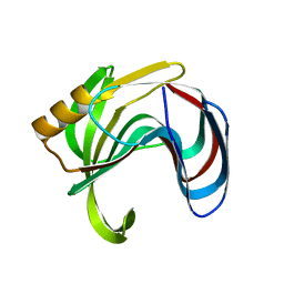
8GMW
 | | Crystal structure of lysozyme | | 分子名称: | CHLORIDE ION, Lysozyme C, SODIUM ION | | 著者 | Nam, K.H. | | 登録日 | 2022-08-22 | | 公開日 | 2022-09-21 | | 最終更新日 | 2023-11-29 | | 実験手法 | X-RAY DIFFRACTION (1.35 Å) | | 主引用文献 | Crystal structure of lysozyme
To Be Published
|
|
8GMV
 | | Crystal structure of lysozyme | | 分子名称: | CHLORIDE ION, Lysozyme C, SODIUM ION | | 著者 | Nam, K.H. | | 登録日 | 2022-08-22 | | 公開日 | 2022-09-21 | | 最終更新日 | 2023-11-29 | | 実験手法 | X-RAY DIFFRACTION (2.2 Å) | | 主引用文献 | Crystal structure of lysozyme
To Be Published
|
|
8HVF
 | |
8HVE
 | |
7E25
 | |


