Loading
PDBj
MenuPDBj@FacebookPDBj@TwitterPDBj@YouTubewwPDB FoundationwwPDB
RCSB PDBPDBeBMRBAdv. SearchSearch help
PDB: 93 results

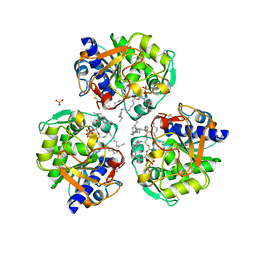
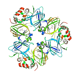
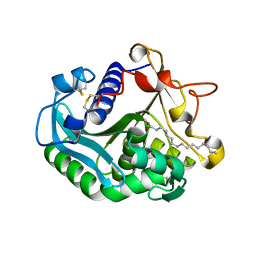
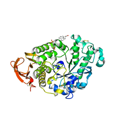
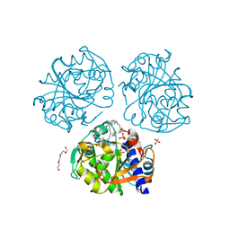
7R66
DownloadVisualize
BU of 7r66 by Molmil
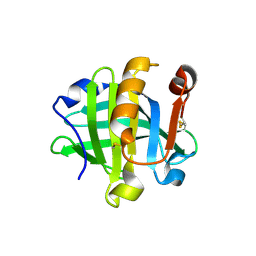
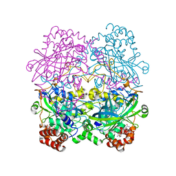
Structure of Pfp1 protease from Thermococcus thioreducens: large unit cell at 1.44 A resolution
Descriptor: (4S)-2-METHYL-2,4-PENTANEDIOL, PHOSPHATE ION, Peptidase
Authors:McPherson, A.
Deposit date:2021-06-22
Release date:2021-09-29
Last modified:2023-11-15
Method:X-RAY DIFFRACTION (1.44 Å)
Cite:Structure of the Pfp1 protease from a hypothermophilic archer, Thermococcus thioreducens, in two crystal forms
Acta Crystallogr., D73, 2017
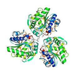
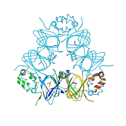
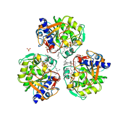
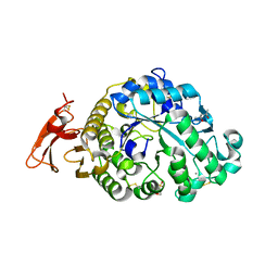
6O8V
DownloadVisualize
BU of 6o8v by Molmil
The structure of lipase from Thermomyces Lanuginosa in complex with 1,3 diacylglycerol: Rhombohedral crystal form
Descriptor: 2-acetamido-2-deoxy-beta-D-glucopyranose, 2-hydroxy-3-(octadecanoyloxy)propyl pentacosanoate, Lipase, ...
Authors:McPherson, A.
Deposit date:2019-03-12
Release date:2024-05-01
Last modified:2024-10-23
Method:X-RAY DIFFRACTION (1.3 Å)
Cite:The Crystal Structures of Thermomyces (Humicola) Lanuginosa Lipase in Complex with Enzymatic Reactants
Curr Enzym Inhib, 16, 2020
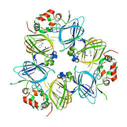
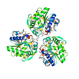
6OSZ
DownloadVisualize
BU of 6osz by Molmil
High Resolution Structure of the Monoclinic Form of Thermomyces Lanuginosa Lipase Complexed with Its Catalytic Products
Descriptor: 2-acetamido-2-deoxy-beta-D-glucopyranose, 2-hydroxy-3-(octadecanoyloxy)propyl pentacosanoate, CALCIUM ION, ...
Authors:McPherson, A.
Deposit date:2019-05-02
Release date:2024-05-01
Last modified:2024-10-23
Method:X-RAY DIFFRACTION (1.43 Å)
Cite:The Crystal Structures of Thermomyces (Humicola) Lanuginosa Lipase in Complex with Enzymatic Reactants
Curr Enzym Inhib, 16, 2020
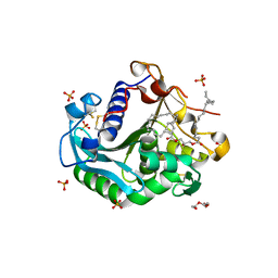
6O9F
DownloadVisualize
BU of 6o9f by Molmil
The structure of Thermomyces Lanuginosa lipase in complex with 1,3 diacylglycerol in a monoclinic crystal form
Descriptor: 2-acetamido-2-deoxy-beta-D-glucopyranose, 2-hydroxy-3-(octadecanoyloxy)propyl pentacosanoate, CALCIUM ION, ...
Authors:McPherson, A.
Deposit date:2019-03-13
Release date:2024-05-01
Method:X-RAY DIFFRACTION (2.48 Å)
Cite:The Crystal Structures of Thermomyces (Humicola) Lanuginosa Lipase in Complex with Enzymatic Reactants
Curr Enzym Inhib, 16, 2020
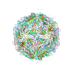
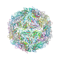
5BKQ
DownloadVisualize
BU of 5bkq by Molmil
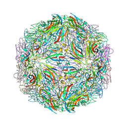
Crystallographic structure of a cubic form of STMV grown from nitrate
Descriptor: Coat protein, MAGNESIUM ION, NITRATE ION
Authors:McPherson, A.
Deposit date:2021-03-21
Release date:2021-12-15
Last modified:2023-09-27
Method:X-RAY DIFFRACTION (3.19 Å)
Cite:Structures of additional crystal forms of Satellite tobacco mosaic virus grown from a variety of salts.
Acta Crystallogr.,Sect.F, 77, 2021
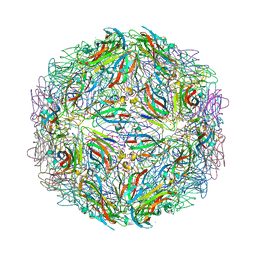
5BKN
DownloadVisualize
BU of 5bkn by Molmil
Crystallographic structure of a cubic crystal form of STMV (84.5 degree rotation) grown from chloride
Descriptor: CHLORIDE ION, Coat protein, MAGNESIUM ION, ...
Authors:McPherson, A.
Deposit date:2021-03-20
Release date:2021-12-15
Last modified:2023-09-27
Method:X-RAY DIFFRACTION (3 Å)
Cite:Structures of additional crystal forms of Satellite tobacco mosaic virus grown from a variety of salts.
Acta Crystallogr.,Sect.F, 77, 2021
5BKL
DownloadVisualize
BU of 5bkl by Molmil
Crystallographic structure of the cubic crystal form of STMV (77.9 degree rotation) grown from NaCl
Descriptor: CHLORIDE ION, Coat protein, MAGNESIUM ION, ...
Authors:McPherson, A.
Deposit date:2021-03-20
Release date:2021-12-15
Last modified:2023-09-27
Method:X-RAY DIFFRACTION (2.94 Å)
Cite:Structures of additional crystal forms of Satellite tobacco mosaic virus grown from a variety of salts.
Acta Crystallogr.,Sect.F, 77, 2021
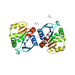
6NF3
DownloadVisualize
BU of 6nf3 by Molmil
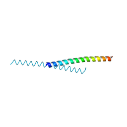
Structure of the Monoclinic-3 (Monocln-3) Crystal Form of Human Apolipoprotein C1
Descriptor: Apolipoprotein C-I
Authors:McPherson, A, Larson, S.B.
Deposit date:2018-12-18
Release date:2018-12-26
Last modified:2023-10-11
Method:X-RAY DIFFRACTION (2.33 Å)
Cite:The structure of human apolipoprotein C-1 in four different crystal forms.
J. Lipid Res., 60, 2019
6NKQ
DownloadVisualize
BU of 6nkq by Molmil
The structure of bovine beta-lactoglobulin in novel crystals grown at pH 3.8
Descriptor: Beta-lactoglobulin, CALCIUM ION
Authors:McPherson, A.
Deposit date:2019-01-07
Release date:2019-01-23
Last modified:2023-10-11
Method:X-RAY DIFFRACTION (2.3 Å)
Cite:The structure of bovine beta-lactoglobulin in crystals grown at pH 3.8 exhibiting novel threefold twinning.
Acta Crystallogr.,Sect.F, 75, 2019
6NTT
DownloadVisualize
BU of 6ntt by Molmil
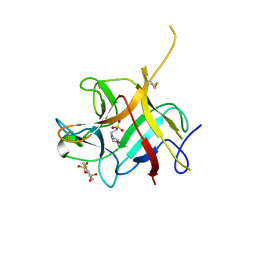
X-ray Crystal Structure of Soybean Trypsin Inhibitor (Kunitz) Complexed with 1,5-Disulfonyl Naphthalene
Descriptor: 2-(N-MORPHOLINO)-ETHANESULFONIC ACID, Trypsin inhibitor A, naphthalene-1,5-disulfonic acid
Authors:McPherson, A.
Deposit date:2019-01-30
Release date:2019-03-20
Last modified:2023-10-11
Method:X-RAY DIFFRACTION (2.4 Å)
Cite:Lattice Interactions in Crystals of Soybean Trypsin Inhibitor (Kunitz) Produced by Inclusion of 1,5-Disulfonylnaphthalene
Cryst.Growth Des., 19, 2019
6V7J
DownloadVisualize
BU of 6v7j by Molmil
The C2221 crystal form of canavalin at 173 K
Descriptor: BENZOIC ACID, CALCIUM ION, Canavalin, ...
Authors:McPherson, A.
Deposit date:2019-12-08
Release date:2020-02-19
Last modified:2023-11-15
Method:X-RAY DIFFRACTION (2 Å)
Cite:Binding of benzoic acid and anions within the cupin domains of the vicilin protein canavalin from jack bean (Canavalia ensiformis): Crystal structures.
Biochem.Biophys.Res.Commun., 524, 2020
6V7G
DownloadVisualize
BU of 6v7g by Molmil
Binding of Benzoic Acid and Anions Within the Cupin Domains of the Vicillin Protein Canavalin from Jack Bean (canavalia ensiformis): Crystal Structures
Descriptor: ACETATE ION, BENZOIC ACID, CITRIC ACID, ...
Authors:McPherson, A.
Deposit date:2019-12-08
Release date:2020-02-19
Last modified:2023-11-15
Method:X-RAY DIFFRACTION (1.4 Å)
Cite:Binding of benzoic acid and anions within the cupin domains of the vicilin protein canavalin from jack bean (Canavalia ensiformis): Crystal structures.
Biochem.Biophys.Res.Commun., 524, 2020
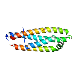
6V7M
DownloadVisualize
BU of 6v7m by Molmil
Crystal structure of a proteolytically cleaved, amino terminal domain of apolipoprotein E3
Descriptor: Apolipoprotein E, PHOSPHATE ION
Authors:McPherson, A.
Deposit date:2019-12-08
Release date:2020-03-11
Last modified:2023-10-11
Method:X-RAY DIFFRACTION (2 Å)
Cite:Crystal structure of a proteolytically cleaved, amino terminal domain of apolipoprotein E3.
Biochem.Biophys.Res.Commun., 2020
6V7L
DownloadVisualize
BU of 6v7l by Molmil
The structure of the P212121 crystal form of canavalin at 173 K
Descriptor: BENZOIC ACID, Canavalin
Authors:McPherson, A.
Deposit date:2019-12-08
Release date:2020-02-19
Last modified:2023-10-11
Method:X-RAY DIFFRACTION (2.8 Å)
Cite:Binding of benzoic acid and anions within the cupin domains of the vicilin protein canavalin from jack bean (Canavalia ensiformis): Crystal structures.
Biochem.Biophys.Res.Commun., 524, 2020
6OR3
DownloadVisualize
BU of 6or3 by Molmil
Structure of an Acyl Intermediate of Thermomyces Lanuginosa Lipase With Palmitic Acid in an Orthorhombic Crystal
Descriptor: 2-acetamido-2-deoxy-beta-D-glucopyranose, CALCIUM ION, Lipase, ...
Authors:McPherson, A.
Deposit date:2019-04-29
Release date:2020-05-20
Last modified:2024-10-23
Method:X-RAY DIFFRACTION (1.45 Å)
Cite:The crystal Structures of Thermomyces (Humicola) lanuginosa lipase in complex with enzymatic reactants
Current Enzyme Inhibition, 16, 2020
6XRV
DownloadVisualize
BU of 6xrv by Molmil
X-ray structure of the monoclinic crystal form at 1.43 A resolution of lipase from Thermomyces (Humicola) lanuginosa at 173 K
Descriptor: 2-acetamido-2-deoxy-beta-D-glucopyranose, 2-hydroxy-3-(octadecanoyloxy)propyl pentacosanoate, CALCIUM ION, ...
Authors:McPherson, A.
Deposit date:2020-07-13
Release date:2020-10-14
Last modified:2023-10-18
Method:X-RAY DIFFRACTION (1.43 Å)
Cite:The crystal Structures of Thermomyces (Humicola) lanuginosa lipase in complex with enzymatic reactants
Current Enzyme Inhibition, 16, 2020
6XS3
DownloadVisualize
BU of 6xs3 by Molmil
X-ray structure of the monoclinic crystal form at 2.48 A resolution of lipase from Thermomyces (Humicola) lanuginosa at 298 K
Descriptor: 2-acetamido-2-deoxy-beta-D-glucopyranose, 2-hydroxy-3-(octadecanoyloxy)propyl pentacosanoate, CALCIUM ION, ...
Authors:McPherson, A.
Deposit date:2020-07-14
Release date:2020-10-14
Last modified:2023-10-18
Method:X-RAY DIFFRACTION (2.48 Å)
Cite:The crystal Structures of Thermomyces (Humicola) lanuginosa lipase in complex with enzymatic reactants
Current Enzyme Inhibition, 16, 2020
6XOK
DownloadVisualize
BU of 6xok by Molmil
X-ray structure of the rhombohedral form of the lipase from Thermomyces lanuginosa at 1.3 A resolution
Descriptor: 2-acetamido-2-deoxy-beta-D-glucopyranose, 2-hydroxy-3-(octadecanoyloxy)propyl pentacosanoate, Lipase, ...
Authors:McPherson, A.
Deposit date:2020-07-07
Release date:2020-10-14
Last modified:2024-10-16
Method:X-RAY DIFFRACTION (1.3 Å)
Cite:The crystal Structures of Thermomyces (Humicola) lanuginosa lipase in complex with enzymatic reactants
Current Enzyme Inhibition, 16, 2020
6P57
DownloadVisualize
BU of 6p57 by Molmil
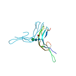
Crystal Structure of the Beta Subunit of Luteinizing Hormone
Descriptor: 2-acetamido-2-deoxy-beta-D-glucopyranose-(1-2)-alpha-D-mannopyranose-(1-6)-beta-D-mannopyranose-(1-4)-2-acetamido-2-deoxy-beta-D-glucopyranose-(1-4)-[beta-L-fucopyranose-(1-6)]2-acetamido-2-deoxy-beta-D-glucopyranose, Lutropin subunit beta, alpha-D-mannopyranose, ...
Authors:McPherson, A.
Deposit date:2019-05-29
Release date:2019-12-11
Last modified:2024-10-23
Method:X-RAY DIFFRACTION (3.16 Å)
Cite:The Crystal Structure of the Beta Subunit of Luteinizing Hormone and a Model for the Intact Hormone
Curr Res Struct Biol, 2019
6P6U
DownloadVisualize
BU of 6p6u by Molmil
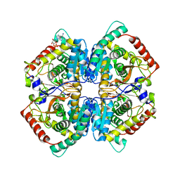
Crystal Structure of Monoclinic Rabbit Muscle Lactate Dehydrogenase with Four Tetramers as the Asymmetric Unit
Descriptor: ADENOSINE MONOPHOSPHATE, L-lactate dehydrogenase A chain
Authors:McPherson, A.
Deposit date:2019-06-04
Release date:2020-06-17
Last modified:2023-10-11
Method:X-RAY DIFFRACTION (2.42 Å)
Cite:Crystal Structure of Monoclinic Rabbit Muscle Lactate Dehydrogenase with Four Tetramers as the Asymmetric Unit
to be published
6PM7
DownloadVisualize
BU of 6pm7 by Molmil
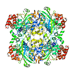
The structure of the triclinic crystal form of beef liver catalase at 1.85 A resolution
Descriptor: Catalase, NADPH DIHYDRO-NICOTINAMIDE-ADENINE-DINUCLEOTIDE PHOSPHATE, PROTOPORPHYRIN IX CONTAINING FE
Authors:McPherson, A.
Deposit date:2019-07-01
Release date:2020-05-20
Last modified:2024-03-13
Method:X-RAY DIFFRACTION (1.85 Å)
Cite:The structure of the triclinic crystal form of beef liver catalase at 1.85 A resolution
to be published
6PO0
DownloadVisualize
BU of 6po0 by Molmil
The structure of the orthorhombic (P212121) crystal form of beef liver catalase at 1.85 A resolution
Descriptor: Catalase, NADPH DIHYDRO-NICOTINAMIDE-ADENINE-DINUCLEOTIDE PHOSPHATE, PROTOPORPHYRIN IX CONTAINING FE
Authors:McPherson, A.
Deposit date:2019-07-03
Release date:2020-05-20
Last modified:2024-03-13
Method:X-RAY DIFFRACTION (1.75 Å)
Cite:The structure of the triclinic crystal form of beef liver catalase at 1.85 A resolution
To be published
6XSV
DownloadVisualize
BU of 6xsv by Molmil
X-ray structure of a tetragonal crystal form of alpha amylase from Aspergillus oryzae (Tala-Amylase) at 1.65 A resolution
Descriptor: 2-(N-MORPHOLINO)-ETHANESULFONIC ACID, 2-acetamido-2-deoxy-beta-D-glucopyranose, Alpha-amylase, ...
Authors:McPherson, A.
Deposit date:2020-07-16
Release date:2020-10-14
Last modified:2024-10-23
Method:X-RAY DIFFRACTION (1.65 Å)
Cite:Structures of two novel crystal forms of Aspergillus oryzae alpha amylase (taka-amylase).
J.Biosci.Bioeng., 131, 2021
6XSJ
DownloadVisualize
BU of 6xsj by Molmil
X-ray structure of a monoclinic form of alpha amylase from Aspergillus at 1.4 A resolution
Descriptor: (4S)-2-METHYL-2,4-PENTANEDIOL, 2-(N-MORPHOLINO)-ETHANESULFONIC ACID, 2-acetamido-2-deoxy-beta-D-glucopyranose, ...
Authors:McPherson, A.
Deposit date:2020-07-15
Release date:2020-10-14
Last modified:2024-10-30
Method:X-RAY DIFFRACTION (1.4 Å)
Cite:Structures of two novel crystal forms of Aspergillus oryzae alpha amylase (taka-amylase).
J.Biosci.Bioeng., 131, 2021
7M3R
DownloadVisualize
BU of 7m3r by Molmil
Crystallographic Structure of the Rhombohedral Crystal Form of STMV Grown from Bromide
Descriptor: BROMIDE ION, Coat protein, MAGNESIUM ION
Authors:McPherson, A.
Deposit date:2021-03-18
Release date:2021-12-15
Last modified:2023-10-18
Method:X-RAY DIFFRACTION (2.1 Å)
Cite:Structures of additional crystal forms of Satellite tobacco mosaic virus grown from a variety of salts.
Acta Crystallogr.,Sect.F, 77, 2021


1234>

226707

数据于2024-10-30公开中

PDB statisticsPDBj update infoContact PDBjnumon