1RA4
 
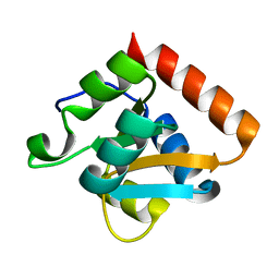 | |
1XBI
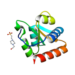 | | High resolution structure of Methanocaldococcus jannaschii L7AE | | 分子名称: | 4-(2-HYDROXYETHYL)-1-PIPERAZINE ETHANESULFONIC ACID, 50S ribosomal protein L7Ae | | 著者 | Brown II, B.A, Suryadi, J, Lieberman, D.V, Tran, E.J, Maxwell, E.S. | | 登録日 | 2004-08-30 | | 公開日 | 2005-08-09 | | 最終更新日 | 2023-08-23 | | 実験手法 | X-RAY DIFFRACTION (1.45 Å) | | 主引用文献 | The Crystal Structure of the Methanocaldococcus jannaschii Multifunctional L7Ae RNA-Binding Protein Reveals an Induced-Fit Interaction with the Box C/D RNAs.
Biochemistry, 44, 2005
|
|
3PAF
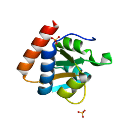 | | M. jannaschii L7Ae mutant | | 分子名称: | 50S ribosomal protein L7Ae, ACETATE ION, SULFATE ION | | 著者 | Biswas, S, Maxwell, E.S. | | 登録日 | 2010-10-19 | | 公開日 | 2011-11-02 | | 最終更新日 | 2023-09-06 | | 実験手法 | X-RAY DIFFRACTION (1.7 Å) | | 主引用文献 | Structure and stability of M.jannaschii L7Ae El9 KtoQ mutant
To be Published
|
|
3O85
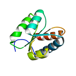 | | Giardia lamblia 15.5kD RNA binding protein | | 分子名称: | Ribosomal protein L7Ae | | 著者 | Biswas, S, Maxwell, E.S. | | 登録日 | 2010-08-02 | | 公開日 | 2011-03-16 | | 最終更新日 | 2023-09-06 | | 実験手法 | X-RAY DIFFRACTION (1.806 Å) | | 主引用文献 | Comparative analysis of the 15.5kD box C/D snoRNP core protein in the primitive eukaryote Giardia lamblia reveals unique structural and functional features.
Biochemistry, 50, 2011
|
|


