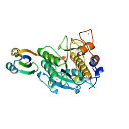
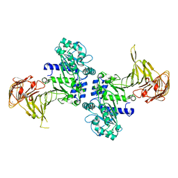
2Z30
 
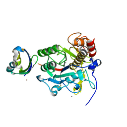 | | Crystal structure of complex form between mat-Tk-subtilisin and Tk-propeptide | | 分子名称: | CALCIUM ION, Tk-subtilisin, ZINC ION | | 著者 | Tanaka, S, Matsumura, H, Koga, Y, Takano, K, Kanaya, S. | | 登録日 | 2007-05-29 | | 公開日 | 2007-12-04 | | 最終更新日 | 2023-11-01 | | 実験手法 | X-RAY DIFFRACTION (1.65 Å) | | 主引用文献 | Four new crystal structures of Tk-subtilisin in unautoprocessed, autoprocessed and mature forms: insight into structural changes during maturation
J.Mol.Biol., 372, 2007
|
|
2Z56
 | | Crystal structure of G56S-propeptide:S324A-subtilisin complex | | 分子名称: | CALCIUM ION, Tk-subtilisin, ZINC ION | | 著者 | Pulido, M.A, Tanaka, S, Sringiew, C, You, D.J, Matsumura, H, Koga, Y, Takano, K, Kanaya, S. | | 登録日 | 2007-06-29 | | 公開日 | 2008-01-01 | | 最終更新日 | 2023-11-01 | | 実験手法 | X-RAY DIFFRACTION (1.9 Å) | | 主引用文献 | Requirement of left-handed glycine residue for high stability of the Tk-subtilisin propeptide as revealed by mutational and crystallographic analyses
J.Mol.Biol., 374, 2007
|
|
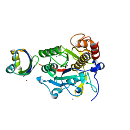
2Z2Z
 | | Crystal structure of unautoprocessed form of Tk-subtilisin soaked by 10mM CaCl2 | | 分子名称: | CALCIUM ION, Tk-subtilisin precursor | | 著者 | Tanaka, S, Matsumura, H, Koga, Y, Takano, K, Kanaya, S. | | 登録日 | 2007-05-29 | | 公開日 | 2007-12-04 | | 最終更新日 | 2023-11-01 | | 実験手法 | X-RAY DIFFRACTION (1.87 Å) | | 主引用文献 | Four new crystal structures of Tk-subtilisin in unautoprocessed, autoprocessed and mature forms: insight into structural changes during maturation
J.Mol.Biol., 372, 2007
|
|
2ZCT
 | | Oxidation of archaeal peroxiredoxin involves a hypervalent sulfur intermediate | | 分子名称: | Probable peroxiredoxin | | 著者 | Nakamura, T, Hagihara, Y, Abe, M, Inoue, T, Yamamoto, T, Matsumura, H. | | 登録日 | 2007-11-12 | | 公開日 | 2008-05-27 | | 最終更新日 | 2021-11-10 | | 実験手法 | X-RAY DIFFRACTION (1.7 Å) | | 主引用文献 | Oxidation of archaeal peroxiredoxin involves a hypervalent sulfur intermediate
Proc.Natl.Acad.Sci.Usa, 105, 2008
|
|
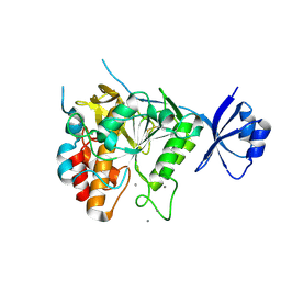
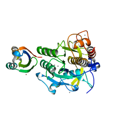
2Z2X
 | | Crystal structure of mature form of Tk-subtilisin | | 分子名称: | CALCIUM ION, Tk-subtilisin | | 著者 | Tanaka, S, Matsumura, H, Koga, Y, Takano, K, Kanaya, S. | | 登録日 | 2007-05-29 | | 公開日 | 2007-12-04 | | 最終更新日 | 2023-11-15 | | 実験手法 | X-RAY DIFFRACTION (1.7 Å) | | 主引用文献 | Four new crystal structures of Tk-subtilisin in unautoprocessed, autoprocessed and mature forms: insight into structural changes during maturation
J.Mol.Biol., 372, 2007
|
|
3A3P
 | | Crystal structure of complex between E201A/SA-subtilisin and Tk-propeptide | | 分子名称: | CALCIUM ION, Tk-subtilisin, ZINC ION | | 著者 | Tanaka, S, Matsumura, H, Koga, Y, Takano, K, Kanaya, S. | | 登録日 | 2009-06-15 | | 公開日 | 2009-08-18 | | 最終更新日 | 2023-11-01 | | 実験手法 | X-RAY DIFFRACTION (1.9 Å) | | 主引用文献 | Identification of the interactions critical for propeptide-catalyzed folding of Tk-subtilisin
J.Mol.Biol., 394, 2009
|
|
3A2X
 | | Peroxiredoxin (C50S) from Aeropyrum pernix K1 (acetate-bound form) | | 分子名称: | ACETATE ION, Probable peroxiredoxin | | 著者 | Nakamura, T, Kado, Y, Yamaguchi, F, Matsumura, H, Ishikawa, K, Inoue, T. | | 登録日 | 2009-06-04 | | 公開日 | 2009-10-27 | | 最終更新日 | 2024-05-29 | | 実験手法 | X-RAY DIFFRACTION (1.9 Å) | | 主引用文献 | Crystal structure of peroxiredoxin from Aeropyrum pernix K1 complexed with its substrate, hydrogen peroxide
J.Biochem., 147, 2010
|
|
3A3O
 | | Crystal structure of complex between SA-subtilisin and Tk-propeptide with deletion of the five C-terminal residues | | 分子名称: | CALCIUM ION, Tk-subtilisin, ZINC ION | | 著者 | Tanaka, S, Matsumura, H, Koga, Y, Takano, K, Kanaya, S. | | 登録日 | 2009-06-15 | | 公開日 | 2009-08-18 | | 最終更新日 | 2023-11-01 | | 実験手法 | X-RAY DIFFRACTION (1.9 Å) | | 主引用文献 | Identification of the interactions critical for propeptide-catalyzed folding of Tk-subtilisin
J.Mol.Biol., 394, 2009
|
|
3A2W
 | | Peroxiredoxin (C50S) from Aeropytum pernix K1 (peroxide-bound form) | | 分子名称: | GLYCEROL, PEROXIDE ION, Probable peroxiredoxin | | 著者 | Nakamura, T, Kado, Y, Yamaguchi, F, Matsumura, H, Ishikawa, K, Inoue, T. | | 登録日 | 2009-06-04 | | 公開日 | 2009-10-27 | | 最終更新日 | 2021-11-10 | | 実験手法 | X-RAY DIFFRACTION (2.3 Å) | | 主引用文献 | Crystal structure of peroxiredoxin from Aeropyrum pernix K1 complexed with its substrate, hydrogen peroxide
J.Biochem., 147, 2010
|
|
3A6Z
 | | Crystal structure of Pseudomonas sp. MIS38 lipase (PML) in the open conformation following dialysis against Ca-free buffer | | 分子名称: | CALCIUM ION, Lipase | | 著者 | Angkawidjaja, C, Matsumura, H, Koga, Y, Takano, K, Kanaya, S. | | 登録日 | 2009-09-10 | | 公開日 | 2010-05-26 | | 最終更新日 | 2023-11-01 | | 実験手法 | X-RAY DIFFRACTION (2.15 Å) | | 主引用文献 | X-ray Crystallographic and MD Simulation Studies on the Mechanism of Interfacial Activation of a Family I.3 Lipase with Two Lids
J.Mol.Biol., 2010
|
|
3WVM
 | | The 0.88 angstrom X-ray structure of the human heart fatty acid-binding protein complexed with stearic acid | | 分子名称: | Fatty acid-binding protein, heart, HEXAETHYLENE GLYCOL, ... | | 著者 | Sugiyama, S, Matsuoka, S, Mizohata, E, Matsuoka, D, Ishida, H, Hirose, M, Kakinouchi, K, Hara, T, Matsumura, H, Murakami, S, Inoue, T, Murata, M. | | 登録日 | 2014-05-25 | | 公開日 | 2015-01-28 | | 最終更新日 | 2024-05-29 | | 実験手法 | X-RAY DIFFRACTION (0.88 Å) | | 主引用文献 | Water-mediated recognition of simple alkyl chains by heart-type fatty-acid-binding protein.
Angew.Chem.Int.Ed.Engl., 54, 2015
|
|
1WSI
 | |
1WSJ
 | |
1WSH
 | |
2CWR
 | |
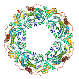
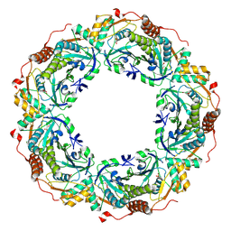
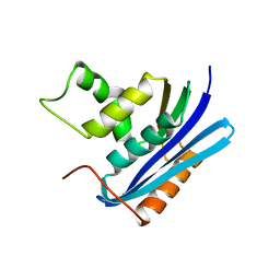
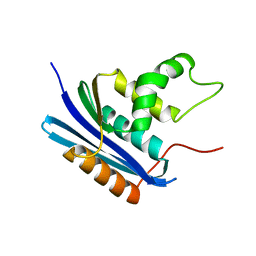
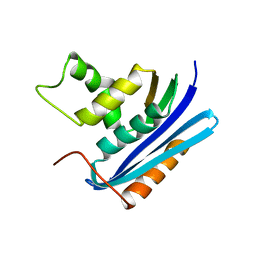
2CZN
 | | Solution structure of the chitin-binding domain of hyperthermophilic chitinase from pyrococcus furiosus | | 分子名称: | chitinase | | 著者 | Uegaki, T, Ikegami, T, Nakamura, T, Hagihara, Y, Mine, S, Inoue, T, Matsumura, H, Ataka, M, Ishikawa, K. | | 登録日 | 2005-07-13 | | 公開日 | 2006-07-18 | | 最終更新日 | 2024-05-29 | | 実験手法 | SOLUTION NMR | | 主引用文献 | Tertiary structure and carbohydrate recognition by the chitin-binding domain of a hyperthermophilic chitinase from Pyrococcus furiosus.
J.Mol.Biol., 381, 2008
|
|
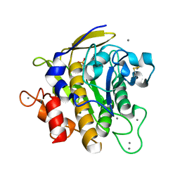
3VHQ
 | | Crystal structure of the Ca6 site mutant of Pro-SA-subtilisin | | 分子名称: | CALCIUM ION, Tk-subtilisin | | 著者 | Uehara, R, Takeuchi, Y, Tanaka, S, Matsumura, H, Koga, Y, Takano, K, Kanaya, S. | | 登録日 | 2011-09-01 | | 公開日 | 2012-07-11 | | 実験手法 | X-RAY DIFFRACTION (2.15 Å) | | 主引用文献 | Requirement of Ca(2+) Ions for the Hyperthermostability of Tk-Subtilisin from Thermococcus kodakarensis
Biochemistry, 51, 2012
|
|


