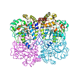
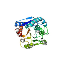
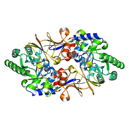
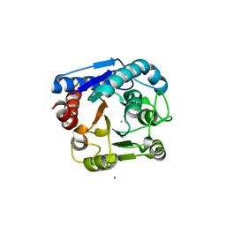
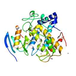
1EWJ
 
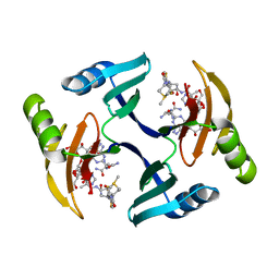 | | CRYSTAL STRUCTURE OF BLEOMYCIN-BINDING PROTEIN COMPLEXED WITH BLEOMYCIN | | 分子名称: | BLEOMYCIN A2, BLEOMYCIN RESISTANCE DETERMINANT | | 著者 | Maruyama, M, Kumagai, T, Matoba, Y, Hata, Y, Sugiyama, M. | | 登録日 | 2000-04-26 | | 公開日 | 2001-04-26 | | 最終更新日 | 2024-02-07 | | 実験手法 | X-RAY DIFFRACTION (2.5 Å) | | 主引用文献 | Crystal structures of the transposon Tn5-carried bleomycin resistance determinant uncomplexed and complexed with bleomycin.
J.Biol.Chem., 276, 2001
|
|
6LDO
 | |
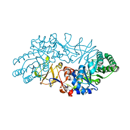
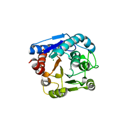
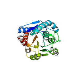
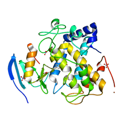
1QTO
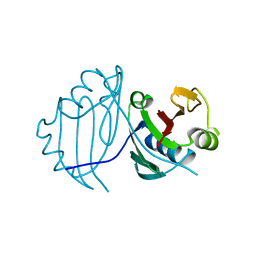 | | 1.5 A CRYSTAL STRUCTURE OF A BLEOMYCIN RESISTANCE DETERMINANT FROM BLEOMYCIN-PRODUCING STREPTOMYCES VERTICILLUS | | 分子名称: | BLEOMYCIN-BINDING PROTEIN | | 著者 | Kawano, Y, Kumagai, T, Muta, K, Matoba, Y, Davies, J, Sugiyama, M. | | 登録日 | 1999-06-28 | | 公開日 | 2000-06-28 | | 最終更新日 | 2024-02-14 | | 実験手法 | X-RAY DIFFRACTION (1.5 Å) | | 主引用文献 | The 1.5 A crystal structure of a bleomycin resistance determinant from bleomycin-producing Streptomyces verticillus.
J.Mol.Biol., 295, 2000
|
|
6LE4
 | |
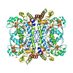
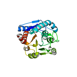
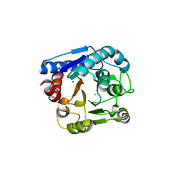
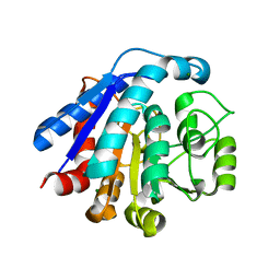
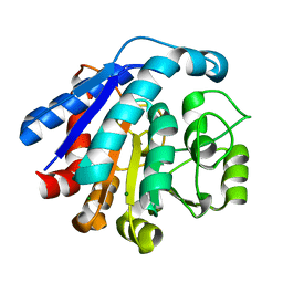
1ECS
 | | THE 1.7 A CRYSTAL STRUCTURE OF A BLEOMYCIN RESISTANCE DETERMINANT ENCODED ON THE TRANSPOSON TN5 | | 分子名称: | BLEOMYCIN RESISTANCE PROTEIN, CALCIUM ION, TETRAETHYLENE GLYCOL | | 著者 | Maruyama, M, Matoba, Y, Kumagai, T, Sugiyama, M. | | 登録日 | 2000-01-25 | | 公開日 | 2001-05-02 | | 最終更新日 | 2023-08-09 | | 実験手法 | X-RAY DIFFRACTION (1.7 Å) | | 主引用文献 | Crystal structures of the transposon Tn5-carried bleomycin resistance determinant uncomplexed and complexed with bleomycin.
J.Biol.Chem., 276, 2001
|
|
8WKO
 | |
8WKR
 | |
6LUG
 | | Crystal structure of N(omega)-hydroxy-L-arginine hydrolase | | 分子名称: | MANGANESE (II) ION, N(omega)-hydroxy-L-arginine amidinohydrolase | | 著者 | Oda, K, Matoba, Y. | | 登録日 | 2020-01-28 | | 公開日 | 2020-06-17 | | 最終更新日 | 2024-03-27 | | 実験手法 | X-RAY DIFFRACTION (1.9 Å) | | 主引用文献 | Crystal structure of an Nomega-hydroxy-L-arginine hydrolase found in the D-cycloserine biosynthetic pathway.
Acta Crystallogr D Struct Biol, 76, 2020
|
|
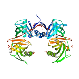
3VVL
 | | Crystal structure of L-serine-O-acetyltransferase found in D-cycloserine biosynthetic pathway | | 分子名称: | Homoserine O-acetyltransferase | | 著者 | Oda, K, Matoba, Y, Kumagai, T, Noda, M, Sugiyama, M. | | 登録日 | 2012-07-26 | | 公開日 | 2013-03-20 | | 最終更新日 | 2023-11-08 | | 実験手法 | X-RAY DIFFRACTION (1.81 Å) | | 主引用文献 | Crystallographic study to determine the substrate specificity of an L-serine-acetylating enzyme found in the D-cycloserine biosynthetic pathway
J.Bacteriol., 195, 2013
|
|
6LUH
 | |
1VFH
 | | Crystal structure of alanine racemase from D-cycloserine producing Streptomyces lavendulae | | 分子名称: | PYRIDOXAL-5'-PHOSPHATE, alanine racemase | | 著者 | Noda, M, Matoba, Y, Kumagai, T, Sugiyama, M. | | 登録日 | 2004-04-13 | | 公開日 | 2004-09-14 | | 最終更新日 | 2023-11-15 | | 実験手法 | X-RAY DIFFRACTION (2 Å) | | 主引用文献 | Structural evidence that alanine racemase from a D-cycloserine-producing microorganism exhibits resistance to its own product.
J.Biol.Chem., 279, 2004
|
|
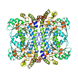
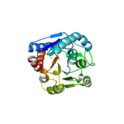
1JIE
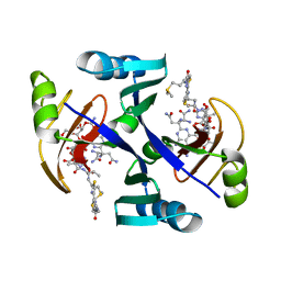 | | Crystal structure of bleomycin-binding protein from bleomycin-producing Streptomyces verticillus complexed with metal-free bleomycin | | 分子名称: | BLEOMYCIN A2, bleomycin-binding protein | | 著者 | Sugiyama, M, Kumagai, T, Hayashida, M, Maruyama, M, Matoba, Y. | | 登録日 | 2001-07-02 | | 公開日 | 2002-02-06 | | 最終更新日 | 2023-10-25 | | 実験手法 | X-RAY DIFFRACTION (1.8 Å) | | 主引用文献 | The 1.6-A crystal structure of the copper(II)-bound bleomycin complexed with the bleomycin-binding protein from bleomycin-producing Streptomyces verticillus.
J.Biol.Chem., 277, 2002
|
|
1JIF
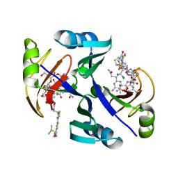 | | Crystal structure of bleomycin-binding protein from bleomycin-producing Streptomyces verticillus complexed with copper(II)-bleomycin | | 分子名称: | BLEOMYCIN A2, CHLORIDE ION, COPPER (II) ION, ... | | 著者 | Sugiyama, M, Kumagai, T, Hayashida, M, Maruyama, M, Matoba, Y. | | 登録日 | 2001-07-02 | | 公開日 | 2002-02-06 | | 最終更新日 | 2024-03-13 | | 実験手法 | X-RAY DIFFRACTION (1.6 Å) | | 主引用文献 | The 1.6-A crystal structure of the copper(II)-bound bleomycin complexed with the bleomycin-binding protein from bleomycin-producing Streptomyces verticillus.
J.Biol.Chem., 277, 2002
|
|
1VFS
 | | Crystal structure of D-cycloserine-bound form of alanine racemase from D-cycloserine-producing Streptomyces lavendulae | | 分子名称: | CHLORIDE ION, D-[3-HYDROXY-2-METHYL-5-PHOSPHONOOXYMETHYL-PYRIDIN-4-YLMETHYL]-N,O-CYCLOSERYLAMIDE, alanine racemase | | 著者 | Noda, M, Matoba, Y, Kumagai, T, Sugiyama, M. | | 登録日 | 2004-04-19 | | 公開日 | 2004-09-14 | | 最終更新日 | 2023-11-15 | | 実験手法 | X-RAY DIFFRACTION (1.9 Å) | | 主引用文献 | Structural evidence that alanine racemase from a D-cycloserine-producing microorganism exhibits resistance to its own product.
J.Biol.Chem., 279, 2004
|
|
7EUN
 | |
7VDY
 | |
8IUS
 | |
8IUU
 | |
8IUV
 | |
8IUT
 | |
8IUW
 | |
7CIT
 | |
7CIY
 | |
7EUQ
 | |
7EUL
 | |


