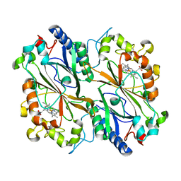
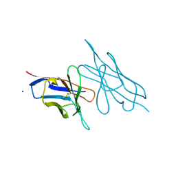
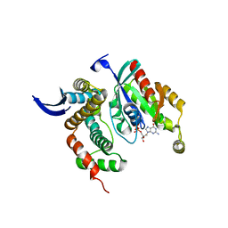
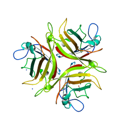
3LJC
 
 | | Crystal structure of Lon N-terminal domain. | | 分子名称: | ATP-dependent protease La | | 著者 | Li, M, Gustchina, A, Dauter, Z, Wlodawer, A. | | 登録日 | 2010-01-26 | | 公開日 | 2010-07-21 | | 最終更新日 | 2017-11-01 | | 実験手法 | X-RAY DIFFRACTION (2.6 Å) | | 主引用文献 | Structure of the N-terminal fragment of Escherichia coli Lon protease
Acta Crystallogr.,Sect.D, 66, 2010
|
|
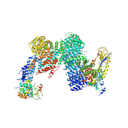
8OLH
 | |
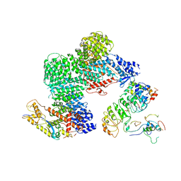
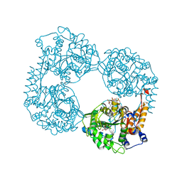
8OR3
 | | CAND1-CUL1-RBX1-SKP1-SKP2-DCNL1 | | 分子名称: | Cullin-1, Cullin-associated NEDD8-dissociated protein 1, DCN1-like protein 1, ... | | 著者 | Shaaban, M, Clapperton, J.A, Ding, S, Maeots, M.E, Enchev, R.I. | | 登録日 | 2023-04-13 | | 公開日 | 2023-06-28 | | 最終更新日 | 2024-07-24 | | 実験手法 | ELECTRON MICROSCOPY (2.9 Å) | | 主引用文献 | Structural and mechanistic insights into the CAND1-mediated SCF substrate receptor exchange.
Mol.Cell, 83, 2023
|
|
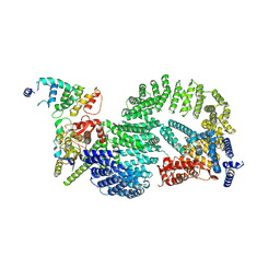
8OR0
 | | CAND1-CUL1-RBX1-SKP1-SKP2-CKS1-CDK2 | | 分子名称: | Cullin-1, Cullin-associated NEDD8-dissociated protein 1, Cyclin-dependent kinase 2, ... | | 著者 | Shaaban, M, Clapperton, J.A, Ding, S, Maeots, M.E, Enchev, R.I. | | 登録日 | 2023-04-12 | | 公開日 | 2023-06-28 | | 最終更新日 | 2024-07-24 | | 実験手法 | ELECTRON MICROSCOPY (3.1 Å) | | 主引用文献 | Structural and mechanistic insights into the CAND1-mediated SCF substrate receptor exchange.
Mol.Cell, 83, 2023
|
|
8OR2
 | | CAND1-CUL1-RBX1-DCNL1 | | 分子名称: | Cullin-1, Cullin-associated NEDD8-dissociated protein 1, DCN1-like protein 1, ... | | 著者 | Shaaban, M, Clapperton, J.A, Ding, S, Maeots, M.E, Enchev, R.I. | | 登録日 | 2023-04-12 | | 公開日 | 2023-06-28 | | 最終更新日 | 2024-07-24 | | 実験手法 | ELECTRON MICROSCOPY (3.2 Å) | | 主引用文献 | Structural and mechanistic insights into the CAND1-mediated SCF substrate receptor exchange.
Mol.Cell, 83, 2023
|
|
3LVE
 | |
8PCH
 | | CRYSTAL STRUCTURE OF PORCINE CATHEPSIN H DETERMINED AT 2.1 ANGSTROM RESOLUTION: LOCATION OF THE MINI-CHAIN C-TERMINAL CARBOXYL GROUP DEFINES CATHEPSIN H AMINOPEPTIDASE FUNCTION | | 分子名称: | CATHEPSIN H, beta-D-mannopyranose-(1-4)-2-acetamido-2-deoxy-beta-D-glucopyranose-(1-4)-2-acetamido-2-deoxy-beta-D-glucopyranose | | 著者 | Guncar, G, Podobnik, M, Pungercar, J, Strukelj, B, Turk, V, Turk, D. | | 登録日 | 1997-11-07 | | 公開日 | 1998-12-09 | | 最終更新日 | 2023-08-09 | | 実験手法 | X-RAY DIFFRACTION (2.1 Å) | | 主引用文献 | Crystal structure of porcine cathepsin H determined at 2.1 A resolution: location of the mini-chain C-terminal carboxyl group defines cathepsin H aminopeptidase function.
Structure, 6, 1998
|
|
6DBJ
 | | Cryo-EM structure of RAG in complex with 12-RSS and 23-RSS nicked DNA intermediates | | 分子名称: | CALCIUM ION, Forward stand of RSS signal end, Forward strand of coding flank, ... | | 著者 | Wu, H, Liao, M, Ru, H, Mi, W. | | 登録日 | 2018-05-03 | | 公開日 | 2018-08-01 | | 最終更新日 | 2024-03-13 | | 実験手法 | ELECTRON MICROSCOPY (3 Å) | | 主引用文献 | DNA melting initiates the RAG catalytic pathway.
Nat. Struct. Mol. Biol., 25, 2018
|
|
6D9K
 | | Ternary RsAgo Complex with Guide RNA and Target DNA Containing A-G Non-canonical Pair | | 分子名称: | (4S)-2-METHYL-2,4-PENTANEDIOL, ACETATE ION, DNA (5'-D(P*TP*CP*GP*TP*CP*AP*CP*CP*TP*GP*GP*GP*CP*AP*GP*TP*AP*AP*C)-3'), ... | | 著者 | Liu, Y, Esyunina, D, Olovnikov, I, Teplova, M, Patel, D.J. | | 登録日 | 2018-04-30 | | 公開日 | 2018-07-25 | | 最終更新日 | 2024-03-13 | | 実験手法 | X-RAY DIFFRACTION (2 Å) | | 主引用文献 | Accommodation of Helical Imperfections in Rhodobacter sphaeroides Argonaute Ternary Complexes with Guide RNA and Target DNA.
Cell Rep, 24, 2018
|
|
3LW8
 | | Shigella IpgB2 in complex with human RhoA, GDP and Mg2+ (complex A) | | 分子名称: | GUANOSINE-5'-DIPHOSPHATE, IpgB2, MAGNESIUM ION, ... | | 著者 | Klink, B.U, Barden, S, Heidler, T.V, Borchers, C, Ladwein, M, Stradal, T.E.B, Rottner, K, Heinz, D.W. | | 登録日 | 2010-02-23 | | 公開日 | 2010-03-31 | | 最終更新日 | 2023-11-01 | | 実験手法 | X-RAY DIFFRACTION (1.85 Å) | | 主引用文献 | Structure of Shigella IPGB2 in complex with human RhoA: Implications for the mechanism of bacterial GEF-mimicry
J.Biol.Chem., 285, 2010
|
|
3LXI
 | | Crystal Structure of Camphor-Bound CYP101D1 | | 分子名称: | CAMPHOR, Cytochrome P450, PHOSPHATE ION, ... | | 著者 | Yang, W, Bell, S.G, Wang, H, Bartlam, M, Wong, L.L, Rao, Z. | | 登録日 | 2010-02-25 | | 公開日 | 2010-06-23 | | 最終更新日 | 2023-11-01 | | 実験手法 | X-RAY DIFFRACTION (2.2 Å) | | 主引用文献 | Molecular characterization of a class I P450 electron transfer system from Novosphingobium aromaticivorans DSM12444
J.Biol.Chem., 285, 2010
|
|
6DDZ
 | | Crystal structure of the double mutant (D52N/R238W) of NT5C2-537X in the active state, Northeast Structural Genomics Target | | 分子名称: | ADENOSINE-5'-TRIPHOSPHATE, Cytosolic purine 5'-nucleotidase, GLYCEROL, ... | | 著者 | Forouhar, F, Dieck, C.L, Tzoneva, G, Carpenter, Z, Ambesi-Impiombato, A, Sanchez-Martin, M, Kirschner-Schwabe, R, Lew, S, Seetharaman, J, Ferrando, A.A, Tong, L, Northeast Structural Genomics Consortium (NESG) | | 登録日 | 2018-05-10 | | 公開日 | 2018-07-04 | | 最終更新日 | 2023-10-11 | | 実験手法 | X-RAY DIFFRACTION (1.97 Å) | | 主引用文献 | Structure and Mechanisms of NT5C2 Mutations Driving Thiopurine Resistance in Relapsed Lymphoblastic Leukemia.
Cancer Cell, 34, 2018
|
|
8OVN
 | | X-ray structure of the SF-iGluSnFR-S72A | | 分子名称: | CITRIC ACID, Putative periplasmic binding transport protein,Green fluorescent protein | | 著者 | Tarnawski, M, Hellweg, L, Bergner, A, Hiblot, J, Leippe, P, Johnsson, K. | | 登録日 | 2023-04-26 | | 公開日 | 2023-05-17 | | 実験手法 | X-RAY DIFFRACTION (2.6 Å) | | 主引用文献 | X-ray structure of the SF-iGluSnFR-S72A
To Be Published
|
|
6DE8
 | | Crystal Structure of Bifunctional Enzyme FolD-Methylenetetrahydrofolate Dehydrogenase/Cyclohydrolase from Campylobacter jejuni | | 分子名称: | Bifunctional protein FolD, CHLORIDE ION, GLYCEROL, ... | | 著者 | Kim, Y, Makowska-Grzyska, M, Zhang, R, Peterson, S.N, Joachimiak, A, Center for Structural Genomics of Infectious Diseases (CSGID) | | 登録日 | 2018-05-11 | | 公開日 | 2018-05-30 | | 最終更新日 | 2019-12-18 | | 実験手法 | X-RAY DIFFRACTION (2.104 Å) | | 主引用文献 | Crystal Structure of Bifunctional Enzyme FolD-Methylenetetrahydrofolate Dehydrogenase/Cyclohydrolase from Campylobacter jejuni
To Be Published
|
|
8OVO
 | | X-ray structure of the SF-iGluSnFR-S72A in complex with L-aspartate | | 分子名称: | ASPARTIC ACID, Putative periplasmic binding transport protein,Green fluorescent protein | | 著者 | Tarnawski, M, Hellweg, L, Bergner, A, Hiblot, J, Leippe, P, Johnsson, K. | | 登録日 | 2023-04-26 | | 公開日 | 2023-05-17 | | 実験手法 | X-RAY DIFFRACTION (1.7 Å) | | 主引用文献 | X-ray structure of the SF-iGluSnFR-S72A in complex with L-aspartate
To Be Published
|
|
8OVP
 | | X-ray structure of the iAspSnFR in complex with L-aspartate | | 分子名称: | ACETATE ION, ASPARTIC ACID, MAGNESIUM ION, ... | | 著者 | Tarnawski, M, Hellweg, L, Bergner, A, Hiblot, J, Leippe, P, Johnsson, K. | | 登録日 | 2023-04-26 | | 公開日 | 2023-05-17 | | 実験手法 | X-RAY DIFFRACTION (1.7 Å) | | 主引用文献 | X-ray structure of the SF-iAspSnFR in complex with L-aspartate
To Be Published
|
|
8OPQ
 | | Structure of Human Solute Carrier 26 family member A6 (SLC26A6) anion transporter in an inward-facing state | | 分子名称: | CHLORIDE ION, Solute carrier family 26 member 6 | | 著者 | Tippett, D.N, Breen, C, Butler, S.J, Sawicka, M, Dutzler, R. | | 登録日 | 2023-04-07 | | 公開日 | 2023-05-17 | | 最終更新日 | 2024-07-24 | | 実験手法 | ELECTRON MICROSCOPY (3.28 Å) | | 主引用文献 | Structural and functional properties of the transporter SLC26A6 reveal mechanism of coupled anion exchange.
Elife, 12, 2023
|
|
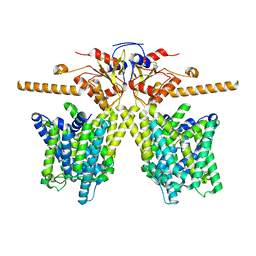
3L8Z
 | | H-Ras wildtype new crystal form | | 分子名称: | CALCIUM ION, GTPase HRas, MAGNESIUM ION, ... | | 著者 | Rosnizeck, I.C, Graf, T, Spoerner, M, Traenkle, J, Filchtinski, D, Herrmann, C, Gremer, L, Vetter, I.R, Wittinghofer, A, Koenig, B, Kalbitzer, H.R. | | 登録日 | 2010-01-04 | | 公開日 | 2011-01-05 | | 最終更新日 | 2023-11-01 | | 実験手法 | X-RAY DIFFRACTION (1.44 Å) | | 主引用文献 | Stabilizing a weak binding state for effectors in the human ras protein by cyclen complexes
Angew.Chem.Int.Ed.Engl., 49, 2010
|
|
6DDO
 | | Crystal structure of the single mutant (D52N) of the full-length NT5C2 in the basal state | | 分子名称: | Cytosolic purine 5'-nucleotidase, PHOSPHATE ION | | 著者 | Forouhar, F, Dieck, C.L, Tzoneva, G, Carpenter, Z, Ambesi-Impiombato, A, Sanchez-Martin, M, Kirschner-Schwabe, R, Lew, S, Seetharaman, J, Ferrando, A.A, Tong, L. | | 登録日 | 2018-05-10 | | 公開日 | 2018-07-04 | | 最終更新日 | 2023-10-11 | | 実験手法 | X-RAY DIFFRACTION (2.48 Å) | | 主引用文献 | Structure and Mechanisms of NT5C2 Mutations Driving Thiopurine Resistance in Relapsed Lymphoblastic Leukemia.
Cancer Cell, 34, 2018
|
|
3L9Z
 | | Crystal Structure of UreE from Helicobacter pylori (apo form) | | 分子名称: | Urease accessory protein ureE | | 著者 | Shi, R, Munger, C, Assinas, A, Matte, A, Cygler, M, Montreal-Kingston Bacterial Structural Genomics Initiative (BSGI) | | 登録日 | 2010-01-06 | | 公開日 | 2010-08-25 | | 最終更新日 | 2024-02-21 | | 実験手法 | X-RAY DIFFRACTION (2.08 Å) | | 主引用文献 | Crystal Structures of Apo and Metal-Bound Forms of the UreE Protein from Helicobacter pylori: Role of Multiple Metal Binding Sites
Biochemistry, 49, 2010
|
|
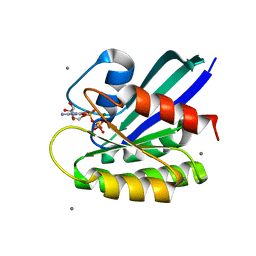
3LQ7
 | | Crystal structure of glutathione s-transferase from agrobacterium tumefaciens str. c58 | | 分子名称: | Glutathione S-transferase | | 著者 | Patskovsky, Y, Toro, R, Gilmore, M, Chang, S, Sauder, J.M, Burley, S.K, Almo, S.C, New York SGX Research Center for Structural Genomics (NYSGXRC) | | 登録日 | 2010-02-08 | | 公開日 | 2010-02-23 | | 最終更新日 | 2024-02-21 | | 実験手法 | X-RAY DIFFRACTION (2.3 Å) | | 主引用文献 | Crystal Structure of Glutathione S-Transferase from Agrobacterium Tumefaciens
To be Published
|
|
6DII
 | | Structure of Arabidopsis Fatty Acid Amide Hydrolase in Complex with methyl linolenyl fluorophosphonate | | 分子名称: | Fatty acid amide hydrolase, methyl-9Z,12Z,15Z-octadecatrienylphosphonofluoridate | | 著者 | Aziz, M, Wang, X, Tripathi, A, Bankaitis, V, Chapman, K.D. | | 登録日 | 2018-05-23 | | 公開日 | 2019-03-27 | | 最終更新日 | 2023-10-11 | | 実験手法 | X-RAY DIFFRACTION (3.2 Å) | | 主引用文献 | Structural analysis of a plant fatty acid amide hydrolase provides insights into the evolutionary diversity of bioactive acylethanolamides.
J.Biol.Chem., 294, 2019
|
|
3L88
 | | Crystal structure of the human Adenovirus type 21 fiber knob | | 分子名称: | CHLORIDE ION, Fiber protein, GLYCEROL, ... | | 著者 | Cupelli, K, Jost, M, Persson, B.D, Stehle, T. | | 登録日 | 2009-12-30 | | 公開日 | 2010-04-14 | | 最終更新日 | 2023-09-06 | | 実験手法 | X-RAY DIFFRACTION (2.5 Å) | | 主引用文献 | Structure of adenovirus type 21 knob in complex with CD46 reveals key differences in receptor contacts among species B adenoviruses.
J.Virol., 84, 2010
|
|
8OMT
 | | X-ray structure of lysozyme obtained upon reaction with [VIVO(empp)2] (Structure C) | | 分子名称: | 1-methyl-2-ethyl-3-hydroxy-4(1H)-pyridinone)V(IV)O4, 4-(2-HYDROXYETHYL)-1-PIPERAZINE ETHANESULFONIC ACID, Lysozyme C, ... | | 著者 | Paolillo, M, Merlino, A, Ferraro, G. | | 登録日 | 2023-03-31 | | 公開日 | 2023-06-07 | | 実験手法 | X-RAY DIFFRACTION (1.097 Å) | | 主引用文献 | Implications of Protein Interaction in the Speciation of Potential V IV O-Pyridinone Drugs.
Inorg.Chem., 62, 2023
|
|
8OM8
 | | X-ray structure of lysozyme obtained upon reaction with [VIVO(empp)2] (Structure A) | | 分子名称: | 1-methyl-2-ethyl-3-hydroxy-4(1H)-pyridinone)V(IV)O4, ACETATE ION, CHLORIDE ION, ... | | 著者 | Paolillo, M, Ferraro, G, Merlino, A. | | 登録日 | 2023-03-31 | | 公開日 | 2023-06-07 | | 実験手法 | X-RAY DIFFRACTION (1.08 Å) | | 主引用文献 | Implications of Protein Interaction in the Speciation of Potential V IV O-Pyridinone Drugs.
Inorg.Chem., 62, 2023
|
|


