5YO4
 
 | |
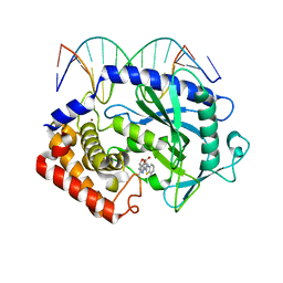
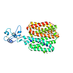
5YKX
 | | The crystal structure of Macrobrachium rosenbergii nodavirus P-domain with Cd ion | | 分子名称: | 4-(2-HYDROXYETHYL)-1-PIPERAZINE ETHANESULFONIC ACID, CADMIUM ION, Capsid protein, ... | | 著者 | Chen, N.C, Yoshimura, M, Lin, C.C, Guan, H.H, Chuankhayan, P, Chen, C.J. | | 登録日 | 2017-10-16 | | 公開日 | 2018-10-24 | | 最終更新日 | 2019-03-13 | | 実験手法 | X-RAY DIFFRACTION (2 Å) | | 主引用文献 | The atomic structures of shrimp nodaviruses reveal new dimeric spike structures and particle polymorphism.
Commun Biol, 2, 2019
|
|
5YRB
 | | Crystal Structure of Oxidized Cypovirus Polyhedra R13A/E73C/Y83C/S193C/A194C Mutant | | 分子名称: | 1,2-ETHANEDIOL, Polyhedrin | | 著者 | Negishi, H, Abe, S, Yamashita, K, Hirata, K, Niwase, K, Boudes, M, Coulibaly, F, Mori, H, Ueno, T. | | 登録日 | 2017-11-09 | | 公開日 | 2018-02-21 | | 最終更新日 | 2018-03-07 | | 実験手法 | X-RAY DIFFRACTION (1.65 Å) | | 主引用文献 | Supramolecular protein cages constructed from a crystalline protein matrix
Chem. Commun. (Camb.), 54, 2018
|
|
6QA3
 | | ERK2 mini-fragment binding | | 分子名称: | Mitogen-activated protein kinase 1, PYRAZOLE, SULFATE ION | | 著者 | O'Reilly, M, Cleasby, A, Davies, T.G, Hall, R, Ludlow, F, Murray, C.W, Tisi, D, Jhoti, H. | | 登録日 | 2018-12-18 | | 公開日 | 2019-06-26 | | 実験手法 | X-RAY DIFFRACTION (1.57 Å) | | 主引用文献 | Crystallographic screening using ultra-low-molecular-weight ligands to guide drug design.
Drug Discov Today, 24, 2019
|
|
5XXM
 | | Crystal structure of GH3 beta-glucosidase from Bacteroides thetaiotaomicron in complex with gluconolactone | | 分子名称: | D-glucono-1,5-lactone, MAGNESIUM ION, Periplasmic beta-glucosidase, ... | | 著者 | Nakajima, M, Ishiguro, R, Tanaka, N, Abe, K, Maeda, T, Miyanaga, A, Takahash, Y, Sugimoto, N, Nakai, H, Taguchi, H. | | 登録日 | 2017-07-04 | | 公開日 | 2017-12-13 | | 最終更新日 | 2024-03-27 | | 実験手法 | X-RAY DIFFRACTION (1.7 Å) | | 主引用文献 | Function and structure relationships of a beta-1,2-glucooligosaccharide-degrading beta-glucosidase.
FEBS Lett., 591, 2017
|
|
5XZE
 | | Mouse cGAS bound to the inhibitor RU332 | | 分子名称: | (3R)-3-[1-(3H-1lambda~4~,3-benzothiazol-2-yl)-5-hydroxy-3-methyl-1H-pyrazol-4-yl]-2-benzofuran-1(3H)-one, Cyclic GMP-AMP synthase, DNA (5'-D(*AP*AP*AP*TP*TP*GP*CP*CP*GP*AP*AP*GP*AP*CP*G)-3'), ... | | 著者 | Vincent, J, Adura, C, Gao, P, Luz, A, Lama, L, Asano, Y, Okamoto, R, Imaeda, T, Aida, J, Rothamel, K, Gogakos, T, Steinberg, J, Reasoner, S, Aso, K, Tuschl, T, Patel, D.J, Glickman, J.F, Ascano, M. | | 登録日 | 2017-07-12 | | 公開日 | 2017-10-11 | | 最終更新日 | 2024-03-13 | | 実験手法 | X-RAY DIFFRACTION (2.177 Å) | | 主引用文献 | Small molecule inhibition of cGAS reduces interferon expression in primary macrophages from autoimmune mice.
Nat Commun, 8, 2017
|
|
5YRA
 | | Crystal Structure of Cypovirus Polyhedra R13A/S193C/A194C Mutant | | 分子名称: | 1,2-ETHANEDIOL, Polyhedrin | | 著者 | Negishi, H, Abe, S, Yamashita, K, Hirata, K, Niwase, K, Boudes, M, Coulibaly, F, Mori, H, Ueno, T. | | 登録日 | 2017-11-09 | | 公開日 | 2018-02-21 | | 最終更新日 | 2024-03-27 | | 実験手法 | X-RAY DIFFRACTION (1.79 Å) | | 主引用文献 | Supramolecular protein cages constructed from a crystalline protein matrix
Chem. Commun. (Camb.), 54, 2018
|
|
5YRK
 | | Crystal structure of PPL3C | | 分子名称: | PPL3-b, SULFATE ION | | 著者 | Nakae, S, Shionyu, M, Ogawa, T, Shirai, T. | | 登録日 | 2017-11-09 | | 公開日 | 2018-08-29 | | 実験手法 | X-RAY DIFFRACTION (1.35 Å) | | 主引用文献 | Structures of jacalin-related lectin PPL3 regulating pearl shell biomineralization
Proteins, 86, 2018
|
|
8SFY
 | | Crystal structure of TuUGT202A2 (Tetur22g00270) in complex with UDP-glucose | | 分子名称: | UDP-glycosyltransferase 202A2, URIDINE-5'-DIPHOSPHATE-GLUCOSE | | 著者 | Arriaza, R.H, Daneshian, L, Dermauw, W, Wybouw, N, Van Leeuwen, T, Chruszcz, M. | | 登録日 | 2023-04-11 | | 公開日 | 2023-09-13 | | 実験手法 | X-RAY DIFFRACTION (2.35 Å) | | 主引用文献 | Crystal structure of TuUGT202A2 (Tetur22g00270) in complex with UDP-glucose
To Be Published
|
|
6PYT
 | | CryoEM Structure of Pyocin R2 - precontracted - trunk | | 分子名称: | Pyocin sheath PA0622, Pyocin tube PA0623 | | 著者 | Ge, P, Avaylon, J, Scholl, D, Shneider, M.M, Browning, C, Buth, S.A, Plattner, M, Ding, K, Leiman, P.G, Miller, J.F, Zhou, Z.H. | | 登録日 | 2019-07-30 | | 公開日 | 2020-04-15 | | 最終更新日 | 2024-03-20 | | 実験手法 | ELECTRON MICROSCOPY (2.9 Å) | | 主引用文献 | Action of a minimal contractile bactericidal nanomachine.
Nature, 580, 2020
|
|
6Q84
 | | Crystal structure of RanGTP-Pdr6-eIF5A export complex | | 分子名称: | Eukaryotic translation initiation factor 5A-1, GTP-binding nuclear protein Ran, GUANOSINE-5'-TRIPHOSPHATE, ... | | 著者 | Aksu, M, Trakhanov, S, Vera-Rodriguez, A, Gorlich, D. | | 登録日 | 2018-12-14 | | 公開日 | 2019-05-01 | | 最終更新日 | 2024-05-15 | | 実験手法 | X-RAY DIFFRACTION (3.7 Å) | | 主引用文献 | Structural basis for the nuclear import and export functions of the biportin Pdr6/Kap122.
J.Cell Biol., 218, 2019
|
|
8SFU
 | | Crystal structure of TuUGT202A2 (Tetur22g00270) in complex with naringin | | 分子名称: | (2S)-5-hydroxy-2-(4-hydroxyphenyl)-4-oxo-3,4-dihydro-2H-1-benzopyran-7-yl 2-O-(6-deoxy-alpha-L-mannopyranosyl)-beta-D-glucopyranoside, UDP-glycosyltransferase 202A2, URIDINE-5'-DIPHOSPHATE | | 著者 | Arriaza, R.H, Dermauw, W, Wybouw, N, Van Leeuwen, T, Chruszcz, M. | | 登録日 | 2023-04-11 | | 公開日 | 2023-09-13 | | 実験手法 | X-RAY DIFFRACTION (1.8 Å) | | 主引用文献 | Crystal structure of TuUGT202A2 (Tetur22g00270) in complex with naringin
To Be Published
|
|
8SSF
 | | Minimal protein-only/RNA-free Ribonuclease P from Hydrogenobacter thermophilus | | 分子名称: | RNA-free ribonuclease P, SULFATE ION | | 著者 | Mendoza, J, Mallik, L, Wilhelm, C.A, Koutmos, M. | | 登録日 | 2023-05-08 | | 公開日 | 2023-10-18 | | 最終更新日 | 2023-11-22 | | 実験手法 | X-RAY DIFFRACTION (2.5 Å) | | 主引用文献 | Bacterial RNA-free RNase P: Structural and functional characterization of multiple oligomeric forms of a minimal protein-only ribonuclease P.
J.Biol.Chem., 299, 2023
|
|
8SC3
 | |
6Q93
 | | MgADP-bound Fe protein of Vanadium nitrogenase from Azotobacter vinelandii | | 分子名称: | ADENOSINE-5'-DIPHOSPHATE, IRON/SULFUR CLUSTER, MAGNESIUM ION, ... | | 著者 | Rohde, M, Gerhardt, S, Einsle, O. | | 登録日 | 2018-12-17 | | 公開日 | 2019-02-27 | | 最終更新日 | 2024-01-24 | | 実験手法 | X-RAY DIFFRACTION (2.2 Å) | | 主引用文献 | Crystal structure of VnfH, the iron protein component of vanadium nitrogenase.
J. Biol. Inorg. Chem., 23, 2018
|
|
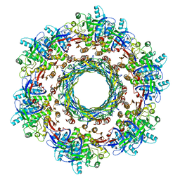
5Y5S
 | | Structure of photosynthetic LH1-RC super-complex at 1.9 angstrom resolution | | 分子名称: | (1R)-2-{[{[(2S)-2,3-DIHYDROXYPROPYL]OXY}(HYDROXY)PHOSPHORYL]OXY}-1-[(PALMITOYLOXY)METHYL]ETHYL (11E)-OCTADEC-11-ENOATE, 1,2-DIPALMITOYL-PHOSPHATIDYL-GLYCEROLE, BACTERIOCHLOROPHYLL A, ... | | 著者 | Yu, L.-J, Suga, M, Wang-Otomo, Z.-Y, Shen, J.-R. | | 登録日 | 2017-08-09 | | 公開日 | 2018-04-11 | | 最終更新日 | 2023-11-22 | | 実験手法 | X-RAY DIFFRACTION (1.9 Å) | | 主引用文献 | Structure of photosynthetic LH1-RC supercomplex at 1.9 angstrom resolution.
Nature, 556, 2018
|
|
8SC4
 | |
5Y8J
 | | Mycobacterium tuberculosis 3-Hydroxyisobutyrate dehydrogenase (MtHIBADH) + (R)-3-hydroxyisobutyrate (R-HIBA) | | 分子名称: | (2R)-3-HYDROXY-2-METHYLPROPANOIC ACID, (2~{S})-2-methylpentanedioic acid, ACRYLIC ACID, ... | | 著者 | Srikalaivani, R, Singh, A, Surolia, A, Vijayan, M. | | 登録日 | 2017-08-21 | | 公開日 | 2018-07-11 | | 最終更新日 | 2023-11-22 | | 実験手法 | X-RAY DIFFRACTION (1.86 Å) | | 主引用文献 | Structure, interactions and action ofMycobacterium tuberculosis3-hydroxyisobutyric acid dehydrogenase.
Biochem. J., 475, 2018
|
|
6QA1
 | | ERK2 mini-fragment binding | | 分子名称: | Mitogen-activated protein kinase 1, SULFATE ION, pyridin-2-amine | | 著者 | O'Reilly, M, Cleasby, A, Davies, T.G, Hall, R, Ludlow, F, Murray, C.W, Tisi, D, Jhoti, H. | | 登録日 | 2018-12-18 | | 公開日 | 2019-06-26 | | 実験手法 | X-RAY DIFFRACTION (1.58 Å) | | 主引用文献 | Crystallographic screening using ultra-low-molecular-weight ligands to guide drug design.
Drug Discov Today, 24, 2019
|
|
8SEB
 | | Cryo-EM structure of a single loaded human UBA7-UBE2L6-ISG15 adenylate complex | | 分子名称: | ADENOSINE MONOPHOSPHATE, Ubiquitin-like modifier-activating enzyme 7, Ubiquitin-like protein ISG15, ... | | 著者 | Afsar, M, Jia, L, Ruben, E.A, Olsen, S.K. | | 登録日 | 2023-04-08 | | 公開日 | 2023-10-11 | | 実験手法 | ELECTRON MICROSCOPY (3.24 Å) | | 主引用文献 | Cryo-EM structures of Uba7 reveal the molecular basis for ISG15 activation and E1-E2 thioester transfer.
Nat Commun, 14, 2023
|
|
6Q3W
 | | Structure of human galactokinase 1 bound with Ethyl 1-(2-pyrazinyl)-4-piperidinecarboxylate | | 分子名称: | 2-(1,3-benzoxazol-2-ylamino)spiro[1,6,7,8-tetrahydroquinazoline-4,1'-cyclohexane]-5-one, Galactokinase, beta-D-galactopyranose, ... | | 著者 | Mackinnon, S.R, Bezerra, G.A, Zhang, M, Foster, W, Krojer, T, Brandao-Neto, J, Douangamath, A, Arrowsmith, C, Edwards, A, Bountra, C, Brennan, P, Lai, K, Yue, W.W. | | 登録日 | 2018-12-04 | | 公開日 | 2019-01-23 | | 最終更新日 | 2024-01-24 | | 実験手法 | X-RAY DIFFRACTION (1.962 Å) | | 主引用文献 | Structure of human galactokinase 1 bound with Ethyl 1-(2-pyrazinyl)-4-piperidinecarboxylate
To Be Published
|
|
8SC1
 | |
5YA6
 | |
6Q4S
 | | Crystal structure of a-eudesmol synthase | | 分子名称: | CHLORIDE ION, Pentalenene synthase | | 著者 | Correia Cordeiro, R.S, Hakansson, M, Logan, D.T, Kourist, R. | | 登録日 | 2018-12-06 | | 公開日 | 2018-12-19 | | 最終更新日 | 2024-01-24 | | 実験手法 | X-RAY DIFFRACTION (1.83 Å) | | 主引用文献 | Discovery of three novel sesquiterpene synthases from Streptomyces chartreusis NRRL 3882 and crystal structure of an alpha-eudesmol synthase.
J.Biotechnol., 297, 2019
|
|
5YBM
 | |


