6TB7
 
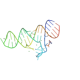 | |
6TF2
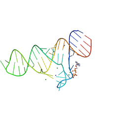 | |
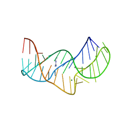
5O69
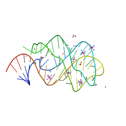
 | | The structure of the thermobifida fusca guanidine III riboswitch with agmatine. | | 分子名称: | AGMATINE, MAGNESIUM ION, RNA (37-MER), ... | | 著者 | Huang, L, Wang, J, Lilley, D.M.J. | | 登録日 | 2017-06-06 | | 公開日 | 2017-10-18 | | 最終更新日 | 2024-05-08 | | 実験手法 | X-RAY DIFFRACTION (2.319 Å) | | 主引用文献 | Structure of the Guanidine III Riboswitch.
Cell Chem Biol, 24, 2017
|
|
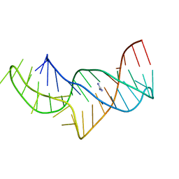
5O62
 | |
5NWQ
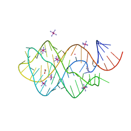
 | | The structure of the thermobifida fusca guanidine III riboswitch with guanidine. | | 分子名称: | GUANIDINE, Guanidine III riboswitch, MAGNESIUM ION, ... | | 著者 | Huang, L, Wang, J, Lilley, D.M.J. | | 登録日 | 2017-05-08 | | 公開日 | 2017-10-18 | | 最終更新日 | 2024-05-08 | | 実験手法 | X-RAY DIFFRACTION (1.91 Å) | | 主引用文献 | Structure of the Guanidine III Riboswitch.
Cell Chem Biol, 24, 2017
|
|
5NY8
 | | The structure of the thermobifida fusca guanidine III riboswitch with aminoguanidine | | 分子名称: | AMINOGUANIDINE, MAGNESIUM ION, RNA (41-MER), ... | | 著者 | Huang, L, Wang, J, Lilley, D.M.J. | | 登録日 | 2017-05-11 | | 公開日 | 2017-10-18 | | 最終更新日 | 2024-05-08 | | 実験手法 | X-RAY DIFFRACTION (2.04 Å) | | 主引用文献 | Structure of the Guanidine III Riboswitch.
Cell Chem Biol, 24, 2017
|
|
5NZ6
 | |
5NS4
 | | Crystal structures of Cy3 cyanine fluorophores stacked onto the end of double-stranded RNA | | 分子名称: | 3-[(2~{Z})-2-[(~{E})-3-[3,3-dimethyl-1-(3-oxidanylpropyl)indol-1-ium-2-yl]prop-2-enylidene]-3,3-dimethyl-indol-1-yl]propan-1-ol, 50S ribosomal protein L5, MAGNESIUM ION, ... | | 著者 | Liu, Y.J, Lilley, D.M.J. | | 登録日 | 2017-04-25 | | 公開日 | 2017-12-20 | | 最終更新日 | 2024-01-17 | | 実験手法 | X-RAY DIFFRACTION (2.4 Å) | | 主引用文献 | Crystal Structures of Cyanine Fluorophores Stacked onto the End of Double-Stranded RNA.
Biophys. J., 113, 2017
|
|
5NS3
 | |
5NZD
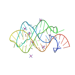
 | | The structure of the thermobifida fusca guanidine III riboswitch in space group P212121. | | 分子名称: | ACETATE ION, MAGNESIUM ION, SODIUM ION, ... | | 著者 | Huang, L, Wang, J, Lilley, D.M.J. | | 登録日 | 2017-05-13 | | 公開日 | 2017-10-18 | | 最終更新日 | 2024-05-08 | | 実験手法 | X-RAY DIFFRACTION (2.007 Å) | | 主引用文献 | Structure of the Guanidine III Riboswitch.
Cell Chem Biol, 24, 2017
|
|
5NZ3
 | | The structure of the thermobifida fusca guanidine III riboswitch with methylguanidine | | 分子名称: | 1-METHYLGUANIDINE, MAGNESIUM ION, RNA (41-MER), ... | | 著者 | Huang, L, Wang, J, Lilley, D.M.J. | | 登録日 | 2017-05-12 | | 公開日 | 2017-10-18 | | 最終更新日 | 2024-05-08 | | 実験手法 | X-RAY DIFFRACTION (2.059 Å) | | 主引用文献 | Structure of the Guanidine III Riboswitch.
Cell Chem Biol, 24, 2017
|
|
2XO1
 | | xpt-pbuX C74U Riboswitch from B. subtilis bound to N6-methyladenine | | 分子名称: | ACETATE ION, COBALT HEXAMMINE(III), Guanine riboswitch, ... | | 著者 | Daldrop, P, Reyes, F.E, Robinson, D.A, Hammond, C.M, Lilley, D.M.J, Batey, R.T, Brenk, R. | | 登録日 | 2010-08-09 | | 公開日 | 2011-04-06 | | 最終更新日 | 2024-05-08 | | 実験手法 | X-RAY DIFFRACTION (1.6 Å) | | 主引用文献 | Novel ligands for a purine riboswitch discovered by RNA-ligand docking.
Chem. Biol., 18, 2011
|
|
2XO0
 | | xpt-pbuX C74U Riboswitch from B. subtilis bound to 24-diamino-1,3,5- triazine identified by virtual screening | | 分子名称: | 1,3,5-TRIAZINE-2,4-DIAMINE, ACETATE ION, COBALT HEXAMMINE(III), ... | | 著者 | Daldrop, P, Reyes, F.E, Robinson, D.A, Hammond, C.M, Lilley, D.M.J, Batey, R.T, Brenk, R. | | 登録日 | 2010-08-09 | | 公開日 | 2011-04-06 | | 最終更新日 | 2023-12-20 | | 実験手法 | X-RAY DIFFRACTION (1.7 Å) | | 主引用文献 | Novel ligands for a purine riboswitch discovered by RNA-ligand docking.
Chem. Biol., 18, 2011
|
|
2XNW
 | | XPT-PBUX C74U RIBOSWITCH FROM B. SUBTILIS BOUND TO A TRIAZOLO- TRIAZOLE-DIAMINE LIGAND IDENTIFIED BY VIRTUAL SCREENING | | 分子名称: | 3,6-diamino-1,5-dihydro[1,2,4]triazolo[4,3-b][1,2,4]triazol-4-ium, ACETATE ION, COBALT HEXAMMINE(III), ... | | 著者 | Daldrop, P, Reyes, F.E, Robinson, D.A, Hammond, C.M, Lilley, D.M.J, Brenk, R. | | 登録日 | 2010-08-06 | | 公開日 | 2011-04-06 | | 最終更新日 | 2023-12-20 | | 実験手法 | X-RAY DIFFRACTION (1.5 Å) | | 主引用文献 | Novel ligands for a purine riboswitch discovered by RNA-ligand docking.
Chem. Biol., 18, 2011
|
|
352D
 | | THE CRYSTAL STRUCTURE OF A PARALLEL-STRANDED PARALLEL-STRANDED GUANINE TETRAPLEX AT 0.95 ANGSTROM RESOLUTION | | 分子名称: | CALCIUM ION, DNA (5'-D(*TP*GP*GP*GP*GP*T)-3'), SODIUM ION | | 著者 | Phillips, K, Dauter, Z, Murchie, A.I.H, Lilley, D.M.J, Luisi, B. | | 登録日 | 1997-09-04 | | 公開日 | 1997-11-10 | | 最終更新日 | 2023-08-02 | | 実験手法 | X-RAY DIFFRACTION (0.95 Å) | | 主引用文献 | The crystal structure of a parallel-stranded guanine tetraplex at 0.95 A resolution.
J.Mol.Biol., 273, 1997
|
|
2XNZ
 | | xpt-pbuX C74U Riboswitch from B. subtilis bound to acetoguanamine identified by virtual screening | | 分子名称: | 6-METHYL-1,3,5-TRIAZINE-2,4-DIAMINE, ACETATE ION, COBALT HEXAMMINE(III), ... | | 著者 | Daldrop, P, Reyes, F.E, Robinson, D.A, Hammond, C.M, Lilley, D.M.J, Batey, R.T, Brenk, R. | | 登録日 | 2010-08-06 | | 公開日 | 2011-04-06 | | 最終更新日 | 2023-12-20 | | 実験手法 | X-RAY DIFFRACTION (1.59 Å) | | 主引用文献 | Novel ligands for a purine riboswitch discovered by RNA-ligand docking.
Chem. Biol., 18, 2011
|
|
5NEF
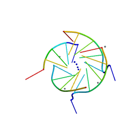
 | | The structure of the G. violaceus guanidine II riboswitch P1 stem-loop with guanidine | | 分子名称: | GUANIDINE, RNA (5'-R(*GP*GP*UP*GP*GP*GP*GP*AP*CP*GP*AP*CP*CP*CP*CP*AP*(CBV)P*C)-3'), SODIUM ION, ... | | 著者 | Huang, L, Wang, J, Lilley, D.M.J. | | 登録日 | 2017-03-10 | | 公開日 | 2017-06-07 | | 最終更新日 | 2024-05-08 | | 実験手法 | X-RAY DIFFRACTION (1.91 Å) | | 主引用文献 | The Structure of the Guanidine-II Riboswitch.
Cell Chem Biol, 24, 2017
|
|
5NDH
 | | The structure of the G. violaceus guanidine II riboswitch P2 stem-loop | | 分子名称: | GUANIDINE, MAGNESIUM ION, RNA (5'-R(*GP*(CBV)P*GP*GP*GP*GP*AP*CP*GP*AP*CP*CP*CP*CP*GP*C)-3'), ... | | 著者 | Huang, L, Wang, J, Lilley, D.M.J. | | 登録日 | 2017-03-08 | | 公開日 | 2017-05-31 | | 最終更新日 | 2024-05-08 | | 実験手法 | X-RAY DIFFRACTION (1.81 Å) | | 主引用文献 | The Structure of the Guanidine-II Riboswitch.
Cell Chem Biol, 24, 2017
|
|
5NOM
 | | The structure of the G. violaceus guanidine II riboswitch P1 stem-loop with guanidine | | 分子名称: | GUANIDINE, RNA (5'-R(*GP*GP*UP*GP*GP*GP*GP*AP*CP*GP*AP*CP*CP*CP*CP*AP*(CBV)P*C)-3'), SODIUM ION, ... | | 著者 | Huang, L, Wang, J, Lilley, D.M.J. | | 登録日 | 2017-04-12 | | 公開日 | 2017-05-31 | | 最終更新日 | 2024-01-17 | | 実験手法 | X-RAY DIFFRACTION (1.93 Å) | | 主引用文献 | The Structure of the Guanidine-II Riboswitch.
Cell Chem Biol, 24, 2017
|
|
5NDI
 | |
5NEP
 | | The structure of the G. violaceus guanidine II riboswitch P1 stem-loop with methylguanidine | | 分子名称: | 1-METHYLGUANIDINE, RNA (5'-R(*GP*GP*UP*GP*GP*GP*GP*AP*CP*GP*AP*CP*CP*CP*CP*AP*(CBV)P*C)-3'), SODIUM ION, ... | | 著者 | Huang, L, Wang, J, Lilley, D.M.J. | | 登録日 | 2017-03-11 | | 公開日 | 2017-05-31 | | 最終更新日 | 2024-05-08 | | 実験手法 | X-RAY DIFFRACTION (1.6 Å) | | 主引用文献 | The Structure of the Guanidine-II Riboswitch.
Cell Chem Biol, 24, 2017
|
|
5NEQ
 | | The structure of the G. violaceus guanidine II riboswitch P1 stem-loop with aminoguanidine | | 分子名称: | AMINOGUANIDINE, RNA (5'-R(*GP*GP*UP*GP*GP*GP*GP*AP*CP*GP*AP*CP*CP*CP*CP*AP*(CBV)P*C)-3'), SODIUM ION, ... | | 著者 | Huang, L, Wang, J, Lilley, D.M.J. | | 登録日 | 2017-03-11 | | 公開日 | 2017-05-31 | | 最終更新日 | 2024-01-17 | | 実験手法 | X-RAY DIFFRACTION (1.69 Å) | | 主引用文献 | The Structure of the Guanidine-II Riboswitch.
Cell Chem Biol, 24, 2017
|
|
5NEO
 | | The structure of the G. violaceus guanidine II riboswitch P1 stem-loop | | 分子名称: | AMMONIUM ION, RNA (5'-R(*GP*GP*UP*GP*GP*GP*GP*AP*CP*GP*AP*CP*CP*CP*CP*AP*(CBV)P*C)-3'), SODIUM ION, ... | | 著者 | Huang, L, Wang, J, Lilley, D.M.J. | | 登録日 | 2017-03-11 | | 公開日 | 2017-05-31 | | 最終更新日 | 2024-05-08 | | 実験手法 | X-RAY DIFFRACTION (1.69 Å) | | 主引用文献 | The Structure of the Guanidine-II Riboswitch.
Cell Chem Biol, 24, 2017
|
|
5NEX
 | |
5FKF
 | |


