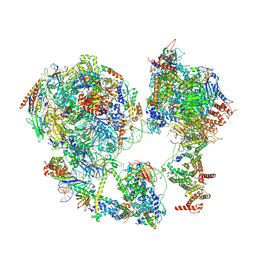
7EGD
 
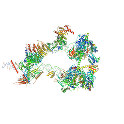 | | SCP promoter-bound TFIID-TFIIA in initial TBP-loading state | | 分子名称: | DNA (72-MER), TATA-box-binding protein, Transcription initiation factor IIA subunit 1, ... | | 著者 | Chen, X, Wu, Z, Li, J, Zhao, D, Xu, Y. | | 登録日 | 2021-03-24 | | 公開日 | 2021-05-12 | | 実験手法 | ELECTRON MICROSCOPY (6.75 Å) | | 主引用文献 | Structural insights into preinitiation complex assembly on core promoters.
Science, 372, 2021
|
|
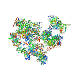
7EGJ
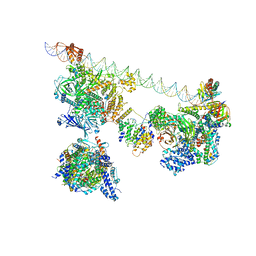 | | SCP promoter-bound TFIID-TFIIA in post TBP-loading state | | 分子名称: | DNA (74-MER), TATA-box-binding protein, Transcription initiation factor IIA subunit 1, ... | | 著者 | Chen, X, Wu, Z, Li, J, Zhao, D, Xu, Y. | | 登録日 | 2021-03-24 | | 公開日 | 2021-05-12 | | 実験手法 | ELECTRON MICROSCOPY (8.64 Å) | | 主引用文献 | Structural insights into preinitiation complex assembly on core promoters.
Science, 372, 2021
|
|
7EGE
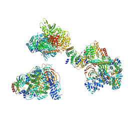 | | TFIID in canonical conformation | | 分子名称: | TATA-box-binding protein, Transcription initiation factor TFIID subunit 1, Transcription initiation factor TFIID subunit 10, ... | | 著者 | Chen, X, Wu, Z, Li, J, Zhao, D, Xu, Y. | | 登録日 | 2021-03-24 | | 公開日 | 2021-05-12 | | 実験手法 | ELECTRON MICROSCOPY (9 Å) | | 主引用文献 | Structural insights into preinitiation complex assembly on core promoters.
Science, 372, 2021
|
|
7EGC
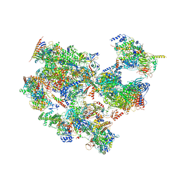 | | p53-bound TFIID-based holo PIC on HDM2 promoter | | 分子名称: | CDK-activating kinase assembly factor MAT1, Cyclin-H, Cyclin-dependent kinase 7, ... | | 著者 | Chen, X, Wu, Z, Hou, H, Qi, Y, Wang, X, Li, J, Xu, Y. | | 登録日 | 2021-03-24 | | 公開日 | 2021-05-12 | | 実験手法 | ELECTRON MICROSCOPY (3.9 Å) | | 主引用文献 | Structural insights into preinitiation complex assembly on core promoters.
Science, 372, 2021
|
|
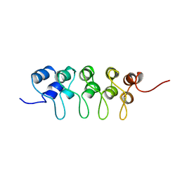
1D3B
 | | CRYSTAL STRUCTURE OF THE D3B SUBCOMPLEX OF THE HUMAN CORE SNRNP DOMAIN AT 2.0A RESOLUTION | | 分子名称: | CITRIC ACID, GLYCEROL, PROTEIN (SMALL NUCLEAR RIBONUCLEOPROTEIN ASSOCIATED PROTEIN B), ... | | 著者 | Kambach, C, Walke, S, Avis, J.M, De La Fortelle, E, Li, J, Nagai, K. | | 登録日 | 1998-12-22 | | 公開日 | 1999-12-22 | | 最終更新日 | 2023-12-27 | | 実験手法 | X-RAY DIFFRACTION (2 Å) | | 主引用文献 | Crystal structures of two Sm protein complexes and their implications for the assembly of the spliceosomal snRNPs.
Cell(Cambridge,Mass.), 96, 1999
|
|
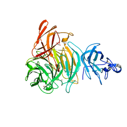
5ZRX
 | | Crystal Structure of EphA2/SHIP2 Complex | | 分子名称: | Phosphatidylinositol 3,4,5-trisphosphate 5-phosphatase 2,Ephrin type-A receptor 2 | | 著者 | Wang, Y, Shang, Y, Li, J, Chen, W, Li, G, Wan, J, Liu, W, Zhang, M. | | 登録日 | 2018-04-25 | | 公開日 | 2018-05-30 | | 最終更新日 | 2023-11-22 | | 実験手法 | X-RAY DIFFRACTION (1.5 Å) | | 主引用文献 | Specific Eph receptor-cytoplasmic effector signaling mediated by SAM-SAM domain interactions.
Elife, 7, 2018
|
|
7C2A
 | |
7C2D
 | | Esterase AlinE4 mutant-S13A | | 分子名称: | ACETATE ION, CADMIUM ION, SGNH-hydrolase family esterase | | 著者 | Li, Z, Li, J. | | 登録日 | 2020-05-07 | | 公開日 | 2020-05-20 | | 最終更新日 | 2023-11-29 | | 実験手法 | X-RAY DIFFRACTION (1.75 Å) | | 主引用文献 | C-terminal swapped dimers revealed a new catalytic mechanism of SGNH-hydrolase family esterases
To Be Published
|
|
7C2C
 | | Esterase AlinE4 mutant, D162A | | 分子名称: | ACETATE ION, CADMIUM ION, GLYCEROL, ... | | 著者 | Li, Z, Li, J. | | 登録日 | 2020-05-07 | | 公開日 | 2020-05-20 | | 最終更新日 | 2023-11-29 | | 実験手法 | X-RAY DIFFRACTION (1.55 Å) | | 主引用文献 | C-terminal swapped dimers revealed a new catalytic mechanism of SGNH-hydrolase family esterases
To Be Published
|
|
7EDX
 | | p53-bound TFIID-based core PIC on HDM2 promoter | | 分子名称: | DNA (84-mer), DNA-directed RNA polymerase II subunit E, DNA-directed RNA polymerase II subunit F, ... | | 著者 | Chen, X, Qi, Y, Hou, H, Wang, X, Wu, Z, Li, J, Xu, Y. | | 登録日 | 2021-03-17 | | 公開日 | 2021-05-05 | | 最終更新日 | 2021-05-19 | | 実験手法 | ELECTRON MICROSCOPY (4.5 Å) | | 主引用文献 | Structural insights into preinitiation complex assembly on core promoters.
Science, 372, 2021
|
|
7EGB
 | | TFIID-based holo PIC on SCP promoter | | 分子名称: | CDK-activating kinase assembly factor MAT1, Cyclin-H, Cyclin-dependent kinase 7, ... | | 著者 | Chen, X, Wu, Z, Hou, H, Qi, Y, Wang, X, Li, J, Xu, Y. | | 登録日 | 2021-03-24 | | 公開日 | 2021-05-05 | | 最終更新日 | 2023-07-26 | | 実験手法 | ELECTRON MICROSCOPY (3.3 Å) | | 主引用文献 | Structural insights into preinitiation complex assembly on core promoters.
Science, 372, 2021
|
|
3QML
 | | The structural analysis of Sil1-Bip complex reveals the mechanism for Sil1 to function as a novel nucleotide exchange factor | | 分子名称: | 78 kDa glucose-regulated protein homolog, MAGNESIUM ION, Nucleotide exchange factor SIL1, ... | | 著者 | Yan, M, Li, J.Z, Sha, B.D. | | 登録日 | 2011-02-04 | | 公開日 | 2011-06-29 | | 最終更新日 | 2024-02-21 | | 実験手法 | X-RAY DIFFRACTION (2.31 Å) | | 主引用文献 | Structural analysis of the Sil1-Bip complex reveals the mechanism for Sil1 to function as a nucleotide-exchange factor.
Biochem.J., 438, 2011
|
|
5ZRZ
 | | Crystal Structure of EphA5/SAMD5 Complex | | 分子名称: | Ephrin type-A receptor 5, Sterile alpha motif domain-containing protein 5 | | 著者 | Wang, Y, Shang, Y, Li, J, Chen, W, Li, G, Wan, J, Liu, W, Zhang, M. | | 登録日 | 2018-04-25 | | 公開日 | 2018-05-30 | | 最終更新日 | 2023-11-22 | | 実験手法 | X-RAY DIFFRACTION (1.89 Å) | | 主引用文献 | Specific Eph receptor-cytoplasmic effector signaling mediated by SAM-SAM domain interactions.
Elife, 7, 2018
|
|
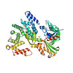
3E2Y
 | | Crystal structure of mouse kynurenine aminotransferase III in complex with glutamine | | 分子名称: | 4'-DEOXY-4'-AMINOPYRIDOXAL-5'-PHOSPHATE, GLUTAMINE, GLYCEROL, ... | | 著者 | Han, Q, Robinson, R, Cai, T, Tagle, D.A, Li, J. | | 登録日 | 2008-08-06 | | 公開日 | 2008-12-30 | | 最終更新日 | 2023-08-30 | | 実験手法 | X-RAY DIFFRACTION (2.26 Å) | | 主引用文献 | Correction for Han et al., "Biochemical and Structural Properties of Mouse Kynurenine Aminotransferase III".
Mol. Cell. Biol., 38, 2018
|
|
1BU9
 | |
6XLT
 | |
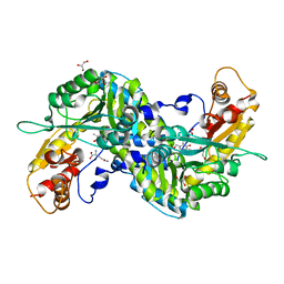
3E2Z
 | | Crystal structure of mouse kynurenine aminotransferase III in complex with kynurenine | | 分子名称: | (2S)-2-amino-4-(2-aminophenyl)-4-oxobutanoic acid, 4'-DEOXY-4'-AMINOPYRIDOXAL-5'-PHOSPHATE, GLYCEROL, ... | | 著者 | Han, Q, Robinson, R, Cai, T, Tagle, D.A, Li, J. | | 登録日 | 2008-08-06 | | 公開日 | 2008-12-30 | | 最終更新日 | 2023-11-15 | | 実験手法 | X-RAY DIFFRACTION (2.81 Å) | | 主引用文献 | Correction for Han et al., "Biochemical and Structural Properties of Mouse Kynurenine Aminotransferase III".
Mol. Cell. Biol., 38, 2018
|
|
5GGZ
 | | Crystal structure of novel inhibitor bound with Hsp90 | | 分子名称: | Heat shock protein HSP 90-alpha, [2,4-bis(oxidanyl)-5-propan-2-yl-phenyl]-(2-ethoxy-7,8-dihydro-5~{H}-pyrido[4,3-d]pyrimidin-6-yl)methanone | | 著者 | Chen, T.T, Li, J, Xu, Y.C. | | 登録日 | 2016-06-16 | | 公開日 | 2017-03-08 | | 最終更新日 | 2024-03-20 | | 実験手法 | X-RAY DIFFRACTION (2.015 Å) | | 主引用文献 | Novel Tetrahydropyrido[4,3-d]pyrimidines as Potent Inhibitors of Chaperone Heat Shock Protein 90
J. Med. Chem., 59, 2016
|
|
4E15
 | |
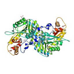
6KUZ
 | | E.coli beta-galactosidase (E537Q) in complex with fluorescent probe KSL01 | | 分子名称: | 3-(1,3-benzothiazol-2-yl)-2-[[4-[(2~{S},3~{R},4~{S},5~{R},6~{R})-6-(hydroxymethyl)-3,4,5-tris(oxidanyl)oxan-2-yl]oxyphenyl]methoxy]-5-methyl-benzaldehyde, Beta-galactosidase, DIMETHYL SULFOXIDE, ... | | 著者 | Chen, X, Hu, Y.L, Li, X.K, Guo, Y, Li, J. | | 登録日 | 2019-09-03 | | 公開日 | 2020-07-08 | | 最終更新日 | 2023-11-22 | | 実験手法 | X-RAY DIFFRACTION (2.83 Å) | | 主引用文献 | First-generation species-selective chemical probes for fluorescence imaging of human senescence-associated beta-galactosidase.
Chem Sci, 11, 2020
|
|
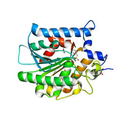
3WLA
 | | Crystal Structure of sOPH Native | | 分子名称: | Oxidized polyvinyl alcohol hydrolase, SULFATE ION | | 著者 | Yang, Y, Ko, T.P, Li, J.H, Liu, L, Huang, C.H, Chan, H.C, Ren, F.F, Jia, D.X, Wang, A.H.-J, Guo, R.T, Chen, J, Du, G.C. | | 登録日 | 2013-11-08 | | 公開日 | 2014-09-24 | | 最終更新日 | 2023-11-08 | | 実験手法 | X-RAY DIFFRACTION (1.9 Å) | | 主引用文献 | Structural insights into enzymatic degradation of oxidized polyvinyl alcohol
Chembiochem, 15, 2014
|
|
2ZJG
 | | Crystal structural of mouse kynurenine aminotransferase III | | 分子名称: | GLYCEROL, Kynurenine-oxoglutarate transaminase 3 | | 著者 | Han, Q, Cai, T, Tagle, D.A, Robinson, H, Li, J. | | 登録日 | 2008-03-07 | | 公開日 | 2009-01-06 | | 最終更新日 | 2023-11-15 | | 実験手法 | X-RAY DIFFRACTION (3 Å) | | 主引用文献 | Structural and functional characterization of mouse kynurenine aminotransferase III
To be Published
|
|
3E2F
 | | Crystal structure of mouse kynurenine aminotransferase III, PLP-bound form | | 分子名称: | GLYCEROL, Kynurenine-oxoglutarate transaminase 3 | | 著者 | Han, Q, Robinson, R, Cai, T, Tagle, D.A, Li, J. | | 登録日 | 2008-08-05 | | 公開日 | 2008-12-30 | | 最終更新日 | 2023-11-15 | | 実験手法 | X-RAY DIFFRACTION (2.59 Å) | | 主引用文献 | Correction for Han et al., "Biochemical and Structural Properties of Mouse Kynurenine Aminotransferase III".
Mol. Cell. Biol., 38, 2018
|
|
4MBS
 | | Crystal Structure of the CCR5 Chemokine Receptor | | 分子名称: | (2R)-2,3-dihydroxypropyl (9Z)-octadec-9-enoate, 4,4-difluoro-N-[(1S)-3-{(3-exo)-3-[3-methyl-5-(propan-2-yl)-4H-1,2,4-triazol-4-yl]-8-azabicyclo[3.2.1]oct-8-yl}-1-phenylpropyl]cyclohexanecarboxamide, Chimera protein of C-C chemokine receptor type 5 and Rubredoxin, ... | | 著者 | Tan, Q, Zhu, Y, Han, G.W, Li, J, Fenalti, G, Liu, H, Cherezov, V, Stevens, R.C, GPCR Network (GPCR), Zhao, Q, Wu, B. | | 登録日 | 2013-08-19 | | 公開日 | 2013-09-11 | | 最終更新日 | 2023-09-20 | | 実験手法 | X-RAY DIFFRACTION (2.71 Å) | | 主引用文献 | Structure of the CCR5 chemokine receptor-HIV entry inhibitor maraviroc complex.
Science, 341, 2013
|
|
5XF7
 | |


