Loading
PDBj
メニューPDBj@FacebookPDBj@TwitterPDBj@YouTubewwPDB FoundationwwPDB
RCSB PDBPDBeBMRBAdv. SearchSearch help
PDB: 10 件

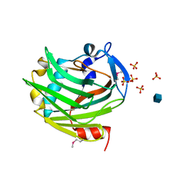
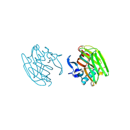
2OUJ
DownloadVisualize
BU of 2ouj by Molmil
The crystal structure of the Thrombospondin-1 N-terminal domain in complex with fractionated Heparin DP8
分子名称: Thrombospondin-1
著者Tan, K, Joachimiak, A, Wang, J, Lawler, J.
登録日2007-02-11
公開日2008-01-08
最終更新日2023-08-30
実験手法X-RAY DIFFRACTION (1.9 Å)
主引用文献Heparin-induced cis- and trans-Dimerization Modes of the Thrombospondin-1 N-terminal Domain.
J.Biol.Chem., 283, 2008
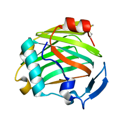
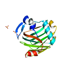
2OUH
DownloadVisualize
BU of 2ouh by Molmil
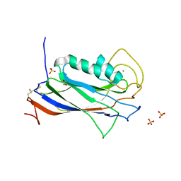
Crystal structure of the Thrombospondin-1 N-terminal domain in complex with fractionated Heparin DP10
分子名称: SULFATE ION, Thrombospondin-1
著者Tan, K, Joachimiak, A, Wang, J, Lawler, J.
登録日2007-02-11
公開日2008-01-08
最終更新日2024-10-09
実験手法X-RAY DIFFRACTION (2.4 Å)
主引用文献Heparin-induced cis- and trans-Dimerization Modes of the Thrombospondin-1 N-terminal Domain.
J.Biol.Chem., 283, 2008
1LSL
DownloadVisualize
BU of 1lsl by Molmil
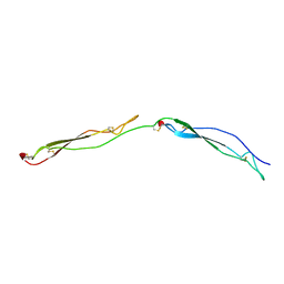
Crystal Structure of the Thrombospondin-1 Type 1 Repeats
分子名称: Thrombospondin 1, alpha-L-fucopyranose, beta-L-fucopyranose
著者Tan, K, Duquette, M, Liu, J, Dong, Y, Zhang, R, Joachimiak, A, Lawler, J, Wang, J.-H.
登録日2002-05-17
公開日2002-12-18
最終更新日2024-10-30
実験手法X-RAY DIFFRACTION (1.9 Å)
主引用文献Crystal structure of the TSP-1 type 1 repeats: a novel layered fold and its biological implication.
J.Cell Biol., 159, 2002
3Q13
DownloadVisualize
BU of 3q13 by Molmil
The Structure of the Ca2+-binding, Glycosylated F-spondin Domain of F-spondin, A C2-domain Variant from Extracellular Matrix
分子名称: ACETATE ION, CALCIUM ION, SULFATE ION, ...
著者Tan, K, Lawler, J.
登録日2010-12-16
公開日2011-06-29
最終更新日2023-09-13
実験手法X-RAY DIFFRACTION (1.95 Å)
主引用文献The structure of the Ca2+-binding, glycosylated F-spondin domain of F-spondin - A C2-domain variant in an extracellular matrix protein.
Bmc Struct.Biol., 11, 2011
3FBY
DownloadVisualize
BU of 3fby by Molmil
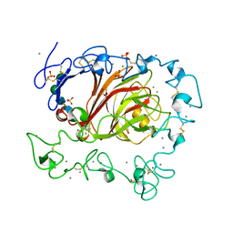
The crystal structure of the signature domain of cartilage oligomeric matrix protein.
分子名称: CALCIUM ION, Cartilage oligomeric matrix protein, SULFATE ION, ...
著者Tan, K, Lawler, J.
登録日2008-11-20
公開日2009-06-23
最終更新日2023-09-06
実験手法X-RAY DIFFRACTION (3.15 Å)
主引用文献The crystal structure of the signature domain of cartilage oligomeric matrix protein: implications for collagen, glycosaminoglycan and integrin binding.
Faseb J., 23, 2009
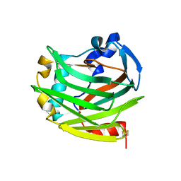
1Z78
DownloadVisualize
BU of 1z78 by Molmil
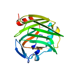
Crystal Structure of the Thrombospondin-1 N-terminal domain
分子名称: Thrombospondin 1
著者Tan, K, Wang, J, Lawler, J.
登録日2005-03-24
公開日2006-01-17
最終更新日2024-04-03
実験手法X-RAY DIFFRACTION (1.8 Å)
主引用文献The structures of the thrombospondin-1 N-terminal domain and its complex with a synthetic pentameric heparin.
Structure, 14, 2006
1ZA4
DownloadVisualize
BU of 1za4 by Molmil
Crystal Structure of the Thrombospondin-1 N-terminal Domain in Complex with Arixtra
分子名称: 2-deoxy-6-O-sulfo-2-(sulfoamino)-alpha-D-glucopyranose, SULFATE ION, Thrombospondin 1
著者Tan, K, Wang, J.H, Lawler, J.
登録日2005-04-05
公開日2006-01-24
最終更新日2024-11-06
実験手法X-RAY DIFFRACTION (1.9 Å)
主引用文献The Structures of the Thrombospondin-1 N-terminal Domain and its Complex with a Synthetic Pentameric Heparin
Structure, 14, 2006
2ERF
DownloadVisualize
BU of 2erf by Molmil
Crystal Structure of the Thrombospondin-1 N-terminal Domain at 1.45A Resolution
分子名称: Thrombospondin-1
著者Tan, K, Wang, J, Lawler, J.
登録日2005-10-24
公開日2006-01-17
最終更新日2024-11-06
実験手法X-RAY DIFFRACTION (1.45 Å)
主引用文献The structures of the thrombospondin-1 N-terminal domain and its complex with a synthetic pentameric heparin.
Structure, 14, 2006
2ES3
DownloadVisualize
BU of 2es3 by Molmil
Crystal Structure of Thrombospondin-1 N-terminal Domain in P1 Form at 1.85A Resolution
分子名称: Thrombospondin-1
著者Tan, K, Wang, J, Lawler, J.
登録日2005-10-25
公開日2006-10-31
最終更新日2024-10-30
実験手法X-RAY DIFFRACTION (1.85 Å)
主引用文献Heparin-induced cis- and trans-Dimerization Modes of the Thrombospondin-1 N-terminal Domain.
J.Biol.Chem., 283, 2008
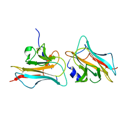
3COO
DownloadVisualize
BU of 3coo by Molmil
The crystal structure of Reelin-N domain of F-spondin
分子名称: Spondin-1
著者Tan, K, Lawler, J, Wang, J.H.
登録日2008-03-28
公開日2008-08-26
最終更新日2024-10-09
実験手法X-RAY DIFFRACTION (2 Å)
主引用文献The crystal structure of the heparin-binding reelin-N domain of f-spondin.
J.Mol.Biol., 381, 2008

227111

件を2024-11-06に公開中

PDB statisticsPDBj update infoContact PDBjnumon