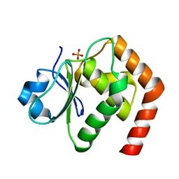
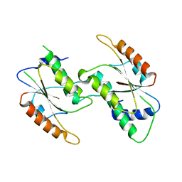
7Y39
 
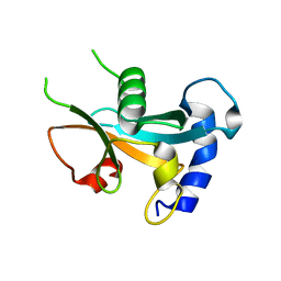 | | Ubiquitin-like domain of human ZFAND1 | | 分子名称: | AN1-type zinc finger protein 1 | | 著者 | Lai, C.H, Ko, K.T, Fan, P.J, Yu, T.A, Chang, C.F, Draczkowski, P, Hsu, S.T.D. | | 登録日 | 2022-06-10 | | 公開日 | 2022-08-10 | | 最終更新日 | 2024-04-03 | | 実験手法 | X-RAY DIFFRACTION (1.88 Å) | | 主引用文献 | Structural Insight into ZFAND1 and p97 Interaction
To Be Published
|
|
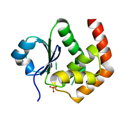
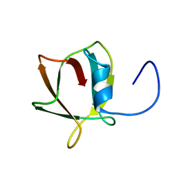
8HW9
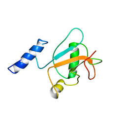 | | Solution structure of ubiquitin-like domain (UBL) of human ZFAND1 | | 分子名称: | AN1-type zinc finger protein 1 | | 著者 | Lai, C.H, Ko, K.T, Fan, P.J, Yu, T.A, Chang, C.F, Hsu, S.T.D. | | 登録日 | 2022-12-29 | | 公開日 | 2024-01-31 | | 最終更新日 | 2024-05-15 | | 実験手法 | SOLUTION NMR | | 主引用文献 | Solution structure of ubiquitin-like domain (UBL) of human ZFAND1
To Be Published
|
|
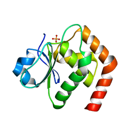
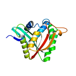
7C8S
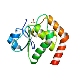 | | Crystal structure of DUSP22 mutant_N128A | | 分子名称: | Dual specificity protein phosphatase 22, SULFATE ION | | 著者 | Lai, C.H, Lyu, P.C. | | 登録日 | 2020-06-03 | | 公開日 | 2020-10-28 | | 最終更新日 | 2023-11-29 | | 実験手法 | X-RAY DIFFRACTION (1.31 Å) | | 主引用文献 | Structural Insights into the Active Site Formation of DUSP22 in N-loop-containing Protein Tyrosine Phosphatases.
Int J Mol Sci, 21, 2020
|
|
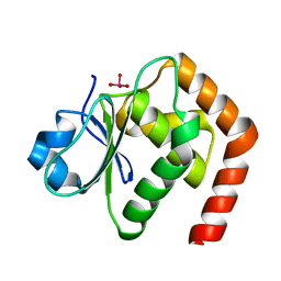
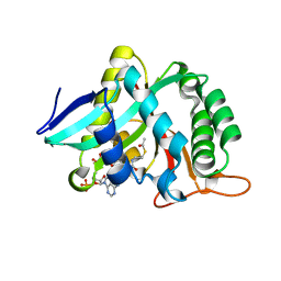
6L1S
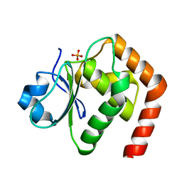 | | Crystal structure of DUSP22 mutant_C88S | | 分子名称: | Dual specificity protein phosphatase 22, PHOSPHATE ION | | 著者 | Lai, C.H, Chang, C.C, Lyu, P.C. | | 登録日 | 2019-09-30 | | 公開日 | 2020-10-28 | | 最終更新日 | 2023-11-22 | | 実験手法 | X-RAY DIFFRACTION (1.3611 Å) | | 主引用文献 | Structural Insights into the Active Site Formation of DUSP22 in N-loop-containing Protein Tyrosine Phosphatases.
Int J Mol Sci, 21, 2020
|
|
6LMY
 | | Crystal structure of DUSP22 mutant_C88S/S93A | | 分子名称: | Dual specificity protein phosphatase 22, PHOSPHATE ION | | 著者 | Lai, C.H, Lyu, P.C. | | 登録日 | 2019-12-27 | | 公開日 | 2020-10-28 | | 最終更新日 | 2023-11-22 | | 実験手法 | X-RAY DIFFRACTION (1.5 Å) | | 主引用文献 | Structural Insights into the Active Site Formation of DUSP22 in N-loop-containing Protein Tyrosine Phosphatases.
Int J Mol Sci, 21, 2020
|
|
6LOT
 | | Crystal structure of DUSP22 mutant_N128D | | 分子名称: | Dual specificity protein phosphatase 22, SULFATE ION | | 著者 | Lai, C.H, Lyu, P.C. | | 登録日 | 2020-01-07 | | 公開日 | 2020-10-28 | | 最終更新日 | 2023-11-29 | | 実験手法 | X-RAY DIFFRACTION (1.69 Å) | | 主引用文献 | Structural Insights into the Active Site Formation of DUSP22 in N-loop-containing Protein Tyrosine Phosphatases.
Int J Mol Sci, 21, 2020
|
|
6LOU
 | | Crystal structure of DUSP22 mutant_C88S/S93N | | 分子名称: | Dual specificity protein phosphatase 22, PHOSPHATE ION | | 著者 | Lai, C.H, Lyu, P.C. | | 登録日 | 2020-01-07 | | 公開日 | 2020-10-28 | | 最終更新日 | 2023-11-29 | | 実験手法 | X-RAY DIFFRACTION (1.5301 Å) | | 主引用文献 | Structural Insights into the Active Site Formation of DUSP22 in N-loop-containing Protein Tyrosine Phosphatases.
Int J Mol Sci, 21, 2020
|
|
6LVQ
 | | Crystal structure of DUSP22_VO4 | | 分子名称: | Dual specificity protein phosphatase 22, VANADATE ION | | 著者 | Lai, C.H, Lyu, P.C. | | 登録日 | 2020-02-04 | | 公開日 | 2020-10-28 | | 最終更新日 | 2023-11-29 | | 実験手法 | X-RAY DIFFRACTION (1.38 Å) | | 主引用文献 | Structural Insights into the Active Site Formation of DUSP22 in N-loop-containing Protein Tyrosine Phosphatases.
Int J Mol Sci, 21, 2020
|
|
7Y7L
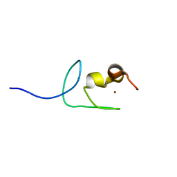 | | Solution structure of zinc finger domain 2 of human ZFAND1 | | 分子名称: | AN1-type zinc finger protein 1, ZINC ION | | 著者 | Fang, P.J, Lai, C.H, Ko, K.T, Chang, C.F, Hsu, S.T.D. | | 登録日 | 2022-06-22 | | 公開日 | 2023-06-28 | | 最終更新日 | 2024-05-15 | | 実験手法 | SOLUTION NMR | | 主引用文献 | Structural basis of p97 recognition by human ZFAND1
To Be Published
|
|
7YAB
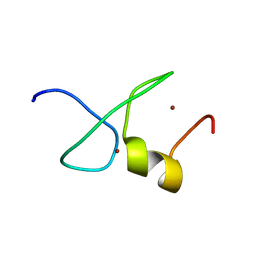 | | Solution structure of zinc finger domain 1 of human ZFAND1 | | 分子名称: | AN1-type zinc finger protein 1, ZINC ION | | 著者 | Fang, P.J, Lai, C.H, Ko, K.T, Chang, C.F, Hsu, S.T.D. | | 登録日 | 2022-06-27 | | 公開日 | 2023-06-28 | | 最終更新日 | 2024-05-15 | | 実験手法 | SOLUTION NMR | | 主引用文献 | Structural basis of p97 recognition by human ZFAND1
To Be Published
|
|
7CEM
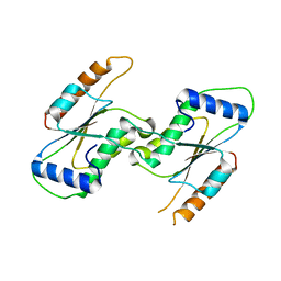 | | Crystal Structure of YbeA CP74 W7F | | 分子名称: | Ribosomal RNA large subunit methyltransferase H,Ribosomal RNA large subunit methyltransferase H | | 著者 | Liu, C.Y, Wu, C.Y, Lai, C.H, Hsu, S.T.D, Lyu, P.C. | | 登録日 | 2020-06-23 | | 公開日 | 2021-06-23 | | 最終更新日 | 2023-11-29 | | 実験手法 | X-RAY DIFFRACTION (2.4251 Å) | | 主引用文献 | Crystal Structure of YbeA CP74 W7F
To Be Published
|
|
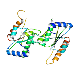
7CF7
 | |
7CFY
 | |
7CIU
 | |
7CIV
 | |
8JRM
 | |


