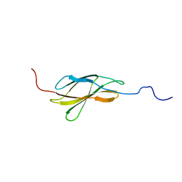
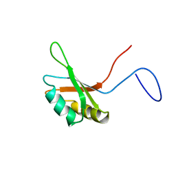
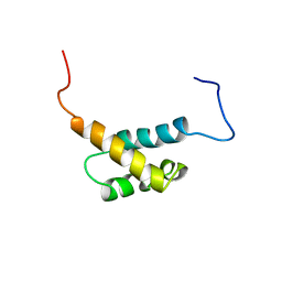
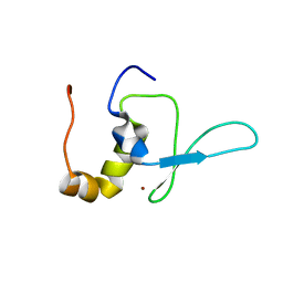
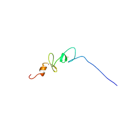
2DOA
 
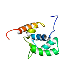 | | Solution structure of the helical domain in human Eleven-nineteen lysine-rich leukemia protein ELL | | 分子名称: | RNA polymerase II elongation factor ELL | | 著者 | Saito, K, Koshiba, S, Inoue, M, Kigawa, T, Yokoyama, S, RIKEN Structural Genomics/Proteomics Initiative (RSGI) | | 登録日 | 2006-04-28 | | 公開日 | 2007-05-22 | | 最終更新日 | 2024-05-29 | | 実験手法 | SOLUTION NMR | | 主引用文献 | Solution structure of the helical domain in human Eleven-nineteen lysine-rich leukemia protein ELL
To be Published
|
|
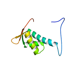
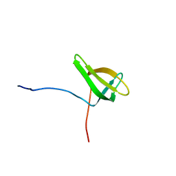
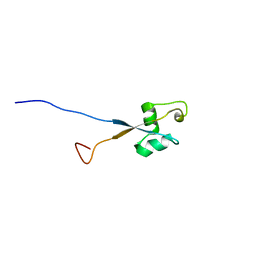
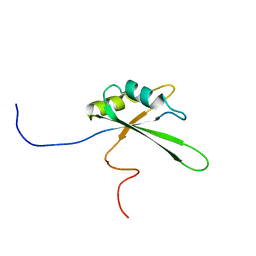
2DJQ
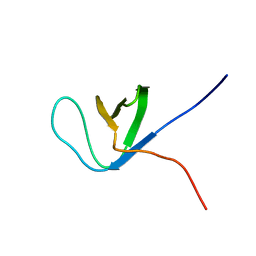 | | The solution structure of the first SH3 domain of mouse SH3 domain containing ring finger 2 | | 分子名称: | SH3 domain containing ring finger 2 | | 著者 | Sasagawa, A, Tochio, N, Koshiba, S, Inoue, M, Kigawa, T, Yokoyama, S, RIKEN Structural Genomics/Proteomics Initiative (RSGI) | | 登録日 | 2006-04-05 | | 公開日 | 2006-10-05 | | 最終更新日 | 2024-05-29 | | 実験手法 | SOLUTION NMR | | 主引用文献 | The solution structure of the first SH3 domain of mouse SH3 domain containing ring finger 2
To be Published
|
|
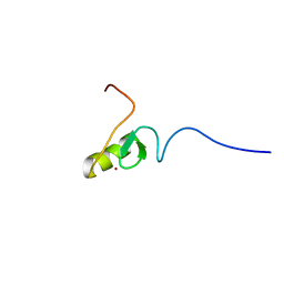
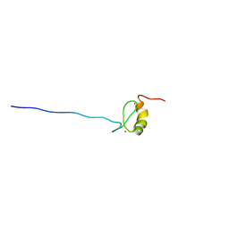
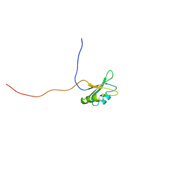
2DHJ
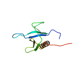 | | Solution structure of the PH domain of Rho GTPase activating protein 21 from human | | 分子名称: | Rho GTPase activating protein 21 | | 著者 | Li, H, Tochio, N, Koshiba, S, Inoue, M, Kigawa, T, Yokoyama, S, RIKEN Structural Genomics/Proteomics Initiative (RSGI) | | 登録日 | 2006-03-24 | | 公開日 | 2006-09-24 | | 最終更新日 | 2024-05-29 | | 実験手法 | SOLUTION NMR | | 主引用文献 | Solution structure of the PH domain of Rho GTPase activating protein 21 from human
To be Published
|
|
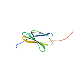
2DKX
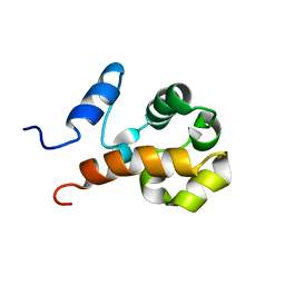 | | Solution structure of the SAM_PNT-domain of ETS transcription factor PDEF (Prostate ets) | | 分子名称: | SAM pointed domain-containing Ets transcription factor | | 著者 | Goroncy, A.K, Sato, M, Koshiba, S, Inoue, M, Kigawa, T, Yokoyama, S, RIKEN Structural Genomics/Proteomics Initiative (RSGI) | | 登録日 | 2006-04-14 | | 公開日 | 2006-10-14 | | 最終更新日 | 2024-05-29 | | 実験手法 | SOLUTION NMR | | 主引用文献 | Solution structure of the SAM_PNT-domain of ETS transcription factor PDEF (Prostate ets)
To be Published
|
|
2DOC
 | | Solution structure of the Fibronectin type-III domain of human Neural cell adhesion molecule 2 | | 分子名称: | Neural cell adhesion molecule 2 | | 著者 | Yoneyama, M, Tochio, N, Koshiba, S, Inoue, M, Kigawa, T, Yokoyama, S, RIKEN Structural Genomics/Proteomics Initiative (RSGI) | | 登録日 | 2006-04-28 | | 公開日 | 2006-10-28 | | 最終更新日 | 2024-05-29 | | 実験手法 | SOLUTION NMR | | 主引用文献 | Solution structure of the Fibronectin type-III domain of human Neural cell adhesion molecule 2
To be Published
|
|
2DO7
 | | Solution structure of the winged helix-turn-helix motif of human CUL-4B | | 分子名称: | Cullin-4B | | 著者 | Suzuki, S, Muto, Y, Inoue, M, Kigawa, T, Terada, T, Shirouzu, M, Yokoyama, S, RIKEN Structural Genomics/Proteomics Initiative (RSGI) | | 登録日 | 2006-04-27 | | 公開日 | 2007-04-17 | | 最終更新日 | 2024-05-29 | | 実験手法 | SOLUTION NMR | | 主引用文献 | Solution structure of the winged helix-turn-helix motif of human CUL-4B
To be Published
|
|
2DIF
 | | One sequence two fold ? : Miss fold of the zf-B-box domain from human tripartite motif protein 39 | | 分子名称: | Tripartite motif protein 39, ZINC ION | | 著者 | Tomizawa, T, Koshiba, S, Inoue, M, Kigawa, T, Yokoyama, S, RIKEN Structural Genomics/Proteomics Initiative (RSGI) | | 登録日 | 2006-03-29 | | 公開日 | 2006-09-29 | | 最終更新日 | 2024-05-29 | | 実験手法 | SOLUTION NMR | | 主引用文献 | One sequence two fold ? : Miss fold of the zf-B-box domain from human tripartite motif protein 39
To be Published
|
|
2DN7
 | | Solution structures of the 6th fn3 domain of human receptor-type tyrosine-protein phosphatase F | | 分子名称: | Receptor-type tyrosine-protein phosphatase F | | 著者 | Sato, M, Tochio, N, Koshiba, S, Inoue, M, Kigawa, T, Yokoyama, S, RIKEN Structural Genomics/Proteomics Initiative (RSGI) | | 登録日 | 2006-04-25 | | 公開日 | 2006-10-25 | | 最終更新日 | 2024-05-29 | | 実験手法 | SOLUTION NMR | | 主引用文献 | Solution structures of the 6th fn3 domain of human receptor-type tyrosine-protein phosphatase F
To be Published
|
|
2DO4
 | | Solution structure of the RNA binding domain of squamous cell carcinoma antigen recognized by T cells 3 | | 分子名称: | Squamous cell carcinoma antigen recognized by T-cells 3 | | 著者 | Tanabe, W, Suzuki, S, Muto, Y, Inoue, M, Kigawa, T, Terada, T, Shirouzu, M, Yokoyama, S, RIKEN Structural Genomics/Proteomics Initiative (RSGI) | | 登録日 | 2006-04-27 | | 公開日 | 2007-04-17 | | 最終更新日 | 2024-05-29 | | 実験手法 | SOLUTION NMR | | 主引用文献 | Solution structure of the RNA binding domain of squamous cell carcinoma antigen recognized by T cells 3
To be Published
|
|
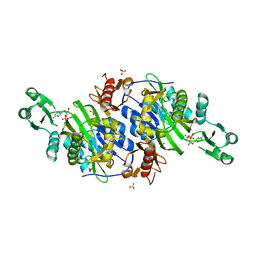
2DEJ
 | | Crystal Structure of galaktokinase from Pyrococcus horikoshii with AMP-PN and galactose | | 分子名称: | Probable galactokinase, alpha-D-galactopyranose, {5'-O-[(R)-{[(S)-AMINO(HYDROXY-KAPPAO)PHOSPHORYL]OXY}(HYDROXY-KAPPAO)PHOSPHORYL]ADENOSINATO(2-)}MAGNESIUM | | 著者 | Inagaki, E, Sakamoto, K, Shinkai, A, Yokoyama, S, RIKEN Structural Genomics/Proteomics Initiative (RSGI) | | 登録日 | 2006-02-10 | | 公開日 | 2007-05-01 | | 最終更新日 | 2023-10-25 | | 実験手法 | X-RAY DIFFRACTION (1.5 Å) | | 主引用文献 | Crystal Structure of galaktokinase from Pyrococcus horikoshii
To be Published
|
|
2DIG
 | | Solusion structure of the Todor domain of human Lamin-B receptor | | 分子名称: | Lamin-B receptor | | 著者 | Yoneyama, M, Tochio, N, Koshiba, S, Inoue, M, Kigawa, T, Yokoyama, S, RIKEN Structural Genomics/Proteomics Initiative (RSGI) | | 登録日 | 2006-03-30 | | 公開日 | 2006-09-30 | | 最終更新日 | 2024-05-29 | | 実験手法 | SOLUTION NMR | | 主引用文献 | Solusion structure of the Todor domain of human Lamin-B receptor
To be Published
|
|
2DJ8
 | | Solution Structure of zf-MYND Domain of Protein CBFA2TI (Protein MTG8) | | 分子名称: | Protein CBFA2T1, ZINC ION | | 著者 | Niraula, T.N, Sasagawa, A, Koshiba, S, Inoue, M, Kigawa, T, Yokoyama, S, RIKEN Structural Genomics/Proteomics Initiative (RSGI) | | 登録日 | 2006-03-31 | | 公開日 | 2006-10-01 | | 最終更新日 | 2024-05-29 | | 実験手法 | SOLUTION NMR | | 主引用文献 | Solution Structure of zf-MYND Domain of Protein CBFA2TI (Protein MTG8)
To be published
|
|
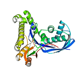
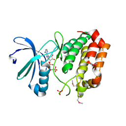
2DUC
 | | Crystal structure of SARS coronavirus main proteinase(3CLPRO) | | 分子名称: | Replicase polyprotein 1ab | | 著者 | Wang, H, Kim, Y.T, Muramatsu, T, Takemoto, C, Shirouzu, M, Yokoyama, S, RIKEN Structural Genomics/Proteomics Initiative (RSGI) | | 登録日 | 2006-07-21 | | 公開日 | 2007-07-24 | | 最終更新日 | 2023-10-25 | | 実験手法 | X-RAY DIFFRACTION (1.7 Å) | | 主引用文献 | SARS-CoV 3CL protease cleaves its C-terminal autoprocessing site by novel subsite cooperativity
Proc. Natl. Acad. Sci. U.S.A., 113, 2016
|
|
2DOD
 | | Solution structure of the first FF domain of human transcription factor CA150 | | 分子名称: | Transcription elongation regulator 1 | | 著者 | Suzuki, S, Muto, Y, Inoue, M, Kigawa, T, Terada, T, Shirouzu, M, Yokoyama, S, RIKEN Structural Genomics/Proteomics Initiative (RSGI) | | 登録日 | 2006-04-28 | | 公開日 | 2006-10-28 | | 最終更新日 | 2024-05-29 | | 実験手法 | SOLUTION NMR | | 主引用文献 | Solution structure of the first FF domain of human transcription factor CA150
To be Published
|
|
2DWK
 | | Crystal structure of the RUN domain of mouse Rap2 interacting protein x | | 分子名称: | Protein RUFY3 | | 著者 | Kukimoto-Niino, M, Murayama, K, Shirouzu, M, Yokoyama, S, RIKEN Structural Genomics/Proteomics Initiative (RSGI) | | 登録日 | 2006-08-15 | | 公開日 | 2006-08-29 | | 最終更新日 | 2011-07-13 | | 実験手法 | X-RAY DIFFRACTION (2 Å) | | 主引用文献 | Crystal Structure of the RUN Domain of the RAP2-interacting Protein x
J.Biol.Chem., 281, 2006
|
|
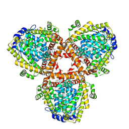
2DWB
 | | Aurora-A kinase complexed with AMPPNP | | 分子名称: | PHOSPHOAMINOPHOSPHONIC ACID-ADENYLATE ESTER, SULFATE ION, Serine/threonine-protein kinase 6 | | 著者 | Kukimoto-Niino, M, Murayama, K, Shirouzu, S, Yokoyama, S, RIKEN Structural Genomics/Proteomics Initiative (RSGI) | | 登録日 | 2006-08-10 | | 公開日 | 2007-07-31 | | 最終更新日 | 2023-11-15 | | 実験手法 | X-RAY DIFFRACTION (2.5 Å) | | 主引用文献 | Aurora-A kinase complexed with AMPPNP
To be Published
|
|
2DHM
 | |
2DJR
 | | Solution structures of the C2H2 type zinc finger domain of human zinc finger BED domain containing protein 2 | | 分子名称: | ZINC ION, Zinc finger BED domain-containing protein 2 | | 著者 | Sato, M, Sasagawa, A, Tochio, N, Koshiba, S, Inoue, M, Kigawa, T, Yokoyama, S, RIKEN Structural Genomics/Proteomics Initiative (RSGI) | | 登録日 | 2006-04-05 | | 公開日 | 2006-10-05 | | 最終更新日 | 2024-05-29 | | 実験手法 | SOLUTION NMR | | 主引用文献 | Solution structures of the C2H2 type zinc finger domain of human zinc finger BED domain containing protein 2
To be Published
|
|
2DJP
 | | The solution structure of the LysM domain of human hypothetical protein SB145 | | 分子名称: | Hypothetical protein SB145 | | 著者 | Sasagawa, A, Tochio, N, Saito, K, Koshiba, S, Inoue, M, Kigawa, T, Yokoyama, S, RIKEN Structural Genomics/Proteomics Initiative (RSGI) | | 登録日 | 2006-04-05 | | 公開日 | 2006-10-05 | | 最終更新日 | 2024-05-29 | | 実験手法 | SOLUTION NMR | | 主引用文献 | The solution structure of the LysM domain of human hypothetical protein SB145
To be Published
|
|
2DO0
 | | Solution structure of the RNA binding domain of heterogeneous nuclear ribonucleoprotein M | | 分子名称: | Heterogeneous nuclear ribonucleoprotein M | | 著者 | Suzuki, S, Muto, Y, Inoue, M, Kigawa, T, Terada, T, Shirouzu, M, Yokoyama, S, RIKEN Structural Genomics/Proteomics Initiative (RSGI) | | 登録日 | 2006-04-27 | | 公開日 | 2006-10-27 | | 最終更新日 | 2024-05-29 | | 実験手法 | SOLUTION NMR | | 主引用文献 | Solution structure of the RNA binding domain of heterogeneous nuclear ribonucleoprotein M
To be Published
|
|
2DWC
 | | Crystal structure of Probable phosphoribosylglycinamide formyl transferase from Pyrococcus horikoshii OT3 complexed with ADP | | 分子名称: | 433aa long hypothetical phosphoribosylglycinamide formyl transferase, ADENOSINE-5'-DIPHOSPHATE, SULFATE ION | | 著者 | Yoshikawa, S, Arai, R, Kamo-Uchikubo, T, Shirouzu, M, Yokoyama, S, RIKEN Structural Genomics/Proteomics Initiative (RSGI) | | 登録日 | 2006-08-10 | | 公開日 | 2007-02-10 | | 最終更新日 | 2023-10-25 | | 実験手法 | X-RAY DIFFRACTION (1.7 Å) | | 主引用文献 | Crystal structure of Probable phosphoribosylglycinamide formyl transferase from Pyrococcus horikoshii OT3 complexed with ADP
To be Published
|
|
2DJ7
 | | Solution Structure of 3rd LIM Domain of Actin-binding LIM Protein 3 | | 分子名称: | Actin-binding LIM protein 3, ZINC ION | | 著者 | Niraula, T.N, Sasagawa, A, Koshiba, S, Inoue, M, Kigawa, T, Yokoyama, S, RIKEN Structural Genomics/Proteomics Initiative (RSGI) | | 登録日 | 2006-03-31 | | 公開日 | 2006-10-01 | | 最終更新日 | 2024-05-29 | | 実験手法 | SOLUTION NMR | | 主引用文献 | Solution Structure of 3rd LIM Domain of Actin-binding LIM Protein 3
To be Published
|
|
2DL0
 | | Solution structure of the SAM-domain of the SAM and SH3 domain containing protein 1 | | 分子名称: | SAM and SH3 domain-containing protein 1 | | 著者 | Goroncy, A.K, Sato, M, Koshiba, S, Inoue, M, Kigawa, T, Yokoyama, S, RIKEN Structural Genomics/Proteomics Initiative (RSGI) | | 登録日 | 2006-04-14 | | 公開日 | 2006-10-14 | | 最終更新日 | 2024-05-29 | | 実験手法 | SOLUTION NMR | | 主引用文献 | Solution structure of the SAM-domain of the SAM and SH3 domain containing protein 1
To be Published
|
|
2DNY
 | | Solution structure of the third RNA binding domain of FBP-interacting repressor, SIAHBP1 | | 分子名称: | Fuse-binding protein-interacting repressor, isoform b | | 著者 | Suzuki, S, Nagata, T, Muto, Y, Inoue, M, Kigawa, T, Terada, T, Shirouzu, M, Yokoyama, S, RIKEN Structural Genomics/Proteomics Initiative (RSGI) | | 登録日 | 2006-04-27 | | 公開日 | 2007-04-17 | | 最終更新日 | 2024-05-29 | | 実験手法 | SOLUTION NMR | | 主引用文献 | Solution structure of the third RNA binding domain of FBP-interacting repressor, SIAHBP1
To be Published
|
|
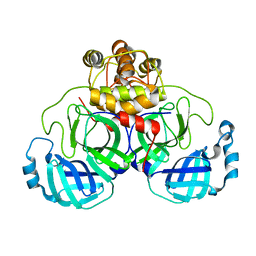
2DQB
 | | Crystal structure of dNTP triphosphohydrolase from Thermus thermophilus HB8, which is homologous to dGTP triphosphohydrolase | | 分子名称: | Deoxyguanosinetriphosphate triphosphohydrolase, putative, MAGNESIUM ION | | 著者 | Kondo, N, Nakagawa, N, Ebihara, A, Chen, L, Liu, Z.-J, Wang, B.-C, Yokoyama, S, Kuramitsu, S, Masui, R, RIKEN Structural Genomics/Proteomics Initiative (RSGI) | | 登録日 | 2006-05-25 | | 公開日 | 2007-01-23 | | 最終更新日 | 2021-11-10 | | 実験手法 | X-RAY DIFFRACTION (2.2 Å) | | 主引用文献 | Structure of dNTP-inducible dNTP triphosphohydrolase: insight into broad specificity for dNTPs and triphosphohydrolase-type hydrolysis
ACTA CRYSTALLOGR.,SECT.D, 63, 2007
|
|


