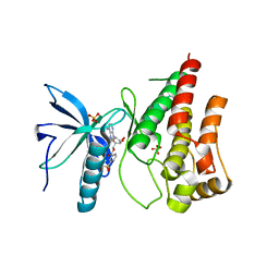
1JR8
 
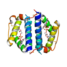 | | Crystal Structure of Erv2p | | 分子名称: | Erv2 PROTEIN, mitochondrial, FLAVIN-ADENINE DINUCLEOTIDE | | 著者 | Gross, E, Sevier, C.S, Vala, A, Kaiser, C.A, Fass, D. | | 登録日 | 2001-08-13 | | 公開日 | 2001-12-28 | | 最終更新日 | 2011-07-13 | | 実験手法 | X-RAY DIFFRACTION (1.5 Å) | | 主引用文献 | A new FAD-binding fold and intersubunit disulfide shuttle in the thiol oxidase Erv2p.
Nat.Struct.Biol., 9, 2002
|
|
6T1R
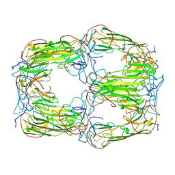 | | Pseudo-atomic model of a 16-mer assembly of reduced recombinant human alphaA-crystallin (non domain swapped configuration) | | 分子名称: | Alpha-crystallin A chain | | 著者 | Peters, C, Kaiser, C.J.O, Weinkauf, S, Zacharias, M, Buchner, J. | | 登録日 | 2019-10-05 | | 公開日 | 2019-12-11 | | 最終更新日 | 2024-05-15 | | 実験手法 | ELECTRON MICROSCOPY (9.8 Å) | | 主引用文献 | The structure and oxidation of the eye lens chaperone alpha A-crystallin.
Nat.Struct.Mol.Biol., 26, 2019
|
|
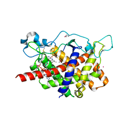
1JRA
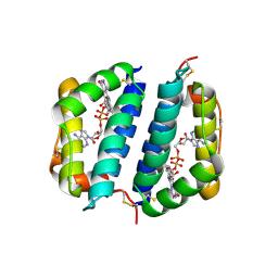 | | Crystal Structure of Erv2p | | 分子名称: | ERV2 PROTEIN, MITOCHONDRIAL, FLAVIN-ADENINE DINUCLEOTIDE | | 著者 | Gross, E, Sevier, C.S, Vala, A, Kaiser, C.A, Fass, D. | | 登録日 | 2001-08-13 | | 公開日 | 2001-12-28 | | 最終更新日 | 2023-08-16 | | 実験手法 | X-RAY DIFFRACTION (2 Å) | | 主引用文献 | A new FAD-binding fold and intersubunit disulfide shuttle in the thiol oxidase Erv2p.
Nat.Struct.Biol., 9, 2002
|
|
5O4A
 | | Human FGF in complex with a covalent inhibitor | | 分子名称: | Fibroblast growth factor receptor 1, GLYCEROL, SULFATE ION, ... | | 著者 | Debreczeni, J, Breed, J, Mukherjee, H, Aquila, B, Kaiser, C, Tentarelli, S, Whitty, A, Grimster, N. | | 登録日 | 2017-05-26 | | 公開日 | 2018-02-07 | | 実験手法 | X-RAY DIFFRACTION (2.01 Å) | | 主引用文献 | A study of the reactivity of S(VI)-F containing warheads with nucleophilic amino-acid side chains under physiological conditions.
Org. Biomol. Chem., 15, 2017
|
|
5O49
 | | Human FGF in complex with a covalent inhibitor | | 分子名称: | Fibroblast growth factor receptor 1, SULFATE ION, [(2~{R},3~{S},4~{R},5~{R})-5-(6-aminopurin-9-yl)-3,4-bis(oxidanyl)oxolan-2-yl]methyl 3-fluorosulfonylbenzoate | | 著者 | Debreczeni, J, Breed, J, Mukherjee, H, Aquila, B, Kaiser, C, Tentarelli, S, Whitty, A, Grimster, N. | | 登録日 | 2017-05-26 | | 公開日 | 2018-02-07 | | 実験手法 | X-RAY DIFFRACTION (1.91 Å) | | 主引用文献 | A study of the reactivity of S(VI)-F containing warheads with nucleophilic amino-acid side chains under physiological conditions.
Org. Biomol. Chem., 15, 2017
|
|
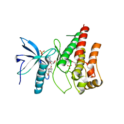
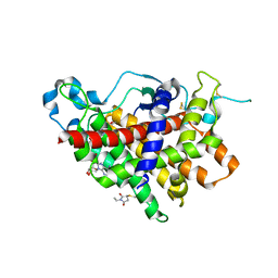
1RP4
 | | Structure of Ero1p, Source of Disulfide Bonds for Oxidative Protein Folding in the Cell | | 分子名称: | 1-ETHYL-PYRROLIDINE-2,5-DIONE, CADMIUM ION, FLAVIN-ADENINE DINUCLEOTIDE, ... | | 著者 | Gross, E, Kastner, D.B, Kaiser, C.A, Fass, D. | | 登録日 | 2003-12-03 | | 公開日 | 2004-06-08 | | 最終更新日 | 2018-01-31 | | 実験手法 | X-RAY DIFFRACTION (2.2 Å) | | 主引用文献 | Structure of ero1p, source of disulfide bonds for oxidative protein folding in the cell.
Cell(Cambridge,Mass.), 117, 2004
|
|
1RQ1
 | | Structure of Ero1p, Source of Disulfide Bonds for Oxidative Protein Folding in the Cell | | 分子名称: | 1-ETHYL-PYRROLIDINE-2,5-DIONE, CADMIUM ION, FLAVIN-ADENINE DINUCLEOTIDE, ... | | 著者 | Gross, E, Kastner, D.B, Kaiser, C.A, Fass, D. | | 登録日 | 2003-12-04 | | 公開日 | 2004-06-08 | | 最終更新日 | 2011-07-13 | | 実験手法 | X-RAY DIFFRACTION (2.8 Å) | | 主引用文献 | Structure of ero1p, source of disulfide bonds for oxidative protein folding in the cell.
Cell(Cambridge,Mass.), 117, 2004
|
|


