Loading
PDBj
MenuPDBj@FacebookPDBj@X(formerly Twitter)PDBj@BlueSkyPDBj@YouTubewwPDB FoundationwwPDB
RCSB PDBPDBeBMRBAdv. SearchSearch help
PDB: 10 results

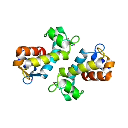
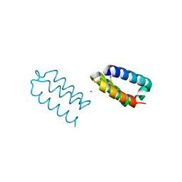
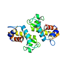
8FNR
DownloadVisualize
BU of 8fnr by Molmil
X-ray crystal structure of Hansschlegelia quercus lanmodulin (LanM) with dysprosium (III) bound at pH 7
Descriptor: DYSPROSIUM ION, EF-hand domain-containing protein
Authors:Jung, J.J, Lin, C.-Y, Boal, A.K.
Deposit date:2022-12-28
Release date:2023-06-07
Last modified:2024-05-22
Method:X-RAY DIFFRACTION (1.4 Å)
Cite:Enhanced rare-earth separation with a metal-sensitive lanmodulin dimer.
Nature, 618, 2023
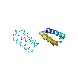
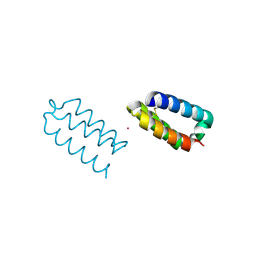
8FNS
DownloadVisualize
BU of 8fns by Molmil
X-ray crystal structure of Methylorubrum extorquens AM1 lanmodulin (LanM) with neodymium (III) bound at pH 7
Descriptor: EF-hand domain-containing protein, Neodymium Ion
Authors:Jung, J.J, Lin, C.-Y, Boal, A.K.
Deposit date:2022-12-28
Release date:2023-06-07
Last modified:2024-05-22
Method:X-RAY DIFFRACTION (1.01 Å)
Cite:Enhanced rare-earth separation with a metal-sensitive lanmodulin dimer.
Nature, 618, 2023
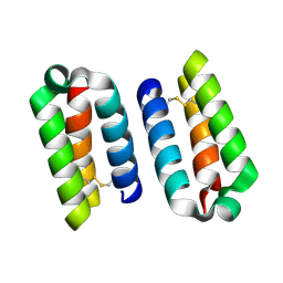
8DQ2
DownloadVisualize
BU of 8dq2 by Molmil
X-ray crystal structure of Hansschlegelia quercus lanmodulin (LanM) with lanthanum (III) bound at pH 7
Descriptor: CITRIC ACID, EF-hand domain-containing protein, LANTHANUM (III) ION, ...
Authors:Jung, J.J, Lin, C.-Y, Boal, A.K.
Deposit date:2022-07-18
Release date:2023-06-07
Last modified:2024-05-22
Method:X-RAY DIFFRACTION (1.8 Å)
Cite:Enhanced rare-earth separation with a metal-sensitive lanmodulin dimer.
Nature, 618, 2023
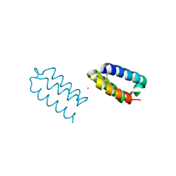
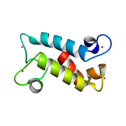
9C8X
DownloadVisualize
BU of 9c8x by Molmil
X-ray crystal structure of Methylorubrum extorquens La(III)-bound LanD
Descriptor: LANTHANUM (III) ION, landiscernin
Authors:Jung, J.J, Lin, C.-Y, Boal, A.K.
Deposit date:2024-06-13
Release date:2024-10-16
Last modified:2024-11-13
Method:X-RAY DIFFRACTION (1.9 Å)
Cite:Modulating metal-centered dimerization of a lanthanide chaperone protein for separation of light lanthanides.
Proc.Natl.Acad.Sci.USA, 121, 2024
9C8W
DownloadVisualize
BU of 9c8w by Molmil
X-ray crystal structure of Methylorubrum extorquens apo LanD
Descriptor: landiscernin
Authors:Jung, J.J, Lin, C.-Y, Boal, A.K.
Deposit date:2024-06-13
Release date:2024-10-16
Last modified:2024-11-13
Method:X-RAY DIFFRACTION (1.65 Å)
Cite:Modulating metal-centered dimerization of a lanthanide chaperone protein for separation of light lanthanides.
Proc.Natl.Acad.Sci.USA, 121, 2024
9C90
DownloadVisualize
BU of 9c90 by Molmil
X-ray crystal structure of Methylorubrum extorquens Ho(III)-bound LanD
Descriptor: HOLMIUM (III) ATOM, landiscernin
Authors:Jung, J.J, Lin, C.-Y, Boal, A.K.
Deposit date:2024-06-13
Release date:2024-10-16
Last modified:2024-11-13
Method:X-RAY DIFFRACTION (1.38 Å)
Cite:Modulating metal-centered dimerization of a lanthanide chaperone protein for separation of light lanthanides.
Proc.Natl.Acad.Sci.USA, 121, 2024
9C8Y
DownloadVisualize
BU of 9c8y by Molmil
X-ray crystal structure of Methylorubrum extorquens Ce(III)-bound LanD
Descriptor: CERIUM (III) ION, landiscernin
Authors:Jung, J.J, Lin, C.-Y, Boal, A.K.
Deposit date:2024-06-13
Release date:2024-10-16
Last modified:2024-11-13
Method:X-RAY DIFFRACTION (1.8 Å)
Cite:Modulating metal-centered dimerization of a lanthanide chaperone protein for separation of light lanthanides.
Proc.Natl.Acad.Sci.USA, 121, 2024
9C8Z
DownloadVisualize
BU of 9c8z by Molmil
X-ray crystal structure of Methylorubrum extorquens Eu(III)-bound LanD
Descriptor: EUROPIUM ION, landiscernin
Authors:Jung, J.J, Lin, C.-Y, Boal, A.K.
Deposit date:2024-06-13
Release date:2024-10-16
Last modified:2024-11-13
Method:X-RAY DIFFRACTION (1.41 Å)
Cite:Modulating metal-centered dimerization of a lanthanide chaperone protein for separation of light lanthanides.
Proc.Natl.Acad.Sci.USA, 121, 2024
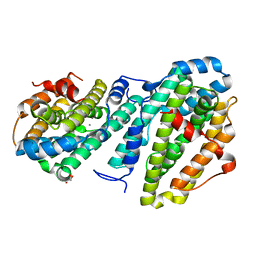
8DQ4
DownloadVisualize
BU of 8dq4 by Molmil
X-ray crystal structure of Flavobacterium johnsoniae dimanganese(II) class Id ribonucleotide reductase beta subunit K71R variant
Descriptor: GLYCEROL, MANGANESE (II) ION, Ribonucleoside-diphosphate reductase
Authors:Rose, H.R, Maggiolo, A.O, Jung, J.J, Boal, A.K.
Deposit date:2022-07-18
Release date:2023-07-26
Last modified:2024-05-22
Method:X-RAY DIFFRACTION (2.35 Å)
Cite:X-ray crystal structure of Flavobacterium johnsoniae dimanganese(II) class Id ribonucleotide reductase beta subunit K71R variant
To Be Published
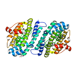
8DQ3
DownloadVisualize
BU of 8dq3 by Molmil
X-ray crystal structure of Aggregatibacter actinomycetemcomitans dimanganese(II) class Id ribonucleotide reductase beta subunit
Descriptor: 1,2-ETHANEDIOL, FORMIC ACID, MAGNESIUM ION, ...
Authors:Rose, H.R, Jung, J.J, Boal, A.K.
Deposit date:2022-07-18
Release date:2023-07-26
Last modified:2024-05-22
Method:X-RAY DIFFRACTION (1.67 Å)
Cite:X-ray crystal structure of Aggregatibacter actinomycetemcomitans dimanganese(II) class Id ribonucleotide reductase beta subunit
To Be Published

227344

数据于2024-11-13公开中

PDB statisticsPDBj update infoContact PDBjnumon