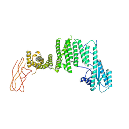
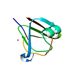
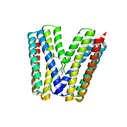
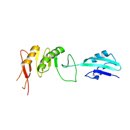
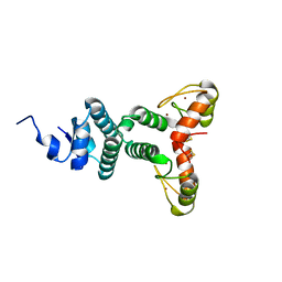
7B6E
 
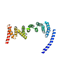 | | Drosophila melanogaster TRAPP C8 subunits region 355 to 661 | | 分子名称: | FI18195p1 | | 著者 | Galindo, A, Munro, S, Planelles-Herrero, V.J, Degliesposti, G. | | 登録日 | 2020-12-07 | | 公開日 | 2021-06-09 | | 最終更新日 | 2024-05-01 | | 実験手法 | ELECTRON MICROSCOPY (4.5 Å) | | 主引用文献 | Cryo-EM structure of metazoan TRAPPIII, the multi-subunit complex that activates the GTPase Rab1.
Embo J., 40, 2021
|
|
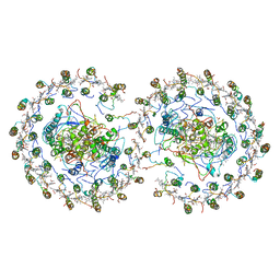
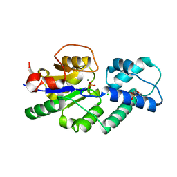
7B6R
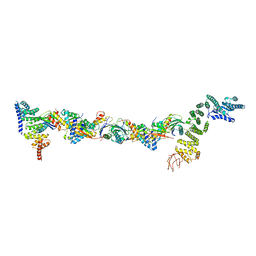 | | Drosophila melanogaster TRAPPIII partial complex: core plus C8 and C11 attached region | | 分子名称: | FI18195p1, GEO08327p1, Probable trafficking protein particle complex subunit 2, ... | | 著者 | Galindo, A, Munro, S, Planelles-Herrero, V.J, Degliesposti, G. | | 登録日 | 2020-12-08 | | 公開日 | 2021-06-09 | | 最終更新日 | 2021-07-28 | | 実験手法 | ELECTRON MICROSCOPY (5.8 Å) | | 主引用文献 | Cryo-EM structure of metazoan TRAPPIII, the multi-subunit complex that activates the GTPase Rab1.
Embo J., 40, 2021
|
|
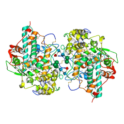
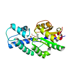
7B70
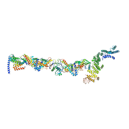 | | TRAPPCore plus C8 (355-596) and C11 (1-718) from MiniTRAPPIII | | 分子名称: | FI18195p1, GEO08327p1, Probable trafficking protein particle complex subunit 2, ... | | 著者 | Galindo, A, Munro, S, Planelles-Herrero, V.J. | | 登録日 | 2020-12-09 | | 公開日 | 2021-06-09 | | 最終更新日 | 2024-05-01 | | 実験手法 | ELECTRON MICROSCOPY (4 Å) | | 主引用文献 | Cryo-EM structure of metazoan TRAPPIII, the multi-subunit complex that activates the GTPase Rab1.
Embo J., 40, 2021
|
|
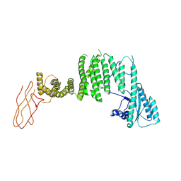
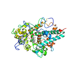
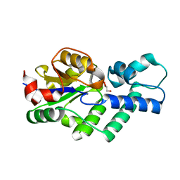
7B6D
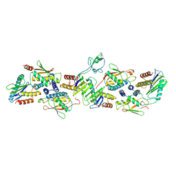 | | Drosophila melanogaster TRAPPCore (C1, C2, C2L, C3a/b, C4, C5, C6 subunits) | | 分子名称: | GEO08327p1, Probable trafficking protein particle complex subunit 2, TRAPPC2L, ... | | 著者 | Galindo, A, Munro, S, Planelles-Herrero, V.J, Degliesposti, G. | | 登録日 | 2020-12-07 | | 公開日 | 2021-06-09 | | 最終更新日 | 2024-05-01 | | 実験手法 | ELECTRON MICROSCOPY (4.27 Å) | | 主引用文献 | Cryo-EM structure of metazoan TRAPPIII, the multi-subunit complex that activates the GTPase Rab1.
Embo J., 40, 2021
|
|
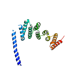
7B6H
 | | Drosophila melanogaster TRAPP C11 subunits region 1 to 718 | | 分子名称: | Trafficking protein particle complex subunit 11 | | 著者 | Galindo, A, Munro, S, Planelles-Herrero, V.J, Degliesposti, G. | | 登録日 | 2020-12-07 | | 公開日 | 2021-06-09 | | 最終更新日 | 2024-05-01 | | 実験手法 | ELECTRON MICROSCOPY (5.4 Å) | | 主引用文献 | Cryo-EM structure of metazoan TRAPPIII, the multi-subunit complex that activates the GTPase Rab1.
Embo J., 40, 2021
|
|
7B6X
 | |
7B6Y
 | |
7B6Z
 | |
5HT8
 | | Crystal structure of clostrillin double mutant (S17H,S19H) in complex with nickel | | 分子名称: | Beta and gamma crystallin, NICKEL (II) ION | | 著者 | Jamkhindikar, A, Srivastava, S.S, Sankaranarayanan, R. | | 登録日 | 2016-01-26 | | 公開日 | 2017-02-01 | | 最終更新日 | 2023-11-08 | | 実験手法 | X-RAY DIFFRACTION (2.01 Å) | | 主引用文献 | A Transition Metal-Binding, Trimeric beta gamma-Crystallin from Methane-Producing Thermophilic Archaea, Methanosaeta thermophila
Biochemistry, 56, 2017
|
|
5HT9
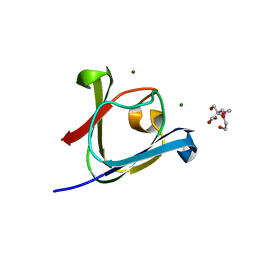 | |
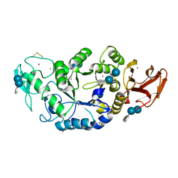
5EE5
 | | Structure of human ARL1 in complex with the DCB domain of BIG1 | | 分子名称: | ACETATE ION, ADP-ribosylation factor-like protein 1, Brefeldin A-inhibited guanine nucleotide-exchange protein 1, ... | | 著者 | Galindo, A, Soler, N, Munro, S. | | 登録日 | 2015-10-22 | | 公開日 | 2016-07-06 | | 最終更新日 | 2016-07-27 | | 実験手法 | X-RAY DIFFRACTION (2.279 Å) | | 主引用文献 | Structural Insights into Arl1-Mediated Targeting of the Arf-GEF BIG1 to the trans-Golgi.
Cell Rep, 16, 2016
|
|
5FGO
 | |
2GS4
 | | The crystal structure of the E.coli stress protein YciF. | | 分子名称: | Protein yciF | | 著者 | Hindupur, A, Liu, D, Zhao, Y, Bellamy, H.D, White, M.A, Fox, R.O. | | 登録日 | 2006-04-25 | | 公開日 | 2006-10-17 | | 最終更新日 | 2011-07-13 | | 実験手法 | X-RAY DIFFRACTION (2 Å) | | 主引用文献 | The crystal structure of the E. coli stress protein YciF.
Protein Sci., 15, 2006
|
|
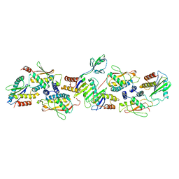
1MXD
 | | Structure of a (Ca,Zn)-dependent alpha-amylase from the hyperthermophilic archaeon Pyrococcus woesei | | 分子名称: | 4,6-dideoxy-4-{[(1S,4R,5S,6S)-4,5,6-trihydroxy-3-(hydroxymethyl)cyclohex-2-en-1-yl]amino}-alpha-D-glucopyranose-(1-4)-alpha-D-glucopyranose-(1-4)-alpha-D-glucopyranose, ACETATE ION, CALCIUM ION, ... | | 著者 | Linden, A, Mayans, O, Meyer-Klaucke, W, Antranikian, G, Wilmanns, M. | | 登録日 | 2002-10-02 | | 公開日 | 2003-06-10 | | 最終更新日 | 2020-07-29 | | 実験手法 | X-RAY DIFFRACTION (2 Å) | | 主引用文献 | Differential Regulation of a Hyperthermophilic alpha-Amylase with a Novel (Ca,Zn) Two-metal Center by Zinc
J.Biol.Chem., 278, 2003
|
|
1MXG
 | | Crystal Structure of a (Ca,Zn)-dependent alpha-amylase from the hyperthermophilic archaeon Pyrococcus woesei in complex with acarbose | | 分子名称: | 2-AMINO-2-HYDROXYMETHYL-PROPANE-1,3-DIOL, 2-{2-[2-2-(METHOXY-ETHOXY)-ETHOXY]-ETHOXY}-ETHANOL, 4,6-dideoxy-4-{[(1S,4R,5S,6S)-4,5,6-trihydroxy-3-(hydroxymethyl)cyclohex-2-en-1-yl]amino}-alpha-D-glucopyranose-(1-4)-alpha-D-glucopyranose-(1-4)-alpha-D-glucopyranose, ... | | 著者 | Linden, A, Mayans, O, Meyer-Klaucke, W, Antranikian, G, Wilmanns, M. | | 登録日 | 2002-10-02 | | 公開日 | 2003-06-10 | | 最終更新日 | 2020-09-09 | | 実験手法 | X-RAY DIFFRACTION (1.6 Å) | | 主引用文献 | Differential Regulation of a Hyperthermophilic alpha-Amylase with a Novel (Ca,Zn) Two-metal Center by Zinc
J.Biol.Chem., 278, 2003
|
|
1MWO
 | | Crystal Structure Analysis of the Hyperthermostable Pyrocoocus woesei alpha-amylase | | 分子名称: | CALCIUM ION, ZINC ION, alpha amylase | | 著者 | Linden, A, Mayans, O, Meyer-Klaucke, W, Antranikian, G, Wilmanns, M. | | 登録日 | 2002-09-30 | | 公開日 | 2003-06-10 | | 最終更新日 | 2011-07-13 | | 実験手法 | X-RAY DIFFRACTION (2.2 Å) | | 主引用文献 | Differential Regulation of a Hyperthermophilic alpha-Amylase with a Novel (Ca,Zn) Two-metal Center by Zinc
J.Biol.Chem., 278, 2003
|
|
4BQL
 | | Crystal structure of archaeal actin | | 分子名称: | ACTIN/ACTIN FAMILY PROTEIN, ADENOSINE-5'-DIPHOSPHATE, MAGNESIUM ION | | 著者 | Lindaas, A.-C, Chruszsz, M, Bernander, R, Valegard, K. | | 登録日 | 2013-05-31 | | 公開日 | 2014-02-12 | | 最終更新日 | 2024-05-08 | | 実験手法 | X-RAY DIFFRACTION (3.34 Å) | | 主引用文献 | Structure of Crenactin, an Archaeal Actin Homologue Active at 90Degc.
Acta Crystallogr.,Sect.D, 70, 2014
|
|
2VH2
 | | Crystal structure of cell divison protein FtsQ from Yersinia enterecolitica | | 分子名称: | CELL DIVISION PROTEIN FTSQ | | 著者 | van den Ent, F, Vinkenvleugel, T, Ind, A, West, P, Veprintsev, D, Nanninga, N, den Blaauwen, T, Lowe, J. | | 登録日 | 2007-11-16 | | 公開日 | 2008-03-11 | | 最終更新日 | 2023-12-13 | | 実験手法 | X-RAY DIFFRACTION (3.4 Å) | | 主引用文献 | Structural and Mutational Analysis of Cell Division Protein Ftsq
Mol.Microbiol., 68, 2008
|
|
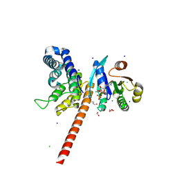
4V9G
 | | RC-LH1-PufX dimer complex from Rhodobacter sphaeroides | | 分子名称: | BACTERIOCHLOROPHYLL A, BACTERIOPHEOPHYTIN A, FE (II) ION, ... | | 著者 | Qian, P, Papiz, M.Z, Jackson, P.J, Brindley, A.A, Ng, I.W, Olsen, J.D, Dickman, M.J, Bullough, P.A, Hunter, C.N. | | 登録日 | 2013-02-21 | | 公開日 | 2014-07-09 | | 最終更新日 | 2024-02-28 | | 実験手法 | X-RAY DIFFRACTION (7.78 Å) | | 主引用文献 | Three-Dimensional Structure of the Rhodobacter sphaeroides RC-LH1-PufX Complex: Dimerization and Quinone Channels Promoted by PufX.
Biochemistry, 52, 2013
|
|
3ZS0
 | | Human Myeloperoxidase inactivated by TX2 | | 分子名称: | 2-acetamido-2-deoxy-beta-D-glucopyranose, 3-(4-FLUOROBENZYL)-2-THIOXO-1,2,3,7-TETRAHYDRO-6H-PURIN-6-ONE, ACETATE ION, ... | | 著者 | Tiden, A.K, Sjogren, T, Svensson, M, Bernlind, A, Senthilmohan, R, Auchere, F, Norman, H, Markgren, P.O, Gustavsson, S, Schmidt, S, Lundquist, S, Forbes, L.V, Magon, N.J, Jameson, G.N, Eriksson, H, Kettle, A.J. | | 登録日 | 2011-06-21 | | 公開日 | 2011-08-31 | | 最終更新日 | 2023-12-20 | | 実験手法 | X-RAY DIFFRACTION (2.3 Å) | | 主引用文献 | 2-Thioxanthines are Mechanism-Based Inactivators of Myeloperoxidase that Block Oxidative Stress During Inflammation.
J.Biol.Chem., 286, 2011
|
|
3ZS1
 | | Human Myeloperoxidase inactivated by TX5 | | 分子名称: | 2-acetamido-2-deoxy-beta-D-glucopyranose, 3-(2-METHOXYETHYL)-2-THIOXO-1,2,3,7-TETRAHYDRO-6H-PURIN-6-ONE, ACETATE ION, ... | | 著者 | Tiden, A.K, Sjogren, T, Svensson, M, Bernlind, A, Senthilmohan, R, Auchere, F, Norman, H, Markgren, P.O, Gustavsson, S, Schmidt, S, Lundquist, S, Forbes, L.V, Magon, N.J, Jameson, G.N, Eriksson, H, Kettle, A.J. | | 登録日 | 2011-06-21 | | 公開日 | 2011-08-31 | | 最終更新日 | 2023-12-20 | | 実験手法 | X-RAY DIFFRACTION (2.6 Å) | | 主引用文献 | 2-Thioxanthines are Mechanism-Based Inactivators of Myeloperoxidase that Block Oxidative Stress During Inflammation.
J.Biol.Chem., 286, 2011
|
|
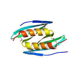
2WBT
 | | The Structure of a Double C2H2 Zinc Finger Protein from a Hyperthermophilic Archaeal Virus in the Absence of DNA | | 分子名称: | B-129, ZINC ION | | 著者 | Eilers, B.J, Menon, S, Windham, A.B, Kraft, P, Dlakic, M, Young, M.J, Lawrence, C.M. | | 登録日 | 2009-03-03 | | 公開日 | 2010-03-31 | | 最終更新日 | 2019-05-08 | | 実験手法 | X-RAY DIFFRACTION (2.7 Å) | | 主引用文献 | The Structure of a Double C2H2 Zinc Finger Protein from a Hyperthermophilic Archaeal Virus in the Absence of DNA
To be Published
|
|
4UAS
 | | Crystal structure of CbbY from Rhodobacter sphaeroides in complex with phosphate | | 分子名称: | 2-(N-MORPHOLINO)-ETHANESULFONIC ACID, CHLORIDE ION, MAGNESIUM ION, ... | | 著者 | Bracher, A, Sharma, A, Starling-Windhof, A, Hartl, F.U, Hayer-Hartl, M. | | 登録日 | 2014-08-11 | | 公開日 | 2014-12-31 | | 最終更新日 | 2023-12-20 | | 実験手法 | X-RAY DIFFRACTION (1.2 Å) | | 主引用文献 | Degradation of potent Rubisco inhibitor by selective sugar phosphatase.
Nat.Plants, 1, 2015
|
|
4UAT
 | | Crystal structure of CbbY (mutant D10N) from Rhodobacter sphaeroides in complex with Xylulose-(1,5)bisphosphate, crystal form I | | 分子名称: | 2-(N-MORPHOLINO)-ETHANESULFONIC ACID, MAGNESIUM ION, Protein CbbY, ... | | 著者 | Bracher, A, Sharma, A, Starling-Windhof, A, Hartl, F.U, Hayer-Hartl, M. | | 登録日 | 2014-08-11 | | 公開日 | 2014-12-31 | | 最終更新日 | 2023-12-20 | | 実験手法 | X-RAY DIFFRACTION (1.3 Å) | | 主引用文献 | Degradation of potent Rubisco inhibitor by selective sugar phosphatase.
Nat.Plants, 1, 2015
|
|
4UAR
 | | Crystal structure of apo-CbbY from Rhodobacter sphaeroides | | 分子名称: | GLYCEROL, Protein CbbY | | 著者 | Bracher, A, Sharma, A, Starling-Windhof, A, Hartl, F.U, Hayer-Hartl, M. | | 登録日 | 2014-08-11 | | 公開日 | 2014-12-31 | | 最終更新日 | 2024-05-08 | | 実験手法 | X-RAY DIFFRACTION (1.9 Å) | | 主引用文献 | Degradation of potent Rubisco inhibitor by selective sugar phosphatase.
Nat.Plants, 1, 2015
|
|


