Loading
PDBj
メニューPDBj@FacebookPDBj@TwitterPDBj@YouTubewwPDB FoundationwwPDB
RCSB PDBPDBeBMRBAdv. SearchSearch help
PDB: 179 件

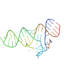
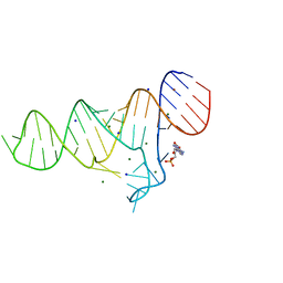
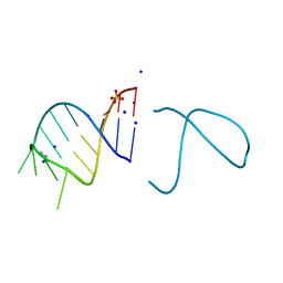
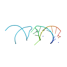
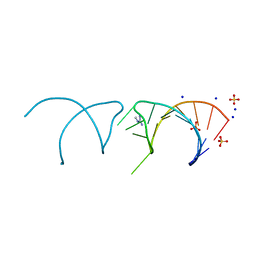
6TFG
DownloadVisualize
BU of 6tfg by Molmil
Crystal structure of the ADP-binding domain of the NAD+ riboswitch with Adenosine 3-phosphate 5-phosphosulfate (APPS)
分子名称: 3'-PHOSPHATE-ADENOSINE-5'-PHOSPHATE SULFATE, Chains: A, MAGNESIUM ION, ...
著者Huang, L, Lilley, D.M.J.
登録日2019-11-14
公開日2020-09-23
最終更新日2024-01-24
実験手法X-RAY DIFFRACTION (2.45 Å)
主引用文献Structure and ligand binding of the ADP-binding domain of the NAD+ riboswitch.
Rna, 26, 2020
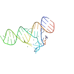
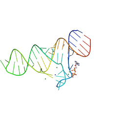
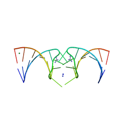
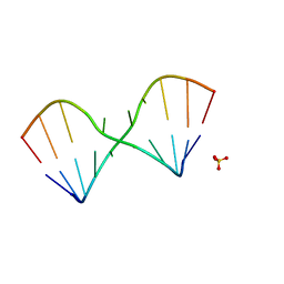
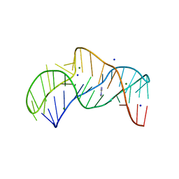
6TF1
DownloadVisualize
BU of 6tf1 by Molmil
Crystal structure of the ADP-binding domain of the NAD+ riboswitch with Adenosine diphosphate (ADP)
分子名称: ADENOSINE-5'-DIPHOSPHATE, Chains: A, MAGNESIUM ION, ...
著者Huang, L, Lilley, D.M.J.
登録日2019-11-12
公開日2020-09-23
最終更新日2024-01-24
実験手法X-RAY DIFFRACTION (2.4 Å)
主引用文献Structure and ligand binding of the ADP-binding domain of the NAD+ riboswitch.
Rna, 26, 2020
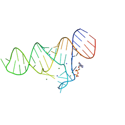
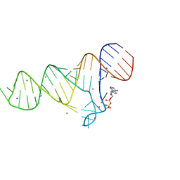
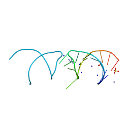
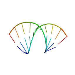
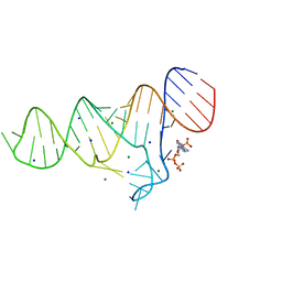
6TFF
DownloadVisualize
BU of 6tff by Molmil
Crystal structure of the ADP-binding domain of the NAD+ riboswitch with Nicotinamide adenine dinucleotide (NAD+)
分子名称: BROMIDE ION, Chains: A, MAGNESIUM ION, ...
著者Huang, L, Lilley, D.M.J.
登録日2019-11-14
公開日2020-09-23
最終更新日2024-01-24
実験手法X-RAY DIFFRACTION (2.52 Å)
主引用文献Structure and ligand binding of the ADP-binding domain of the NAD+ riboswitch.
Rna, 26, 2020
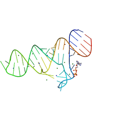
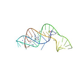
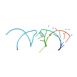
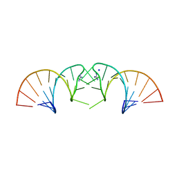
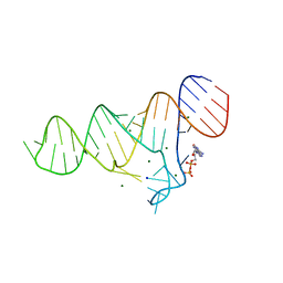
6TF0
DownloadVisualize
BU of 6tf0 by Molmil
Crystal structure of the ADP-binding domain of the NAD+ riboswitch with Nicotinamide adenine dinucleotide, reduced (NADH)
分子名称: 1,4-DIHYDRONICOTINAMIDE ADENINE DINUCLEOTIDE, Chains: A, MAGNESIUM ION, ...
著者Huang, L, Lilley, D.M.J.
登録日2019-11-12
公開日2020-09-23
最終更新日2024-05-15
実験手法X-RAY DIFFRACTION (2.1 Å)
主引用文献Structure and ligand binding of the ADP-binding domain of the NAD+ riboswitch.
Rna, 26, 2020
6TF3
DownloadVisualize
BU of 6tf3 by Molmil
Crystal structure of the ADP-binding domain of the NAD+ riboswitch with Cordycepin 5-triphosphate (3-dATP)
分子名称: 3'-DEOXYADENOSINE-5'-TRIPHOSPHATE, Chains: A, MAGNESIUM ION, ...
著者Huang, L, Lilley, D.M.J.
登録日2019-11-13
公開日2020-09-23
最終更新日2024-01-24
実験手法X-RAY DIFFRACTION (2.66 Å)
主引用文献Structure and ligand binding of the ADP-binding domain of the NAD+ riboswitch.
Rna, 26, 2020
6TFE
DownloadVisualize
BU of 6tfe by Molmil
Crystal structure of the ADP-binding domain of the NAD+ riboswitch with N6-Methyl-adenosine-5'-triphosphate (m6ATP)
分子名称: BROMIDE ION, Chains: A, MAGNESIUM ION, ...
著者Huang, L, Lilley, D.M.J.
登録日2019-11-14
公開日2020-09-23
最終更新日2024-05-15
実験手法X-RAY DIFFRACTION (2.3 Å)
主引用文献Structure and ligand binding of the ADP-binding domain of the NAD+ riboswitch.
Rna, 26, 2020
6TB7
DownloadVisualize
BU of 6tb7 by Molmil
Crystal structure of the ADP-binding domain of the NAD+ riboswitch with Adenosine monophosphate (AMP)
分子名称: ADENOSINE MONOPHOSPHATE, BROMIDE ION, MAGNESIUM ION, ...
著者Huang, L, Lilley, D.M.J.
登録日2019-11-01
公開日2020-09-30
最終更新日2024-05-15
実験手法X-RAY DIFFRACTION (2.53 Å)
主引用文献Structure and ligand binding of the ADP-binding domain of the NAD + riboswitch.
Rna, 26, 2020
6TF2
DownloadVisualize
BU of 6tf2 by Molmil
Crystal structure of the ADP-binding domain of the NAD+ riboswitch with Adenosine 5-triphosphate (ATP)
分子名称: ADENOSINE-5'-TRIPHOSPHATE, BROMIDE ION, Chains: A, ...
著者Huang, L, Lilley, D.M.J.
登録日2019-11-12
公開日2020-09-23
最終更新日2024-05-15
実験手法X-RAY DIFFRACTION (2.55 Å)
主引用文献Structure and ligand binding of the ADP-binding domain of the NAD+ riboswitch.
Rna, 26, 2020
6TFH
DownloadVisualize
BU of 6tfh by Molmil
Crystal structure of the ADP-binding domain of the NAD+ riboswitch with Nicotinamide adenine dinucleotide, reduced (NADH); soaking with Manganese(II) (Mn2+)
分子名称: Chains: A, MANGANESE (II) ION, NICOTINAMIDE-ADENINE-DINUCLEOTIDE, ...
著者Huang, L, Lilley, D.M.J.
登録日2019-11-14
公開日2020-09-23
最終更新日2024-01-24
実験手法X-RAY DIFFRACTION (2.95 Å)
主引用文献Structure and ligand binding of the ADP-binding domain of the NAD+ riboswitch.
Rna, 26, 2020
6FZ0
DownloadVisualize
BU of 6fz0 by Molmil
Crystal structure of the metY SAM V riboswitch
分子名称: MAGNESIUM ION, S-ADENOSYLMETHIONINE, SODIUM ION, ...
著者Huang, L, Lilley, D.M.J.
登録日2018-03-13
公開日2018-07-04
最終更新日2024-05-08
実験手法X-RAY DIFFRACTION (2.499 Å)
主引用文献Structure and ligand binding of the SAM-V riboswitch.
Nucleic Acids Res., 46, 2018
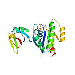
1LFD
DownloadVisualize
BU of 1lfd by Molmil
CRYSTAL STRUCTURE OF THE ACTIVE RAS PROTEIN COMPLEXED WITH THE RAS-INTERACTING DOMAIN OF RALGDS
分子名称: MAGNESIUM ION, PHOSPHOAMINOPHOSPHONIC ACID-GUANYLATE ESTER, RALGDS, ...
著者Huang, L, Hofer, F, Martin, G.S, Kim, S.-H.
登録日1998-04-29
公開日1999-05-04
最終更新日2024-05-22
実験手法X-RAY DIFFRACTION (2.1 Å)
主引用文献Structural basis for the interaction of Ras with RalGDS.
Nat.Struct.Biol., 5, 1998
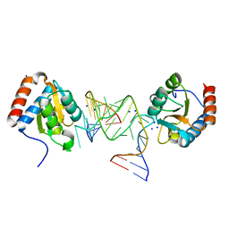
6Q8U
DownloadVisualize
BU of 6q8u by Molmil
Structure of the standard kink turn HmKt-7 variant A2bm6A bound with AfL7Ae protein
分子名称: 50S ribosomal protein L7Ae, RNA (5'-R(*CP*GP*GP*CP*GP*AP*AP*GP*(6MZ)P*AP*CP*CP*GP*GP*GP*GP*AP*GP*CP*CP*G)-3'), SODIUM ION
著者Huang, L, Lilley, D.M.J.
登録日2018-12-16
公開日2019-07-03
最終更新日2024-01-24
実験手法X-RAY DIFFRACTION (1.99 Å)
主引用文献Effect of methylation of adenine N6on kink turn structure depends on location.
Rna Biol., 16, 2019
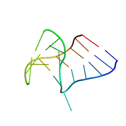
6Q8V
DownloadVisualize
BU of 6q8v by Molmil
Structure of the standard kink turn HmKt-7 variant A2bm6A.
分子名称: RNA (5'-R(*GP*GP*CP*GP*AP*AP*GP*(6MZ)P*AP*CP*CP*GP*GP*GP*GP*AP*GP*CP*C)-3')
著者Huang, L, Lilley, D.M.J.
登録日2018-12-16
公開日2019-07-03
最終更新日2024-01-24
実験手法X-RAY DIFFRACTION (2.09 Å)
主引用文献Effect of methylation of adenine N6on kink turn structure depends on location.
Rna Biol., 16, 2019
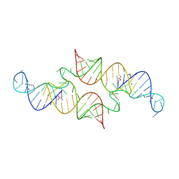
6QN3
DownloadVisualize
BU of 6qn3 by Molmil
Structure of the Glutamine II Riboswitch
分子名称: BROMIDE ION, GLUTAMINE, MAGNESIUM ION, ...
著者Huang, L, Lilley, D.M.J.
登録日2019-02-08
公開日2019-06-12
最終更新日2024-05-15
実験手法X-RAY DIFFRACTION (2.3 Å)
主引用文献Structure and ligand binding of the glutamine-II riboswitch.
Nucleic Acids Res., 47, 2019
5NEF
DownloadVisualize
BU of 5nef by Molmil
The structure of the G. violaceus guanidine II riboswitch P1 stem-loop with guanidine
分子名称: GUANIDINE, RNA (5'-R(*GP*GP*UP*GP*GP*GP*GP*AP*CP*GP*AP*CP*CP*CP*CP*AP*(CBV)P*C)-3'), SODIUM ION, ...
著者Huang, L, Wang, J, Lilley, D.M.J.
登録日2017-03-10
公開日2017-06-07
最終更新日2024-05-08
実験手法X-RAY DIFFRACTION (1.91 Å)
主引用文献The Structure of the Guanidine-II Riboswitch.
Cell Chem Biol, 24, 2017
5NDH
DownloadVisualize
BU of 5ndh by Molmil
The structure of the G. violaceus guanidine II riboswitch P2 stem-loop
分子名称: GUANIDINE, MAGNESIUM ION, RNA (5'-R(*GP*(CBV)P*GP*GP*GP*GP*AP*CP*GP*AP*CP*CP*CP*CP*GP*C)-3'), ...
著者Huang, L, Wang, J, Lilley, D.M.J.
登録日2017-03-08
公開日2017-05-31
最終更新日2024-05-08
実験手法X-RAY DIFFRACTION (1.81 Å)
主引用文献The Structure of the Guanidine-II Riboswitch.
Cell Chem Biol, 24, 2017
5NEO
DownloadVisualize
BU of 5neo by Molmil
The structure of the G. violaceus guanidine II riboswitch P1 stem-loop
分子名称: AMMONIUM ION, RNA (5'-R(*GP*GP*UP*GP*GP*GP*GP*AP*CP*GP*AP*CP*CP*CP*CP*AP*(CBV)P*C)-3'), SODIUM ION, ...
著者Huang, L, Wang, J, Lilley, D.M.J.
登録日2017-03-11
公開日2017-05-31
最終更新日2024-05-08
実験手法X-RAY DIFFRACTION (1.69 Å)
主引用文献The Structure of the Guanidine-II Riboswitch.
Cell Chem Biol, 24, 2017
5NEQ
DownloadVisualize
BU of 5neq by Molmil
The structure of the G. violaceus guanidine II riboswitch P1 stem-loop with aminoguanidine
分子名称: AMINOGUANIDINE, RNA (5'-R(*GP*GP*UP*GP*GP*GP*GP*AP*CP*GP*AP*CP*CP*CP*CP*AP*(CBV)P*C)-3'), SODIUM ION, ...
著者Huang, L, Wang, J, Lilley, D.M.J.
登録日2017-03-11
公開日2017-05-31
最終更新日2024-01-17
実験手法X-RAY DIFFRACTION (1.69 Å)
主引用文献The Structure of the Guanidine-II Riboswitch.
Cell Chem Biol, 24, 2017
5NEX
DownloadVisualize
BU of 5nex by Molmil
The structure of the G. violaceus guanidine II riboswitch P1 stem-loop with agmatine
分子名称: AGMATINE, RNA (5'-R(*GP*GP*UP*GP*GP*GP*GP*AP*CP*GP*AP*CP*CP*CP*CP*AP*(CBV)P*C)-3'), SODIUM ION
著者Huang, L, Wang, J, Lilley, D.M.J.
登録日2017-03-13
公開日2017-05-31
最終更新日2024-05-08
実験手法X-RAY DIFFRACTION (1.72 Å)
主引用文献The Structure of the Guanidine-II Riboswitch.
Cell Chem Biol, 24, 2017
5NOM
DownloadVisualize
BU of 5nom by Molmil
The structure of the G. violaceus guanidine II riboswitch P1 stem-loop with guanidine
分子名称: GUANIDINE, RNA (5'-R(*GP*GP*UP*GP*GP*GP*GP*AP*CP*GP*AP*CP*CP*CP*CP*AP*(CBV)P*C)-3'), SODIUM ION, ...
著者Huang, L, Wang, J, Lilley, D.M.J.
登録日2017-04-12
公開日2017-05-31
最終更新日2024-01-17
実験手法X-RAY DIFFRACTION (1.93 Å)
主引用文献The Structure of the Guanidine-II Riboswitch.
Cell Chem Biol, 24, 2017
5LR3
DownloadVisualize
BU of 5lr3 by Molmil
RNA duplex has central consecutive GA pairs flanked by G-U basepairs
分子名称: RNA (5'-R(*GP*(CBV)P*CP*GP*GP*AP*UP*GP*GP*C)-3'), SULFATE ION
著者Huang, L, Lilley, D.M.J.
登録日2016-08-18
公開日2017-06-21
最終更新日2024-05-08
実験手法X-RAY DIFFRACTION (1.65 Å)
主引用文献Control of box C/D snoRNP assembly by N(6)-methylation of adenine.
EMBO Rep., 18, 2017
5LR5
DownloadVisualize
BU of 5lr5 by Molmil
N6-methyladenine is accommodated in a conventional A-U basepair
分子名称: RNA (5'-R(*GP*GP*(6MZ)P*CP*UP*AP*GP*UP*(CBV)P*C)-3'), SULFATE ION
著者Huang, L, Lilley, D.M.J.
登録日2016-08-18
公開日2017-06-21
最終更新日2024-05-08
実験手法X-RAY DIFFRACTION (2.4 Å)
主引用文献Control of box C/D snoRNP assembly by N(6)-methylation of adenine.
EMBO Rep., 18, 2017
5NDI
DownloadVisualize
BU of 5ndi by Molmil
The structure of the E.coli guanidine II riboswitch P1 stem-loop
分子名称: GUANIDINE, RNA (5'-R(*UP*UP*UP*GP*CP*AP*GP*GP*AP*CP*GP*AP*CP*CP*UP*GP*(CBV)P*AP*AP*A)-3'), SODIUM ION
著者Huang, L, Wang, J, Lilley, D.M.J.
登録日2017-03-08
公開日2017-05-31
最終更新日2024-05-08
実験手法X-RAY DIFFRACTION (2.57 Å)
主引用文献The Structure of the Guanidine-II Riboswitch.
Cell Chem Biol, 24, 2017
5NEP
DownloadVisualize
BU of 5nep by Molmil
The structure of the G. violaceus guanidine II riboswitch P1 stem-loop with methylguanidine
分子名称: 1-METHYLGUANIDINE, RNA (5'-R(*GP*GP*UP*GP*GP*GP*GP*AP*CP*GP*AP*CP*CP*CP*CP*AP*(CBV)P*C)-3'), SODIUM ION, ...
著者Huang, L, Wang, J, Lilley, D.M.J.
登録日2017-03-11
公開日2017-05-31
最終更新日2024-05-08
実験手法X-RAY DIFFRACTION (1.6 Å)
主引用文献The Structure of the Guanidine-II Riboswitch.
Cell Chem Biol, 24, 2017
5NZ3
DownloadVisualize
BU of 5nz3 by Molmil
The structure of the thermobifida fusca guanidine III riboswitch with methylguanidine
分子名称: 1-METHYLGUANIDINE, MAGNESIUM ION, RNA (41-MER), ...
著者Huang, L, Wang, J, Lilley, D.M.J.
登録日2017-05-12
公開日2017-10-18
最終更新日2024-05-08
実験手法X-RAY DIFFRACTION (2.059 Å)
主引用文献Structure of the Guanidine III Riboswitch.
Cell Chem Biol, 24, 2017

226707

件を2024-10-30に公開中

PDB statisticsPDBj update infoContact PDBjnumon