4KFV
 
 | |
4L1M
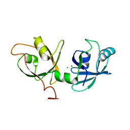
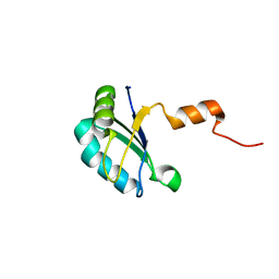
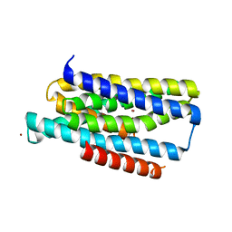
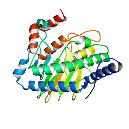
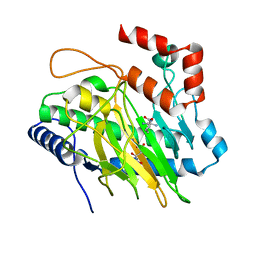
 | | Structure of the first RCC1-like domain of HERC2 | | 分子名称: | E3 ubiquitin-protein ligase HERC2, SULFATE ION, UNKNOWN ATOM OR ION | | 著者 | Tempel, W, Khan, M.B, Dong, A, Hu, J, Li, Y, Bountra, C, Arrowsmith, C.H, Edwards, A.M, Tong, Y, Structural Genomics Consortium (SGC) | | 登録日 | 2013-06-03 | | 公開日 | 2013-07-03 | | 最終更新日 | 2023-09-20 | | 実験手法 | X-RAY DIFFRACTION (2.6 Å) | | 主引用文献 | Structure of the first RCC1-like domain of HERC2
TO BE PUBLISHED
|
|
4FO9
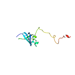
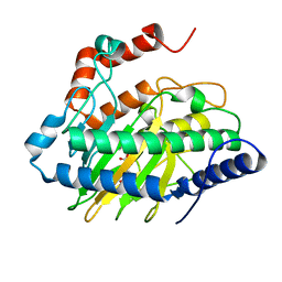
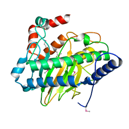
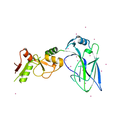 | | Crystal structure of the E3 SUMO Ligase PIAS2 | | 分子名称: | E3 SUMO-protein ligase PIAS2, UNKNOWN ATOM OR ION, ZINC ION | | 著者 | Dong, A, Hu, J, Dobrovetsky, E, Tempel, W, Bountra, C, Arrowsmith, C.H, Edwards, A.M, Tong, Y, Structural Genomics Consortium (SGC) | | 登録日 | 2012-06-20 | | 公開日 | 2012-07-18 | | 最終更新日 | 2024-02-28 | | 実験手法 | X-RAY DIFFRACTION (2.39 Å) | | 主引用文献 | Crystal structure of the E3 SUMO Ligase PIAS2
to be published
|
|
3NYJ
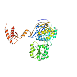
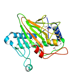
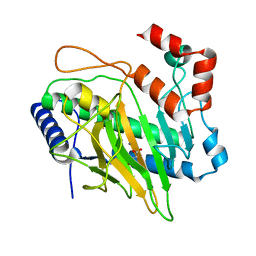
 | | Crystal Structure Analysis of APP E2 domain | | 分子名称: | Amyloid beta A4 protein, OSMIUM ION | | 著者 | Ha, Y, Hu, J, Lee, S, Liu, X. | | 登録日 | 2010-07-15 | | 公開日 | 2011-06-01 | | 最終更新日 | 2017-11-08 | | 実験手法 | X-RAY DIFFRACTION (3.2 Å) | | 主引用文献 | The E2 Domains of APP and APLP1 Share a Conserved Mode of Dimerization.
Biochemistry, 50, 2011
|
|
4JUY
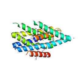
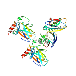
 | | Crystal structure of the PUB domain of E3 ubiquitin ligase RNF31 | | 分子名称: | E3 ubiquitin-protein ligase RNF31, UNKNOWN ATOM OR ION | | 著者 | Dong, A, Hu, J, Li, Y, Wernimont, A, Bountra, C, Arrowsmith, C.H, Edwards, A.M, Tong, Y, Structural Genomics Consortium (SGC) | | 登録日 | 2013-03-25 | | 公開日 | 2013-04-10 | | 最終更新日 | 2024-02-28 | | 実験手法 | X-RAY DIFFRACTION (2.4 Å) | | 主引用文献 | Crystal structure of the PUB domain of E3 ubiquitin ligase RNF31
To be Published
|
|
5HUQ
 | | A tethered niacin-derived pincer complex with a nickel-carbon bond in lactate racemase | | 分子名称: | 3-methanethioyl-1-(5-O-phosphono-beta-D-ribofuranosyl)-5-(sulfanylcarbonyl)pyridin-1-ium, Lactate racemization operon protein LarA, NICKEL (II) ION, ... | | 著者 | Desguin, B, Zhang, T, Soumillion, P, Hols, P, Hu, J, Hausinger, R.P. | | 登録日 | 2016-01-27 | | 公開日 | 2016-02-10 | | 最終更新日 | 2024-05-01 | | 実験手法 | X-RAY DIFFRACTION (3 Å) | | 主引用文献 | METALLOPROTEINS. A tethered niacin-derived pincer complex with a nickel-carbon bond in lactate racemase.
Science, 349, 2015
|
|
4TTB
 | |
4MVT
 | | Crystal structure of SUMO E3 Ligase PIAS3 | | 分子名称: | CHLORIDE ION, E3 SUMO-protein ligase PIAS3, UNKNOWN ATOM OR ION, ... | | 著者 | Dong, A, Hu, J, Li, Y, Tempel, W, Bountra, C, Arrowsmith, C.H, Edwards, A.M, Tong, Y, Structural Genomics Consortium (SGC) | | 登録日 | 2013-09-24 | | 公開日 | 2013-10-16 | | 最終更新日 | 2023-09-20 | | 実験手法 | X-RAY DIFFRACTION (2.3 Å) | | 主引用文献 | Crystal structure of SUMO E3 Ligase PIAS3
to be published
|
|
6JTG
 | |
4QT6
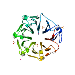
 | | Crystal structure of the SPRY domain of human HERC1 | | 分子名称: | FORMAMIDE, Probable E3 ubiquitin-protein ligase HERC1, UNKNOWN ATOM OR ION | | 著者 | Dong, A, Hu, J, Guan, X, Wernimont, A, Li, Y, Bountra, C, Arrowsmith, C.H, Edwards, A.M, Tong, Y, Structural Genomics Consortium (SGC) | | 登録日 | 2014-07-07 | | 公開日 | 2015-01-07 | | 最終更新日 | 2017-11-22 | | 実験手法 | X-RAY DIFFRACTION (1.64 Å) | | 主引用文献 | Crystal structure of the SPRY domain of human HERC1
To be Published
|
|
5V2X
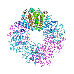
 | | Ethylene forming enzyme in complex with manganese and 2-oxoglutarate | | 分子名称: | 2-OXOGLUTARIC ACID, 2-oxoglutarate-dependent ethylene/succinate-forming enzyme, MANGANESE (II) ION | | 著者 | Fellner, M, Martinez, S, Hu, J, Hausinger, R.P. | | 登録日 | 2017-03-06 | | 公開日 | 2017-08-16 | | 最終更新日 | 2023-10-04 | | 実験手法 | X-RAY DIFFRACTION (1.847 Å) | | 主引用文献 | Structures and Mechanisms of the Non-Heme Fe(II)- and 2-Oxoglutarate-Dependent Ethylene-Forming Enzyme: Substrate Binding Creates a Twist.
J. Am. Chem. Soc., 139, 2017
|
|
3PMR
 | | Crystal Structure of E2 domain of Human Amyloid Precursor-Like Protein 1 | | 分子名称: | Amyloid-like protein 1, PHOSPHATE ION | | 著者 | Lee, S, Xue, Y, Hu, J, Wang, Y, Liu, X, Demeler, B, Ha, Y. | | 登録日 | 2010-11-17 | | 公開日 | 2011-06-01 | | 最終更新日 | 2023-05-31 | | 実験手法 | X-RAY DIFFRACTION (2.11 Å) | | 主引用文献 | The E2 Domains of APP and APLP1 Share a Conserved Mode of Dimerization.
Biochemistry, 50, 2011
|
|
2K3K
 | |
2K4K
 | | Solution structure of GSP13 from Bacillus subtilis | | 分子名称: | General stress protein 13 | | 著者 | Yu, W, Yu, B, Hu, J, Jin, C, Xia, B. | | 登録日 | 2008-06-13 | | 公開日 | 2009-05-12 | | 最終更新日 | 2022-03-16 | | 実験手法 | SOLUTION NMR | | 主引用文献 | Solution structure of GSP13 from Bacillus subtilis exhibits an S1 domain related to cold shock proteins.
J.Biomol.Nmr, 43, 2009
|
|
7WNT
 | | RNase J from Mycobacterium tuberculosis | | 分子名称: | Ribonuclease J, ZINC ION | | 著者 | Li, J, Bao, L, Hu, J, Zhan, B, Li, Z. | | 登録日 | 2022-01-19 | | 公開日 | 2023-01-25 | | 最終更新日 | 2023-11-29 | | 実験手法 | X-RAY DIFFRACTION (2.44 Å) | | 主引用文献 | Structural ans functional study of RNase J from Mycobacterium tuberculosis
To Be Published
|
|
5TSB
 | | Crystal structure of the Zrt-/Irt-like protein from Bordetella bronchiseptica with bound Cd2+ | | 分子名称: | CADMIUM ION, Membrane protein | | 著者 | Zhang, T, Fellner, M, Sui, D, Liu, J, Hu, J. | | 登録日 | 2016-10-28 | | 公開日 | 2017-09-20 | | 最終更新日 | 2020-01-01 | | 実験手法 | X-RAY DIFFRACTION (2.7 Å) | | 主引用文献 | Crystal structures of a ZIP zinc transporter reveal a binuclear metal center in the transport pathway.
Sci Adv, 3, 2017
|
|
7WNU
 | | Mycobacterium tuberculosis Rnase J complex with 7nt RNA | | 分子名称: | RNA (5'-R(P*AP*AP*AP*AP*AP*AP*A)-3'), Ribonuclease J, ZINC ION | | 著者 | Li, J, Hu, J, Bao, L, Zhan, B, Li, Z. | | 登録日 | 2022-01-19 | | 公開日 | 2023-01-25 | | 最終更新日 | 2023-11-29 | | 実験手法 | X-RAY DIFFRACTION (3.2 Å) | | 主引用文献 | Structural ans functional study of RNase J from Mycobacterium tuberculosis
To Be Published
|
|
5TSA
 | | Crystal structure of the Zrt-/Irt-like protein from Bordetella bronchiseptica with bound Zn2+ | | 分子名称: | CADMIUM ION, Membrane protein, ZINC ION | | 著者 | Zhang, T, Fellner, M, Sui, D, Liu, J, Hu, J. | | 登録日 | 2016-10-28 | | 公開日 | 2017-09-20 | | 最終更新日 | 2024-04-03 | | 実験手法 | X-RAY DIFFRACTION (2.4 Å) | | 主引用文献 | Crystal structures of a ZIP zinc transporter reveal a binuclear metal center in the transport pathway.
Sci Adv, 3, 2017
|
|
5V2Z
 | | Ethylene forming enzyme in complex with manganese, 2-oxoadipic acid and L-arginine | | 分子名称: | 2-OXOADIPIC ACID, 2-oxoglutarate-dependent ethylene/succinate-forming enzyme, ARGININE, ... | | 著者 | Fellner, M, Martinez, S, Hu, J, Hausinger, R.P. | | 登録日 | 2017-03-06 | | 公開日 | 2017-08-16 | | 最終更新日 | 2023-10-04 | | 実験手法 | X-RAY DIFFRACTION (1.23 Å) | | 主引用文献 | Structures and Mechanisms of the Non-Heme Fe(II)- and 2-Oxoglutarate-Dependent Ethylene-Forming Enzyme: Substrate Binding Creates a Twist.
J. Am. Chem. Soc., 139, 2017
|
|
5V2V
 | | Ethylene forming enzyme in complex with nickel | | 分子名称: | 2-oxoglutarate-dependent ethylene/succinate-forming enzyme, NICKEL (II) ION | | 著者 | Fellner, M, Martinez, S, Hu, J, Hausinger, R.P. | | 登録日 | 2017-03-06 | | 公開日 | 2017-08-16 | | 最終更新日 | 2023-11-15 | | 実験手法 | X-RAY DIFFRACTION (3.04 Å) | | 主引用文献 | Structures and Mechanisms of the Non-Heme Fe(II)- and 2-Oxoglutarate-Dependent Ethylene-Forming Enzyme: Substrate Binding Creates a Twist.
J. Am. Chem. Soc., 139, 2017
|
|
5V2Y
 | | Ethylene forming enzyme in complex with manganese, 2-oxoglutarate and L-arginine | | 分子名称: | 2-OXOGLUTARIC ACID, 2-oxoglutarate-dependent ethylene/succinate-forming enzyme, ARGININE, ... | | 著者 | Fellner, M, Martinez, S, Hu, J, Hausinger, R.P. | | 登録日 | 2017-03-06 | | 公開日 | 2017-08-16 | | 最終更新日 | 2023-10-04 | | 実験手法 | X-RAY DIFFRACTION (1.428 Å) | | 主引用文献 | Structures and Mechanisms of the Non-Heme Fe(II)- and 2-Oxoglutarate-Dependent Ethylene-Forming Enzyme: Substrate Binding Creates a Twist.
J. Am. Chem. Soc., 139, 2017
|
|
5V2U
 | | Ethylene forming enzyme apo form | | 分子名称: | 2-oxoglutarate-dependent ethylene/succinate-forming enzyme | | 著者 | Fellner, M, Martinez, S, Hu, J, Hausinger, R.P. | | 登録日 | 2017-03-06 | | 公開日 | 2017-08-16 | | 最終更新日 | 2023-11-15 | | 実験手法 | X-RAY DIFFRACTION (2.058 Å) | | 主引用文献 | Structures and Mechanisms of the Non-Heme Fe(II)- and 2-Oxoglutarate-Dependent Ethylene-Forming Enzyme: Substrate Binding Creates a Twist.
J. Am. Chem. Soc., 139, 2017
|
|
5V32
 | | Ethylene forming enzyme in complex with manganese and malic acid | | 分子名称: | 2-oxoglutarate-dependent ethylene/succinate-forming enzyme, D-MALATE, MANGANESE (II) ION | | 著者 | Fellner, M, Martinez, S, Hu, J, Hausinger, R.P. | | 登録日 | 2017-03-06 | | 公開日 | 2017-08-16 | | 最終更新日 | 2023-10-04 | | 実験手法 | X-RAY DIFFRACTION (1.486 Å) | | 主引用文献 | Structures and Mechanisms of the Non-Heme Fe(II)- and 2-Oxoglutarate-Dependent Ethylene-Forming Enzyme: Substrate Binding Creates a Twist.
J. Am. Chem. Soc., 139, 2017
|
|
7YGI
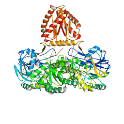
 | | Crystal structure of p53 DBD domain in complex with azurin | | 分子名称: | Azurin, Cellular tumor antigen p53, PHOSPHATE ION, ... | | 著者 | Jiang, W.X, Zuo, J.Q, Hu, J.J, Chen, X.Q, Ma, L.X, Liu, Z, Xing, Q. | | 登録日 | 2022-07-11 | | 公開日 | 2023-02-08 | | 実験手法 | X-RAY DIFFRACTION (2.1 Å) | | 主引用文献 | Structural basis of bacterial effector protein azurin targeting tumor suppressor p53 and inhibiting its ubiquitination.
Commun Biol, 6, 2023
|
|
5V34
 | | Ethylene forming enzyme in complex with manganese, malic acid and L-arginine | | 分子名称: | 2-oxoglutarate-dependent ethylene/succinate-forming enzyme, ARGININE, D-MALATE, ... | | 著者 | Fellner, M, Martinez, S, Hu, J, Hausinger, R.P. | | 登録日 | 2017-03-06 | | 公開日 | 2017-08-16 | | 最終更新日 | 2023-10-04 | | 実験手法 | X-RAY DIFFRACTION (1.48 Å) | | 主引用文献 | Structures and Mechanisms of the Non-Heme Fe(II)- and 2-Oxoglutarate-Dependent Ethylene-Forming Enzyme: Substrate Binding Creates a Twist.
J. Am. Chem. Soc., 139, 2017
|
|


