6L7V
 
 | |
6L7X
 | |
6L7W
 | |
6L7Y
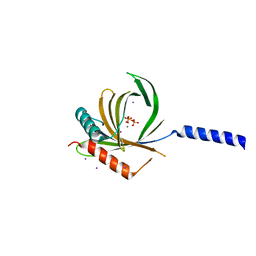
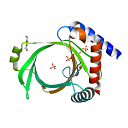 | | Crystal structure of Cet1 from Trypanosoma cruzi in complex with #466 ligand. | | 分子名称: | 3,4,6,7-tetrahydroacridine-1,8(2H,5H)-dione, SULFATE ION, mRNA_triPase domain-containing protein | | 著者 | Kuwabara, N, Ho, K. | | 登録日 | 2019-11-03 | | 公開日 | 2020-06-03 | | 最終更新日 | 2023-11-22 | | 実験手法 | X-RAY DIFFRACTION (2.51 Å) | | 主引用文献 | Crystal structures of the RNA triphosphatase fromTrypanosoma cruziprovide insights into how it recognizes the 5'-end of the RNA substrate.
J.Biol.Chem., 295, 2020
|
|
2JN9
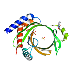
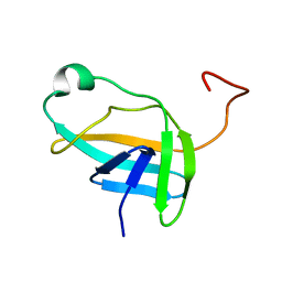 | | NMR solution structure of YkvR protein from Bacillus subtilis: NESG target SR358 | | 分子名称: | YkvR protein | | 著者 | Gurla, S.V.T, Aramini, J.M, Chi Ho, K, Cunningham, K, Ma, L.-C, Xiao, R, Baran, M.C, Acton, T.B, Liu, J, Rost, B, Montelione, G.T, Northeast Structural Genomics Consortium (NESG) | | 登録日 | 2006-12-29 | | 公開日 | 2007-05-01 | | 最終更新日 | 2023-12-20 | | 実験手法 | SOLUTION NMR | | 主引用文献 | NMR solution structure of YkvR protein from Bacillus subtilis: NESG target SR358
To be Published
|
|
4EZ1
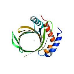
 | | Crystal structure of acetylcholine binding protein (AChBP) from Aplysia Californica in complex with alpha-conotoxin BuIA | | 分子名称: | Alpha-conotoxin BuIA, MANGANESE (II) ION, Soluble acetylcholine receptor | | 著者 | Talley, T.T, Reger, A.S, Kim, C, Sankaran, B, Ho, K, Taylor, P, McIntosh, J.M. | | 登録日 | 2012-05-02 | | 公開日 | 2013-05-08 | | 最終更新日 | 2013-06-19 | | 実験手法 | X-RAY DIFFRACTION (2.49 Å) | | 主引用文献 | Pairwise interaction of alpha-conotoxin BuIA Pro6 with the beta subunit of nicotinic acetylcholine receptor
To be Published
|
|


