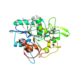
4BVK
 
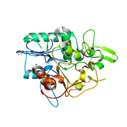 | |
4BVL
 | |
4BVJ
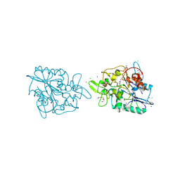
 | | Structure of Y105A mutant of PhaZ7 PHB depolymerase | | 分子名称: | PHB DEPOLYMERASE PHAZ7, SODIUM ION | | 著者 | Hermawan, S, Subedi, B, Papageorgiou, A.C, Jendrossek, D. | | 登録日 | 2013-06-26 | | 公開日 | 2013-09-18 | | 最終更新日 | 2023-12-20 | | 実験手法 | X-RAY DIFFRACTION (1.599 Å) | | 主引用文献 | Biochemical Analysis and Structure Determination of Paucimonas Lemoignei Poly(3-Hydroxybutyrate) (Phb) Depolymerase Phaz7 Muteins Reveal the Phb Binding Site and Details of Substrate-Enzyme Interactions.
Mol.Microbiol., 90, 2013
|
|
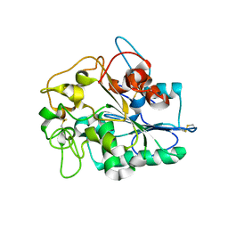
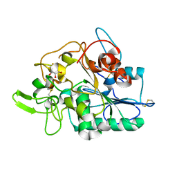
4BRS
 | | Structure of wild type PhaZ7 PHB depolymerase | | 分子名称: | CHLORIDE ION, MAGNESIUM ION, PHB DEPOLYMERASE PHAZ7, ... | | 著者 | Hermawan, S, Subedi, B, Papageorgiou, A.C, Jendrossek, D. | | 登録日 | 2013-06-05 | | 公開日 | 2013-09-18 | | 最終更新日 | 2023-12-20 | | 実験手法 | X-RAY DIFFRACTION (1.6 Å) | | 主引用文献 | Biochemical Analysis and Structure Determination of Paucimonas Lemoignei Poly(3-Hydroxybutyrate) (Phb) Depolymerase Phaz7 Muteins Reveal the Phb Binding Site and Details of Substrate-Enzyme Interactions.
Mol.Microbiol., 90, 2013
|
|
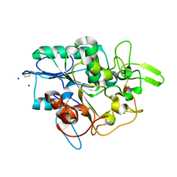
4BYM
 | | Structure of PhaZ7 PHB depolymerase Y105E mutant | | 分子名称: | CHLORIDE ION, PHB DEPOLYMERASE PHAZ7, SODIUM ION | | 著者 | Hermawan, S, Subedi, B, Papageorgiou, A.C, Jendrossek, D. | | 登録日 | 2013-07-20 | | 公開日 | 2013-09-18 | | 最終更新日 | 2023-12-20 | | 実験手法 | X-RAY DIFFRACTION (1.598 Å) | | 主引用文献 | Biochemical Analysis and Structure Determination of Paucimonas Lemoignei Poly(3-Hydroxybutyrate) (Phb) Depolymerase Phaz7 Muteins Reveal the Phb Binding Site and Details of Substrate-Enzyme Interactions.
Mol.Microbiol., 90, 2013
|
|
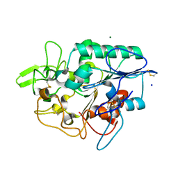
4BTV
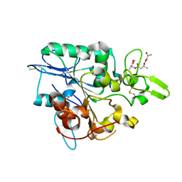 | | Structure of PhaZ7 PHB depolymerase in complex with 3HB trimer | | 分子名称: | (1R)-3-{[(1R)-3-METHOXY-1-METHYL-3-OXOPROPYL]OXY}-1-METHYL-3-OXOPROPYL (3R)-3-HYDROXYBUTANOATE, PHB DEPOLYMERASE PHAZ7 | | 著者 | Hermawan, S, Subedi, B, Papageorgiou, A.C, Jendrossek, D. | | 登録日 | 2013-06-19 | | 公開日 | 2013-09-18 | | 最終更新日 | 2019-07-17 | | 実験手法 | X-RAY DIFFRACTION (1.594 Å) | | 主引用文献 | Biochemical Analysis and Structure Determination of Paucimonas Lemoignei Poly(3-Hydroxybutyrate) (Phb) Depolymerase Phaz7 Muteins Reveal the Phb Binding Site and Details of Substrate-Enzyme Interactions.
Mol.Microbiol., 90, 2013
|
|
2VTV
 | | PhaZ7 depolymerase from Paucimonas lemoignei | | 分子名称: | GLYCEROL, PHB depolymerase PhaZ7 | | 著者 | Papageorgiou, A.C, Hermawan, S, Singh, C.B, Jendrossek, D. | | 登録日 | 2008-05-16 | | 公開日 | 2008-08-26 | | 最終更新日 | 2019-07-24 | | 実験手法 | X-RAY DIFFRACTION (1.9 Å) | | 主引用文献 | Structural basis of poly(3-hydroxybutyrate) hydrolysis by PhaZ7 depolymerase from Paucimonas lemoignei.
J. Mol. Biol., 382, 2008
|
|
2X76
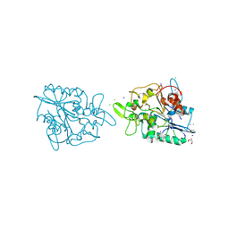 | | The crystal structure of PhaZ7 at atomic (1.2 Angstrom) resolution reveals details of the active site and suggests a substrate binding mode | | 分子名称: | CHLORIDE ION, GLYCEROL, IODIDE ION, ... | | 著者 | Wakadkar, S, Hermawan, S, Jendrossek, D, Papageorgiou, A.C. | | 登録日 | 2010-02-24 | | 公開日 | 2010-06-09 | | 最終更新日 | 2023-12-20 | | 実験手法 | X-RAY DIFFRACTION (1.45 Å) | | 主引用文献 | The structure of PhaZ7 at atomic (1.2 A) resolution reveals details of the active site and suggests a substrate-binding mode.
Acta Crystallogr. Sect. F Struct. Biol. Cryst. Commun., 66, 2010
|
|
2X5X
 | | The crystal structure of PhaZ7 at atomic (1.2 Angstrom) resolution reveals details of the active site and suggests a substrate binding mode | | 分子名称: | CHLORIDE ION, IODIDE ION, PHB DEPOLYMERASE PHAZ7, ... | | 著者 | Wakadkar, S, Hermawan, S, Jendrossek, D, Papageorgiou, A.C. | | 登録日 | 2010-02-11 | | 公開日 | 2010-06-09 | | 最終更新日 | 2023-12-20 | | 実験手法 | X-RAY DIFFRACTION (1.2 Å) | | 主引用文献 | The structure of PhaZ7 at atomic (1.2 A) resolution reveals details of the active site and suggests a substrate-binding mode.
Acta Crystallogr. Sect. F Struct. Biol. Cryst. Commun., 66, 2010
|
|


