6XV2
 
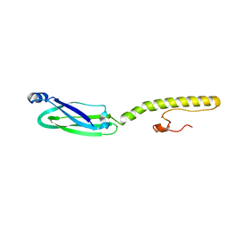 | | Full structure of RYMV P1 protein, derived from crystallographic and NMR data. | | 分子名称: | ZINC ION, p1 | | 著者 | Poignavent, V, Hoh, F, Vignols, F, Demene, H, Yang, Y, Gillet, F.X. | | 登録日 | 2020-01-21 | | 公開日 | 2021-02-03 | | 最終更新日 | 2024-06-19 | | 実験手法 | SOLUTION NMR | | 主引用文献 | A Flexible and Original Architecture of Two Unrelated Zinc Fingers Underlies the Role of the Multitask P1 in RYMV Spread.
J.Mol.Biol., 434, 2022
|
|
2XMZ
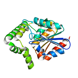 | | Structure of MenH from S. aureus | | 分子名称: | HYDROLASE, ALPHA/BETA HYDROLASE FOLD FAMILY | | 著者 | Dawson, A, Fyfe, P.K, Gillet, F, Hunter, W.N. | | 登録日 | 2010-07-30 | | 公開日 | 2011-05-25 | | 最終更新日 | 2023-12-20 | | 実験手法 | X-RAY DIFFRACTION (1.94 Å) | | 主引用文献 | Exploiting the High-Resolution Crystal Structure of Staphylococcus Aureus Menh to Gain Insight Into Enzyme Activity.
Bmc Struct.Biol., 11, 2011
|
|


