Loading
PDBj
MenuPDBj@FacebookPDBj@TwitterPDBj@YouTubewwPDB FoundationwwPDB
RCSB PDBPDBeBMRBAdv. SearchSearch help
PDB: 187 results

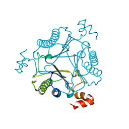
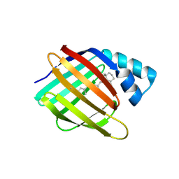
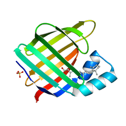
7LHO
DownloadVisualize
BU of 7lho by Molmil
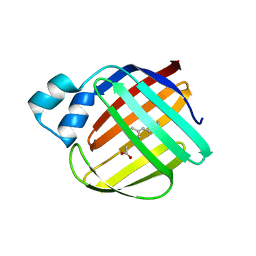
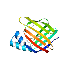
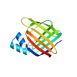
Crystal structure of Q108K:K40L:T51V:T53C:R58W:T29L:Y19W:Q4A mutant of cellular retinol binding protein II complex with all-trans-retinal in the dark
Descriptor: GLYCEROL, RETINAL, Retinol-binding protein 2
Authors:Ehyaei, N, Geiger, J.H, Borhan, B.
Deposit date:2021-01-26
Release date:2021-04-14
Last modified:2023-10-18
Method:X-RAY DIFFRACTION (1.4 Å)
Cite:Crystal structure of Q108K:K40L:T51V:T53C:R58W:T29L:Y19W:Q4A mutant of cellular retinol binding protein II complex with all-trans-retinal in the dark
To Be Published
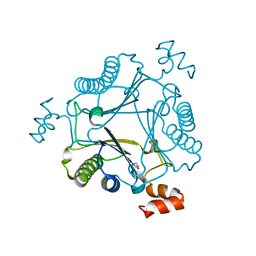
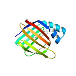
7MS0
DownloadVisualize
BU of 7ms0 by Molmil
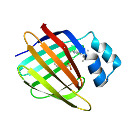
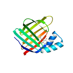
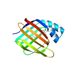
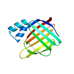
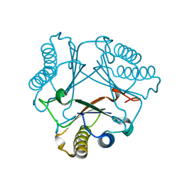
Crystal structure of native Cg10062
Descriptor: 4-oxalocrotonate tautomerase
Authors:Nayebi, G.H, Geiger, J.H, Draths, K.
Deposit date:2021-05-10
Release date:2022-02-02
Last modified:2023-10-18
Method:X-RAY DIFFRACTION (1.37 Å)
Cite:Cg10062 Catalysis Forges a Link between Acetylenecarboxylic Acid and Bacterial Metabolism.
Biochemistry, 60, 2021
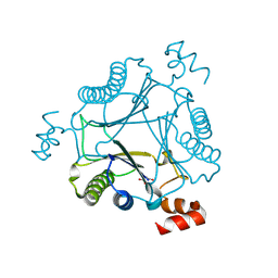
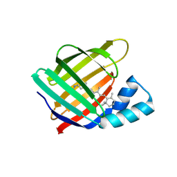
7MS3
DownloadVisualize
BU of 7ms3 by Molmil
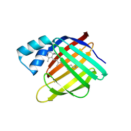
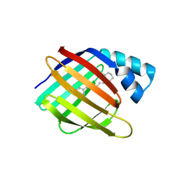
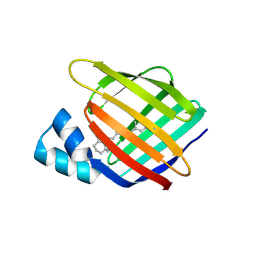
Crystal structure of R73A mutant of Cg10062 with a covalent intermediate of the hydration of acetylenecarboxylic acid
Descriptor: 3-HYDROXY-PROPANOIC ACID, 4-oxalocrotonate tautomerase
Authors:Nayebi, G.H, Geiger, J.H, Draths, K.
Deposit date:2021-05-10
Release date:2022-02-02
Last modified:2023-10-18
Method:X-RAY DIFFRACTION (3 Å)
Cite:Cg10062 Catalysis Forges a Link between Acetylenecarboxylic Acid and Bacterial Metabolism.
Biochemistry, 60, 2021
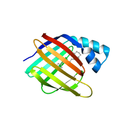
7MS8
DownloadVisualize
BU of 7ms8 by Molmil
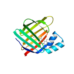
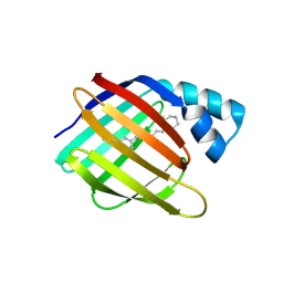
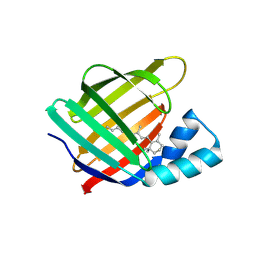
Crystal structure of Y103F mutant of Cg10062 with a covalent intermediate of the hydration of acetylenecarboxylic acid
Descriptor: 3-HYDROXY-PROPANOIC ACID, 4-oxalocrotonate tautomerase
Authors:Nayebi, G.H, Geiger, J.H, Draths, K.
Deposit date:2021-05-10
Release date:2022-02-02
Last modified:2023-10-18
Method:X-RAY DIFFRACTION (2.5 Å)
Cite:Cg10062 Catalysis Forges a Link between Acetylenecarboxylic Acid and Bacterial Metabolism.
Biochemistry, 60, 2021
7MS1
DownloadVisualize
BU of 7ms1 by Molmil
Crystal structure of H28A mutant of Cg10062 with a covalent intermediate of the hydration of acetylenecarboxylic acid
Descriptor: 3-HYDROXY-PROPANOIC ACID, 4-oxalocrotonate tautomerase
Authors:Nayebi, G.H, Geiger, J.H, Draths, K.
Deposit date:2021-05-10
Release date:2022-02-02
Last modified:2023-10-18
Method:X-RAY DIFFRACTION (2.95 Å)
Cite:Cg10062 Catalysis Forges a Link between Acetylenecarboxylic Acid and Bacterial Metabolism.
Biochemistry, 60, 2021
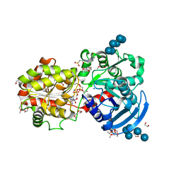
3CX4
DownloadVisualize
BU of 3cx4 by Molmil
Crystal Structure of E.coli GS mutant E377A in complex with ADP and oligosaccharides
Descriptor: (2R)-2-hydroxy-3-[4-(2-hydroxyethyl)piperazin-1-yl]propane-1-sulfonic acid, 2-{2-[2-2-(METHOXY-ETHOXY)-ETHOXY]-ETHOXY}-ETHANOL, ADENOSINE-5'-DIPHOSPHATE, ...
Authors:Sheng, F, Geiger, J.H.
Deposit date:2008-04-23
Release date:2009-04-28
Last modified:2023-08-30
Method:X-RAY DIFFRACTION (2.29 Å)
Cite:Oligosaccharide binding in Escherichia coli glycogen synthase.
Biochemistry, 48, 2009
4ZJ0
DownloadVisualize
BU of 4zj0 by Molmil
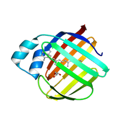
The crystal structure of monomer Q108K:K40L:Y60W CRBPII bound to all-trans-retinal
Descriptor: ACETATE ION, RETINAL, Retinol-binding protein 2
Authors:Nossoni, Z, Assar, Z, Wang, W, Vasileiou, C, Borhan, B, Geiger, J.H.
Deposit date:2015-04-28
Release date:2016-05-11
Last modified:2023-09-27
Method:X-RAY DIFFRACTION (1.5 Å)
Cite:Domain-Swapped Dimers of Intracellular Lipid-Binding Proteins: Evidence for Ordered Folding Intermediates.
Structure, 24, 2016
3FEP
DownloadVisualize
BU of 3fep by Molmil
Crystal structure of the R132K:R111L:L121E:R59W-CRABPII mutant complexed with a synthetic ligand (merocyanin) at 2.60 angstrom resolution.
Descriptor: (2E,4E,6E)-3-methyl-6-(1,3,3-trimethyl-1,3-dihydro-2H-indol-2-ylidene)hexa-2,4-dienal, 2-(N-MORPHOLINO)-ETHANESULFONIC ACID, Cellular retinoic acid-binding protein 2
Authors:Jia, X, Geiger, J.H.
Deposit date:2008-11-30
Release date:2009-11-10
Last modified:2023-09-06
Method:X-RAY DIFFRACTION (2.6 Å)
Cite:"Turn-on" protein fluorescence: in situ formation of cyanine dyes.
J.Am.Chem.Soc., 137, 2015
4YGH
DownloadVisualize
BU of 4ygh by Molmil
Crystal Structure of R111K:Y134F:T54V:R132Q:P39Y:R59Y mutant of human Cellular Retinoic Acid Binding Protein II with Retinal at 2.1 Angstrom resolution - UV irradiated crystal - 2nd cycle
Descriptor: Cellular retinoic acid-binding protein 2, RETINAL
Authors:Nosrati, M, Geiger, J.H.
Deposit date:2015-02-26
Release date:2016-03-02
Last modified:2023-09-27
Method:X-RAY DIFFRACTION (2.1 Å)
Cite:A Photoisomerizing Rhodopsin Mimic Observed at Atomic Resolution.
J.Am.Chem.Soc., 138, 2016
4YH0
DownloadVisualize
BU of 4yh0 by Molmil
Crystal structure of the R111K:Y134F:T54V:R132Q:P39Y:R59Y mutant of human Cellular Retinoic Acid Binding Protein II in complex with Retinal at 2.14 angstrom resolution - UV irradiated crystal - 3rd cycle
Descriptor: Cellular retinoic acid-binding protein 2, RETINAL
Authors:Nosrati, M, Geiger, J.H.
Deposit date:2015-02-26
Release date:2016-03-02
Last modified:2023-09-27
Method:X-RAY DIFFRACTION (2.144 Å)
Cite:A Photoisomerizing Rhodopsin Mimic Observed at Atomic Resolution.
J.Am.Chem.Soc., 138, 2016
4YDA
DownloadVisualize
BU of 4yda by Molmil
Crystal structure of R111K:Y134F:T54V:R132Q:P39Q:R59Y mutant of human cellular retinoic acid binding proteinii with retinal at 1.95 angstrom - after 1hour visible light irradiation - 3rd cycle
Descriptor: Cellular retinoic acid-binding protein 2, RETINAL
Authors:Nosrati, M, Geiger, J.H.
Deposit date:2015-02-21
Release date:2016-02-24
Last modified:2023-09-27
Method:X-RAY DIFFRACTION (1.951 Å)
Cite:A Photoisomerizing Rhodopsin Mimic Observed at Atomic Resolution.
J.Am.Chem.Soc., 138, 2016
4YDB
DownloadVisualize
BU of 4ydb by Molmil
Crystal structure of the R111K:Y134F:T54V:R132Q:P39Q:R59Y mutant of human Cellular Retinoic Acid Binding Protein II in complex with Retinal at 2.03 angstrom -UV irradiated crystal- 3rd cycle
Descriptor: Cellular retinoic acid-binding protein 2, RETINAL
Authors:Nosrati, M, Geiger, J.H.
Deposit date:2015-02-21
Release date:2016-02-24
Last modified:2023-09-27
Method:X-RAY DIFFRACTION (2.03 Å)
Cite:A Photoisomerizing Rhodopsin Mimic Observed at Atomic Resolution.
J.Am.Chem.Soc., 138, 2016
4YFQ
DownloadVisualize
BU of 4yfq by Molmil
CRYSTAL STRUCTURE OF THE R111K:Y134F:T54V:R132Q:P39Y:R59Y MUTANT OF HUMAN CELLULAR RETINOIC ACID BINDING PROTEINII WITH RETINAL AFTER 24 HOURS INCUBATION AT 1.62 ANGSTROM RESOLUTION - Thermodynamic product - 1st cycle
Descriptor: Cellular retinoic acid-binding protein 2, RETINAL
Authors:Nosrati, M, Geiger, J.H.
Deposit date:2015-02-25
Release date:2016-03-02
Last modified:2023-09-27
Method:X-RAY DIFFRACTION (1.62 Å)
Cite:A Photoisomerizing Rhodopsin Mimic Observed at Atomic Resolution.
J.Am.Chem.Soc., 138, 2016
4YCE
DownloadVisualize
BU of 4yce by Molmil
Crystal structure of the R111K:Y134F:T54V:R132Q:P39Q:R59Y mutant of human cellular retinoic acid binding proteinii with retinal at 1.95 angstrom- visible light irradiated crystal for 1 hour - 2nd cycle
Descriptor: Cellular retinoic acid-binding protein 2, RETINAL
Authors:Nosrati, M, Geiger, J.H.
Deposit date:2015-02-19
Release date:2016-02-24
Last modified:2023-09-27
Method:X-RAY DIFFRACTION (1.953 Å)
Cite:A Photoisomerizing Rhodopsin Mimic Observed at Atomic Resolution.
J.Am.Chem.Soc., 138, 2016
4YFR
DownloadVisualize
BU of 4yfr by Molmil
Crystal structure of the R111K:Y134F:T54V:R132Q:P39Y:R59Y mutant of human Cellular Retinoic Acid Binding Protein II with Retinal at 1.95 Angstrom Resolution - UV irradiated crystal for 30 minutes - 1st Cycle
Descriptor: Cellular retinoic acid-binding protein 2, RETINAL
Authors:Nosrati, M, Geiger, J.H.
Deposit date:2015-02-25
Release date:2016-03-02
Last modified:2023-09-27
Method:X-RAY DIFFRACTION (1.952 Å)
Cite:A Photoisomerizing Rhodopsin Mimic Observed at Atomic Resolution.
J.Am.Chem.Soc., 138, 2016
4YKM
DownloadVisualize
BU of 4ykm by Molmil
Crystal structure of the R111K:Y134F:T54V:R132Q:P39Q:R59Y:A32W:F3Q mutant of human Cellular Retinoic Acid Binding Protein II with Retinal at 1.58 angstrom resolution
Descriptor: Cellular retinoic acid-binding protein 2, RETINAL
Authors:Nosrati, M, Geiger, J.H.
Deposit date:2015-03-04
Release date:2016-03-09
Last modified:2023-09-27
Method:X-RAY DIFFRACTION (1.58 Å)
Cite:A Photoisomerizing Rhodopsin Mimic Observed at Atomic Resolution.
J.Am.Chem.Soc., 138, 2016
4YGZ
DownloadVisualize
BU of 4ygz by Molmil
CRYSTAL STRUCTURE OF THE R111K:Y134F:T54V:R132Q:P39Y:R59Y MUTANT OF HUMAN CELLULAR RETINOIC ACID BINDING PROTEIN II WITH RETINAL AT 2.06 ANGSTROM RESOLUTION - VISIBLE LIGHT IRRADIATED CRYSTAL -3RD CYCLE
Descriptor: Cellular retinoic acid-binding protein 2, RETINAL
Authors:Nosrati, M, Geiger, J.H.
Deposit date:2015-02-26
Release date:2016-03-02
Last modified:2023-09-27
Method:X-RAY DIFFRACTION (2.06 Å)
Cite:A Photoisomerizing Rhodopsin Mimic Observed at Atomic Resolution.
J.Am.Chem.Soc., 138, 2016
4YBP
DownloadVisualize
BU of 4ybp by Molmil
Crystal structure of the R111K:Y134F:T54V:R132Q:P39Q:R59Y mutant of human cellular retinoic acid binding proteinii with retinal after 24 hour incubation at 1.83 angstrom resolution - thermodynamic product - 1st cycle
Descriptor: Cellular retinoic acid-binding protein 2, RETINAL
Authors:Nosrati, M, Geiger, J.H.
Deposit date:2015-02-19
Release date:2016-02-24
Last modified:2023-09-27
Method:X-RAY DIFFRACTION (1.831 Å)
Cite:A Photoisomerizing Rhodopsin Mimic Observed at Atomic Resolution.
J.Am.Chem.Soc., 138, 2016
4YFP
DownloadVisualize
BU of 4yfp by Molmil
CRYSTAL STRUCTURE OF THE R111K:Y134F:T54V:R132Q:P39Y:R59Y MUTANT OF HUMAN CELLULAR RETINOIC ACID BINDING PROTEIN II WITH RETINAL AFTER 20 MINUTES INCUBATION AT 1.95 ANGSTROM RESOLUTION-Kinetic Product
Descriptor: Cellular retinoic acid-binding protein 2, RETINAL
Authors:Nosrati, M, Geiger, J.H.
Deposit date:2015-02-25
Release date:2016-03-02
Last modified:2023-09-27
Method:X-RAY DIFFRACTION (1.951 Å)
Cite:A Photoisomerizing Rhodopsin Mimic Observed at Atomic Resolution.
J.Am.Chem.Soc., 138, 2016
4YGG
DownloadVisualize
BU of 4ygg by Molmil
CRYSTAL STRUCTURE OF R111K:Y134F:T54V:R132Q:P39Y:R59Y MUTANT OF HUMAN CELLULAR RETINOIC ACID BINDING PROTEIN II WITH RETINAL AT 1.9 ANGSTROM RESOLUTION - VISIBLE LIGHT IRRADIATED CRYSTAL - 2ND CYCLE
Descriptor: Cellular retinoic acid-binding protein 2, RETINAL
Authors:Nosrati, M, Geiger, J.H.
Deposit date:2015-02-26
Release date:2016-03-02
Last modified:2023-09-27
Method:X-RAY DIFFRACTION (1.9 Å)
Cite:A Photoisomerizing Rhodopsin Mimic Observed at Atomic Resolution.
J.Am.Chem.Soc., 138, 2016
4YBU
DownloadVisualize
BU of 4ybu by Molmil
Crystal structure of the R111K:Y134F:T54V:R132Q:P39Q:R59Y mutant of human Cellular Retinoic Acid Binding ProteinII in complex with Retinal after 24 h incubation and 1 hour UV irradiation at 1.92 angstrom - 1st cycle
Descriptor: Cellular retinoic acid-binding protein 2, RETINAL
Authors:Nosrati, M, Geiger, J.H.
Deposit date:2015-02-19
Release date:2016-02-24
Last modified:2023-09-27
Method:X-RAY DIFFRACTION (1.924 Å)
Cite:A Photoisomerizing Rhodopsin Mimic Observed at Atomic Resolution.
J.Am.Chem.Soc., 138, 2016
4YCH
DownloadVisualize
BU of 4ych by Molmil
Crystal Structure of R111K:Y134F:T54V:R132Q:P39Q:R59Y mutant of human Cellular Retinoic Acid Binding ProteinII with Retinal at 1.96 Angstrom - UV irradiated Crystal for 1 hour - 2nd cycle
Descriptor: Cellular retinoic acid-binding protein 2, RETINAL
Authors:Nosrati, M, Geiger, J.H.
Deposit date:2015-02-20
Release date:2016-02-24
Last modified:2023-09-27
Method:X-RAY DIFFRACTION (1.96 Å)
Cite:A Photoisomerizing Rhodopsin Mimic Observed at Atomic Resolution.
J.Am.Chem.Soc., 138, 2016
3CWK
DownloadVisualize
BU of 3cwk by Molmil
Crystal Structure of the R132K:Y134F:R111L:T54V:L121E Mutant of Cellular Retinoic Acid Binding Protein Type II in Complex with All-trans-Retinoic Acid at 1.57 Angstroms Resolution
Descriptor: Cellular retinoic acid-binding protein 2, RETINOIC ACID, SULFATE ION
Authors:Vaezeslami, S, Geiger, J.H.
Deposit date:2008-04-22
Release date:2008-09-09
Last modified:2024-02-21
Method:X-RAY DIFFRACTION (1.6 Å)
Cite:Structural analysis of site-directed mutants of cellular retinoic acid-binding protein II addresses the relationship between structural integrity and ligand binding.
Acta Crystallogr.,Sect.D, 64, 2008
4ZJZ
DownloadVisualize
BU of 4zjz by Molmil
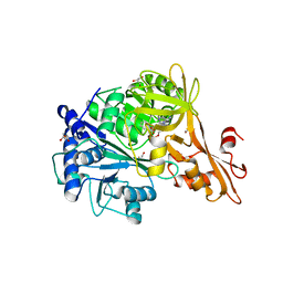
Crystal structure of a benzoate coenzyme A ligase with Benzoyl-AMP
Descriptor: 5'-O-[(R)-(benzoyloxy)(hydroxy)phosphoryl]adenosine, BENZOIC ACID, Benzoate-coenzyme A ligase, ...
Authors:Strom, S, Nosrati, M, Thornburg, C, Walker, K.D, Geiger, J.H.
Deposit date:2015-04-29
Release date:2015-09-30
Last modified:2023-09-27
Method:X-RAY DIFFRACTION (1.7 Å)
Cite:Kinetically and Crystallographically Guided Mutations of a Benzoate CoA Ligase (BadA) Elucidate Mechanism and Expand Substrate Permissivity.
Biochemistry, 54, 2015
3D96
DownloadVisualize
BU of 3d96 by Molmil
Crystal Structure of the R132K:Y134F Mutant of Apo-Cellular Retinoic Acid Binding Protein Type II at 1.71 Angstroms Resolution
Descriptor: ACETATE ION, Cellular retinoic acid-binding protein 2
Authors:Vaezeslami, S, Geiger, J.H.
Deposit date:2008-05-26
Release date:2008-07-22
Last modified:2024-02-21
Method:X-RAY DIFFRACTION (1.71 Å)
Cite:Structural analysis of site-directed mutants of cellular retinoic acid-binding protein II addresses the relationship between structural integrity and ligand binding.
Acta Crystallogr.,Sect.D, 64, 2008

220472

数据于2024-05-29公开中

PDB statisticsPDBj update infoContact PDBjnumon