Loading
PDBj
MenuPDBj@FacebookPDBj@TwitterPDBj@YouTubewwPDB FoundationwwPDB
RCSB PDBPDBeBMRBAdv. SearchSearch help
PDB: 187 results

5FEN
DownloadVisualize
BU of 5fen by Molmil
Crystal structure of the Q108K:K40L:T53C mutant of human Cellular Retinol Binding Protein II in complex with All-trans-Retinal after 24 hours of incubation at 1.55 Angstrom Resolution
Descriptor: ACETATE ION, RETINAL, Retinol-binding protein 2
Authors:Nossoni, Z, Nosrati, M, Geiger, J.H.
Deposit date:2015-12-17
Release date:2016-12-21
Last modified:2023-09-27
Method:X-RAY DIFFRACTION (1.55 Å)
Cite:Crystal structure of the Q108K:K40L:T53C mutant of human Cellular Retinol Binding Protein II in complex with All-trans-Retinal after 24 hours of incubation at 1.55 Angstrom Resolution
To Be Published
5FFH
DownloadVisualize
BU of 5ffh by Molmil
Crystal structure of the Q108K:K40L:T51V:R58W:Y19W mutant of human Cellular Retinol Binding Protein II in complex with All-trans-Retinal at 1.68 Angstrom Resolution
Descriptor: ACETATE ION, RETINAL, Retinol-binding protein 2
Authors:Nossoni, Z, Nosrati, M, Geiger, J.H.
Deposit date:2015-12-18
Release date:2016-12-21
Last modified:2023-09-27
Method:X-RAY DIFFRACTION (1.68 Å)
Cite:Crystal structure of the Q108K:K40L:T51V:R58W:Y19W mutant of human Cellular Retinol Binding ProteinII in complex with All-trans-Retinal at 1.68 Angstrom Resolution
To Be Published
5F7G
DownloadVisualize
BU of 5f7g by Molmil
Crystal structure of the Q108K:K40L:T51V:R58Y:Y19W:Q38L mutant of human Cellular Retinol Binding Protein II in complex with All-trans-Retinal at 1.48 Angstrom Resolution
Descriptor: ACETATE ION, RETINAL, Retinol-binding protein 2
Authors:Nosrati, M, Nossoni, Z, Geiger, J.H.
Deposit date:2015-12-08
Release date:2016-12-14
Last modified:2023-09-27
Method:X-RAY DIFFRACTION (1.48 Å)
Cite:Crystal structure of the Q108K:K40L:T51V:R58F mutant of human Cellular Retinol Binding Protein II in complex with All-trans-Retinal after 24 hours of incubation at 1.54 Angstrom Resolution
To Be Published
4LPC
DownloadVisualize
BU of 4lpc by Molmil
Crystal Structure of E.Coli Branching Enzyme in complex with maltoheptaose
Descriptor: 1,4-alpha-glucan branching enzyme GlgB, GLYCEROL, alpha-D-glucopyranose-(1-4)-alpha-D-glucopyranose, ...
Authors:Feng, L, Geiger, J.H.
Deposit date:2013-07-16
Release date:2015-04-01
Last modified:2024-02-28
Method:X-RAY DIFFRACTION (2.39 Å)
Cite:Crystal Structures of Escherichia coli Branching Enzyme in Complex with Linear Oligosaccharides.
Biochemistry, 54, 2015
4I9S
DownloadVisualize
BU of 4i9s by Molmil
Crystal Structure of the R111K:R132L:Y134F:T54V:R59W Mutant of the Cellular Retinoic Acid Binding Protein Type II in Complex with All-Trans Retinal at 2.58 Angstrom Resolution
Descriptor: Cellular retinoic acid-binding protein 2, RETINAL
Authors:Nosrati, M, Geiger, J.H.
Deposit date:2012-12-05
Release date:2013-10-09
Last modified:2023-09-20
Method:X-RAY DIFFRACTION (2.58 Å)
Cite:Rational Design of a Colorimetric pH Sensor from a Soluble Retinoic Acid Chaperone.
J.Am.Chem.Soc., 135, 2013
4I9R
DownloadVisualize
BU of 4i9r by Molmil
Crystal Structure of the R111K:R132L:Y134F:T54V:R59W:A32W Mutant of the Cellular Retinoic Acid Binding Protein Type II in Complex with All-Trans Retinal at 2.6 Angstrom Resolution
Descriptor: Cellular retinoic acid-binding protein 2, RETINAL
Authors:Nosrati, M, Geiger, J.H.
Deposit date:2012-12-05
Release date:2013-10-09
Last modified:2023-09-20
Method:X-RAY DIFFRACTION (2.6 Å)
Cite:Rational Design of a Colorimetric pH Sensor from a Soluble Retinoic Acid Chaperone.
J.Am.Chem.Soc., 135, 2013
4YGH
DownloadVisualize
BU of 4ygh by Molmil
Crystal Structure of R111K:Y134F:T54V:R132Q:P39Y:R59Y mutant of human Cellular Retinoic Acid Binding Protein II with Retinal at 2.1 Angstrom resolution - UV irradiated crystal - 2nd cycle
Descriptor: Cellular retinoic acid-binding protein 2, RETINAL
Authors:Nosrati, M, Geiger, J.H.
Deposit date:2015-02-26
Release date:2016-03-02
Last modified:2023-09-27
Method:X-RAY DIFFRACTION (2.1 Å)
Cite:A Photoisomerizing Rhodopsin Mimic Observed at Atomic Resolution.
J.Am.Chem.Soc., 138, 2016
4YH0
DownloadVisualize
BU of 4yh0 by Molmil
Crystal structure of the R111K:Y134F:T54V:R132Q:P39Y:R59Y mutant of human Cellular Retinoic Acid Binding Protein II in complex with Retinal at 2.14 angstrom resolution - UV irradiated crystal - 3rd cycle
Descriptor: Cellular retinoic acid-binding protein 2, RETINAL
Authors:Nosrati, M, Geiger, J.H.
Deposit date:2015-02-26
Release date:2016-03-02
Last modified:2023-09-27
Method:X-RAY DIFFRACTION (2.144 Å)
Cite:A Photoisomerizing Rhodopsin Mimic Observed at Atomic Resolution.
J.Am.Chem.Soc., 138, 2016
4YDA
DownloadVisualize
BU of 4yda by Molmil
Crystal structure of R111K:Y134F:T54V:R132Q:P39Q:R59Y mutant of human cellular retinoic acid binding proteinii with retinal at 1.95 angstrom - after 1hour visible light irradiation - 3rd cycle
Descriptor: Cellular retinoic acid-binding protein 2, RETINAL
Authors:Nosrati, M, Geiger, J.H.
Deposit date:2015-02-21
Release date:2016-02-24
Last modified:2023-09-27
Method:X-RAY DIFFRACTION (1.951 Å)
Cite:A Photoisomerizing Rhodopsin Mimic Observed at Atomic Resolution.
J.Am.Chem.Soc., 138, 2016
4YDB
DownloadVisualize
BU of 4ydb by Molmil
Crystal structure of the R111K:Y134F:T54V:R132Q:P39Q:R59Y mutant of human Cellular Retinoic Acid Binding Protein II in complex with Retinal at 2.03 angstrom -UV irradiated crystal- 3rd cycle
Descriptor: Cellular retinoic acid-binding protein 2, RETINAL
Authors:Nosrati, M, Geiger, J.H.
Deposit date:2015-02-21
Release date:2016-02-24
Last modified:2023-09-27
Method:X-RAY DIFFRACTION (2.03 Å)
Cite:A Photoisomerizing Rhodopsin Mimic Observed at Atomic Resolution.
J.Am.Chem.Soc., 138, 2016
4YFQ
DownloadVisualize
BU of 4yfq by Molmil
CRYSTAL STRUCTURE OF THE R111K:Y134F:T54V:R132Q:P39Y:R59Y MUTANT OF HUMAN CELLULAR RETINOIC ACID BINDING PROTEINII WITH RETINAL AFTER 24 HOURS INCUBATION AT 1.62 ANGSTROM RESOLUTION - Thermodynamic product - 1st cycle
Descriptor: Cellular retinoic acid-binding protein 2, RETINAL
Authors:Nosrati, M, Geiger, J.H.
Deposit date:2015-02-25
Release date:2016-03-02
Last modified:2023-09-27
Method:X-RAY DIFFRACTION (1.62 Å)
Cite:A Photoisomerizing Rhodopsin Mimic Observed at Atomic Resolution.
J.Am.Chem.Soc., 138, 2016
4YCE
DownloadVisualize
BU of 4yce by Molmil
Crystal structure of the R111K:Y134F:T54V:R132Q:P39Q:R59Y mutant of human cellular retinoic acid binding proteinii with retinal at 1.95 angstrom- visible light irradiated crystal for 1 hour - 2nd cycle
Descriptor: Cellular retinoic acid-binding protein 2, RETINAL
Authors:Nosrati, M, Geiger, J.H.
Deposit date:2015-02-19
Release date:2016-02-24
Last modified:2023-09-27
Method:X-RAY DIFFRACTION (1.953 Å)
Cite:A Photoisomerizing Rhodopsin Mimic Observed at Atomic Resolution.
J.Am.Chem.Soc., 138, 2016
4YFR
DownloadVisualize
BU of 4yfr by Molmil
Crystal structure of the R111K:Y134F:T54V:R132Q:P39Y:R59Y mutant of human Cellular Retinoic Acid Binding Protein II with Retinal at 1.95 Angstrom Resolution - UV irradiated crystal for 30 minutes - 1st Cycle
Descriptor: Cellular retinoic acid-binding protein 2, RETINAL
Authors:Nosrati, M, Geiger, J.H.
Deposit date:2015-02-25
Release date:2016-03-02
Last modified:2023-09-27
Method:X-RAY DIFFRACTION (1.952 Å)
Cite:A Photoisomerizing Rhodopsin Mimic Observed at Atomic Resolution.
J.Am.Chem.Soc., 138, 2016
4YKM
DownloadVisualize
BU of 4ykm by Molmil
Crystal structure of the R111K:Y134F:T54V:R132Q:P39Q:R59Y:A32W:F3Q mutant of human Cellular Retinoic Acid Binding Protein II with Retinal at 1.58 angstrom resolution
Descriptor: Cellular retinoic acid-binding protein 2, RETINAL
Authors:Nosrati, M, Geiger, J.H.
Deposit date:2015-03-04
Release date:2016-03-09
Last modified:2023-09-27
Method:X-RAY DIFFRACTION (1.58 Å)
Cite:A Photoisomerizing Rhodopsin Mimic Observed at Atomic Resolution.
J.Am.Chem.Soc., 138, 2016
4YBP
DownloadVisualize
BU of 4ybp by Molmil
Crystal structure of the R111K:Y134F:T54V:R132Q:P39Q:R59Y mutant of human cellular retinoic acid binding proteinii with retinal after 24 hour incubation at 1.83 angstrom resolution - thermodynamic product - 1st cycle
Descriptor: Cellular retinoic acid-binding protein 2, RETINAL
Authors:Nosrati, M, Geiger, J.H.
Deposit date:2015-02-19
Release date:2016-02-24
Last modified:2023-09-27
Method:X-RAY DIFFRACTION (1.831 Å)
Cite:A Photoisomerizing Rhodopsin Mimic Observed at Atomic Resolution.
J.Am.Chem.Soc., 138, 2016
4YGZ
DownloadVisualize
BU of 4ygz by Molmil
CRYSTAL STRUCTURE OF THE R111K:Y134F:T54V:R132Q:P39Y:R59Y MUTANT OF HUMAN CELLULAR RETINOIC ACID BINDING PROTEIN II WITH RETINAL AT 2.06 ANGSTROM RESOLUTION - VISIBLE LIGHT IRRADIATED CRYSTAL -3RD CYCLE
Descriptor: Cellular retinoic acid-binding protein 2, RETINAL
Authors:Nosrati, M, Geiger, J.H.
Deposit date:2015-02-26
Release date:2016-03-02
Last modified:2023-09-27
Method:X-RAY DIFFRACTION (2.06 Å)
Cite:A Photoisomerizing Rhodopsin Mimic Observed at Atomic Resolution.
J.Am.Chem.Soc., 138, 2016
4YFP
DownloadVisualize
BU of 4yfp by Molmil
CRYSTAL STRUCTURE OF THE R111K:Y134F:T54V:R132Q:P39Y:R59Y MUTANT OF HUMAN CELLULAR RETINOIC ACID BINDING PROTEIN II WITH RETINAL AFTER 20 MINUTES INCUBATION AT 1.95 ANGSTROM RESOLUTION-Kinetic Product
Descriptor: Cellular retinoic acid-binding protein 2, RETINAL
Authors:Nosrati, M, Geiger, J.H.
Deposit date:2015-02-25
Release date:2016-03-02
Last modified:2023-09-27
Method:X-RAY DIFFRACTION (1.951 Å)
Cite:A Photoisomerizing Rhodopsin Mimic Observed at Atomic Resolution.
J.Am.Chem.Soc., 138, 2016
4YGG
DownloadVisualize
BU of 4ygg by Molmil
CRYSTAL STRUCTURE OF R111K:Y134F:T54V:R132Q:P39Y:R59Y MUTANT OF HUMAN CELLULAR RETINOIC ACID BINDING PROTEIN II WITH RETINAL AT 1.9 ANGSTROM RESOLUTION - VISIBLE LIGHT IRRADIATED CRYSTAL - 2ND CYCLE
Descriptor: Cellular retinoic acid-binding protein 2, RETINAL
Authors:Nosrati, M, Geiger, J.H.
Deposit date:2015-02-26
Release date:2016-03-02
Last modified:2023-09-27
Method:X-RAY DIFFRACTION (1.9 Å)
Cite:A Photoisomerizing Rhodopsin Mimic Observed at Atomic Resolution.
J.Am.Chem.Soc., 138, 2016
4M6S
DownloadVisualize
BU of 4m6s by Molmil
Crystal structure of the R111K:R132Y:Y134F:T54V:R59W:A32W mutant of the Cellular Retinoic Acid Binding Protein Type II in complex with All-Trans Retinal at 2.38 Angstrom Resolution
Descriptor: Cellular retinoic acid-binding protein 2, RETINAL
Authors:Nosrati, M, Geiger, J.H.
Deposit date:2013-08-10
Release date:2013-10-09
Last modified:2023-09-20
Method:X-RAY DIFFRACTION (2.47 Å)
Cite:Rational Design of a Colorimetric pH Sensor from a Soluble Retinoic Acid Chaperone.
J.Am.Chem.Soc., 135, 2013
4M7M
DownloadVisualize
BU of 4m7m by Molmil
Crystal structure of the R111K:R132Q:Y134F:T54V:R59W:A32W mutant of the Cellular Retinoic Acid Binding Protein Type II in complex with All-Trans Retinal at 2.57 Angstrom Resolution
Descriptor: Cellular retinoic acid-binding protein 2, RETINAL
Authors:Nosrati, M, Geiger, J.H.
Deposit date:2013-08-12
Release date:2013-10-09
Last modified:2014-02-19
Method:X-RAY DIFFRACTION (2.571 Å)
Cite:Rational Design of a Colorimetric pH Sensor from a Soluble Retinoic Acid Chaperone.
J.Am.Chem.Soc., 135, 2013
2DPQ
DownloadVisualize
BU of 2dpq by Molmil
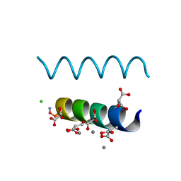
The crystal structures of the calcium-bound con-G and con-T(K7gamma) dimeric peptides demonstrate a novel metal-dependent helix-forming motif
Descriptor: CALCIUM ION, CHLORIDE ION, Conantokin-G
Authors:Cnudde, S.E, Prorok, M, Dai, Q, Castellino, F.J, Geiger, J.H.
Deposit date:2006-05-13
Release date:2007-04-24
Last modified:2024-04-03
Method:X-RAY DIFFRACTION (1.25 Å)
Cite:The crystal structures of the calcium-bound con-G and con-T[K7gamma] dimeric peptides demonstrate a metal-dependent helix-forming motif
J.Am.Chem.Soc., 129, 2007
2DPR
DownloadVisualize
BU of 2dpr by Molmil
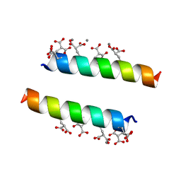
The crystal structures of the calcium-bound con-G and con-T(K7Glu) dimeric peptides demonstrate a novel metal-dependent helix-forming motif
Descriptor: CALCIUM ION, Conantokin-T
Authors:Cnudde, S.E, Prorok, M, Dai, Q, Castellino, F.J, Geiger, J.H.
Deposit date:2006-05-13
Release date:2007-04-24
Last modified:2024-04-03
Method:X-RAY DIFFRACTION (1.7 Å)
Cite:The crystal structures of the calcium-bound con-G and con-T[K7gamma] dimeric peptides demonstrate a metal-dependent helix-forming motif
J.Am.Chem.Soc., 129, 2007
4EFG
DownloadVisualize
BU of 4efg by Molmil
Crystal Structure of the Q108K:K40L:T51V:T53C:Y19W:R58W:T29L Mutant of Cellular Retinol Binding Protein Type II in Complex with All-trans-Retinal at 1.58 Angstrom Resolution
Descriptor: ACETATE ION, RETINAL, Retinol-binding protein 2
Authors:Nossoni, Z, Geiger, J.H.
Deposit date:2012-03-29
Release date:2012-12-26
Last modified:2023-09-13
Method:X-RAY DIFFRACTION (1.58 Å)
Cite:Tuning the electronic absorption of protein-embedded all-trans-retinal.
Science, 338, 2012
4EXZ
DownloadVisualize
BU of 4exz by Molmil
Crystal Structure of the Q108K:K40L Mutant of Cellular Retinol Binding Protein Type II in Complex with All-trans-Retinal at 1.7 Angstrom Resolution
Descriptor: ACETATE ION, RETINAL, Retinol-binding protein 2
Authors:Nossoni, Z, Geiger, J.H.
Deposit date:2012-05-01
Release date:2012-12-26
Last modified:2023-09-13
Method:X-RAY DIFFRACTION (1.608 Å)
Cite:Tuning the electronic absorption of protein-embedded all-trans-retinal.
Science, 338, 2012
4EDE
DownloadVisualize
BU of 4ede by Molmil
Crystal Structure of the Q108K:K40L:T51V:T53C:Y19W:R58W:T29L:A33W Mutant of Cellular Retinol Binding Protein Type II in Complex with All-trans-Retinal at 1.4 Angstrom Resolution
Descriptor: ACETATE ION, RETINAL, Retinol-binding protein 2
Authors:Nossoni, Z, Geiger, J.H.
Deposit date:2012-03-27
Release date:2012-12-26
Last modified:2023-09-13
Method:X-RAY DIFFRACTION (1.4 Å)
Cite:Tuning the electronic absorption of protein-embedded all-trans-retinal.
Science, 338, 2012

219869

数据于2024-05-15公开中

PDB statisticsPDBj update infoContact PDBjnumon