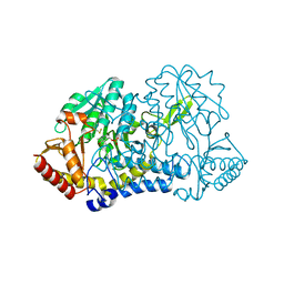
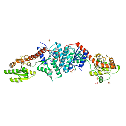
7RUJ
 
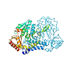 | | E. coli cysteine desulfurase SufS N99A | | 分子名称: | CHLORIDE ION, Cysteine desulfurase, PYRIDOXAL-5'-PHOSPHATE | | 著者 | Dunkle, J.A, Gogar, R, Frantom, P.A. | | 登録日 | 2021-08-17 | | 公開日 | 2023-01-25 | | 最終更新日 | 2023-10-25 | | 実験手法 | X-RAY DIFFRACTION (2.5 Å) | | 主引用文献 | The beta-latch structural element of the SufS cysteine desulfurase mediates active site accessibility and SufE transpersulfurase positioning.
J.Biol.Chem., 299, 2023
|
|
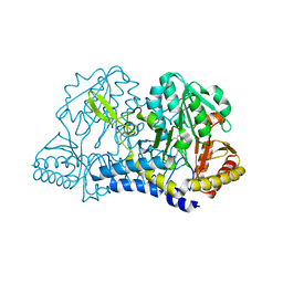
7RW3
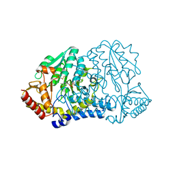 | | E. coli cysteine desulfurase SufS N99D | | 分子名称: | Cysteine desulfurase, PYRIDOXAL-5'-PHOSPHATE | | 著者 | Dunkle, J.A, Gogar, R, Frantom, P.A. | | 登録日 | 2021-08-19 | | 公開日 | 2023-01-25 | | 最終更新日 | 2023-11-15 | | 実験手法 | X-RAY DIFFRACTION (2.3 Å) | | 主引用文献 | The beta-latch structural element of the SufS cysteine desulfurase mediates active site accessibility and SufE transpersulfurase positioning.
J.Biol.Chem., 299, 2023
|
|
7RRN
 | |
6UY5
 | |
6MRH
 | |
6MRE
 | |
6MRI
 | |
6MR2
 | |
6MR6
 | |
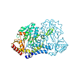
6O12
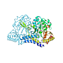
 | | E. coli cysteine desulfurase SufS H123A | | 分子名称: | CHLORIDE ION, Cysteine desulfurase, PYRIDOXAL-5'-PHOSPHATE | | 著者 | Dunkle, J.A, Frantom, P.A. | | 登録日 | 2019-02-17 | | 公開日 | 2019-06-26 | | 最終更新日 | 2023-10-11 | | 実験手法 | X-RAY DIFFRACTION (2.05 Å) | | 主引用文献 | Direct observation of intermediates in the SufS cysteine desulfurase reaction reveals functional roles of conserved active-site residues.
J.Biol.Chem., 294, 2019
|
|
6O13
 | |
6O11
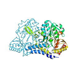 | |
6O10
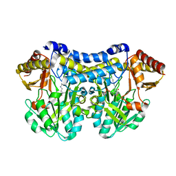
 | | E. coli cysteine desulfurase SufS | | 分子名称: | CHLORIDE ION, Cysteine desulfurase, PYRIDOXAL-5'-PHOSPHATE | | 著者 | Dunkle, J.A, Frantom, P.A. | | 登録日 | 2019-02-17 | | 公開日 | 2019-06-26 | | 最終更新日 | 2023-10-11 | | 実験手法 | X-RAY DIFFRACTION (2 Å) | | 主引用文献 | Direct observation of intermediates in the SufS cysteine desulfurase reaction reveals functional roles of conserved active-site residues.
J.Biol.Chem., 294, 2019
|
|
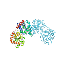
3C4Q
 | | Structure of the retaining glycosyltransferase MshA : The first step in mycothiol biosynthesis. Organism : Corynebacterium glutamicum- Complex with UDP | | 分子名称: | MAGNESIUM ION, Predicted glycosyltransferases, SULFATE ION, ... | | 著者 | Vetting, M.W, Frantom, P.A, Blanchard, J.S. | | 登録日 | 2008-01-30 | | 公開日 | 2008-04-01 | | 最終更新日 | 2023-08-30 | | 実験手法 | X-RAY DIFFRACTION (2.8 Å) | | 主引用文献 | Structural and Enzymatic Analysis of MshA from Corynebacterium glutamicum: SUBSTRATE-ASSISTED CATALYSIS
J.Biol.Chem., 283, 2008
|
|
3C48
 | |
3C4V
 | | Structure of the retaining glycosyltransferase MshA:The first step in mycothiol biosynthesis. Organism: Corynebacterium glutamicum : Complex with UDP and 1L-INS-1-P. | | 分子名称: | L-MYO-INOSITOL-1-PHOSPHATE, MAGNESIUM ION, Predicted glycosyltransferases, ... | | 著者 | Vetting, M.W, Frantom, P.A, Blanchard, J.S. | | 登録日 | 2008-01-30 | | 公開日 | 2008-04-01 | | 最終更新日 | 2023-08-30 | | 実験手法 | X-RAY DIFFRACTION (2.6 Å) | | 主引用文献 | Structural and Enzymatic Analysis of MshA from Corynebacterium glutamicum: SUBSTRATE-ASSISTED CATALYSIS
J.Biol.Chem., 283, 2008
|
|


