5L8W
 
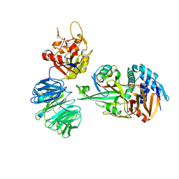 | | Structure of USP12-UB-PRG/UAF1 | | 分子名称: | GLYCEROL, Polyubiquitin-B, Ubiquitin carboxyl-terminal hydrolase 12, ... | | 著者 | Dharadhar, S, Sixma, T. | | 登録日 | 2016-06-08 | | 公開日 | 2016-09-28 | | 最終更新日 | 2016-11-30 | | 実験手法 | X-RAY DIFFRACTION (2.79 Å) | | 主引用文献 | A conserved two-step binding for the UAF1 regulator to the USP12 deubiquitinating enzyme.
J.Struct.Biol., 196, 2016
|
|
5L8E
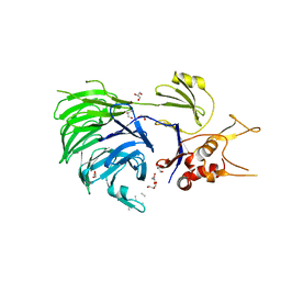 | | Structure of UAF1 | | 分子名称: | GLYCEROL, Unknown, WD repeat-containing protein 48 | | 著者 | Dharadhar, S, Sixma, T. | | 登録日 | 2016-06-07 | | 公開日 | 2016-09-28 | | 最終更新日 | 2024-01-10 | | 実験手法 | X-RAY DIFFRACTION (2.3 Å) | | 主引用文献 | A conserved two-step binding for the UAF1 regulator to the USP12 deubiquitinating enzyme.
J.Struct.Biol., 196, 2016
|
|
6I5F
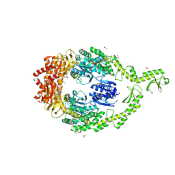 | | Crystal structure of DNA-free E.coli MutS P839E dimer mutant | | 分子名称: | ADENOSINE-5'-DIPHOSPHATE, DNA mismatch repair protein MutS, GLYCEROL, ... | | 著者 | Bhairosing-Kok, D, Groothuizen, F.S, Fish, A, Dharadhar, S, Winterwerp, H.H.K, Sixma, T.K. | | 登録日 | 2018-11-13 | | 公開日 | 2019-08-14 | | 最終更新日 | 2024-01-24 | | 実験手法 | X-RAY DIFFRACTION (2.6 Å) | | 主引用文献 | Sharp kinking of a coiled-coil in MutS allows DNA binding and release.
Nucleic Acids Res., 47, 2019
|
|
5L8H
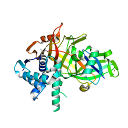 | | Structure of USP46-UbVME | | 分子名称: | Polyubiquitin-B, Ubiquitin carboxyl-terminal hydrolase 46, ZINC ION | | 著者 | Clerici, M, Sixma, T, Dharadhar, S. | | 登録日 | 2016-06-07 | | 公開日 | 2016-09-28 | | 最終更新日 | 2024-01-10 | | 実験手法 | X-RAY DIFFRACTION (1.85 Å) | | 主引用文献 | A conserved two-step binding for the UAF1 regulator to the USP12 deubiquitinating enzyme.
J.Struct.Biol., 196, 2016
|
|


